Potential of Sulforaphane to Ameliorate the Deleterious Effects of Monosodium Glutamate on Spermatic Vitality and the Histological Architecture of Seminiferous Tubules Antioxidant Mediated Pathway
Sulforaphane (Sf) is found abundantly in raw vegetables with a potent antioxidant potential. Several studies had reported the harmful effects of monosodium glutamate (MG) on the histological structure of testis and semen quality. This study was established to study the potential of Sf to protect the spermatic vitality and the histological architecture of seminiferous tubules from the deleterious effects of MG. Forty albino rats were used and were separated into 4 groups that received Sf and MG supplementations. Sf was found to ameliorate the deleterious action of Sf on spermatic vitality and motility. Sf showed its potential to restore the sex hormones levels and to enhance antioxidant markers, in addition, to preserve the histological architecture of the testis after administration of MG. In conclusion, Sf plays a crucial role in protecting the testicles on a functional and histological dimension.
Introduction
Sulforaphane (Sf) is a natural phytocompound found abundantly in raw cabbages, kale, and broccoli [1]. It has an anti-cancerous, anti-diabetic, anti-apoptotic activities in addition to an anti-inflammatory potential [2]. It can activate the nuclear factor erythroid 2–related factor 2 (Nrf2) pathway and inhibit nuclear factor kappa-light-chain- enhancer of activated B cells (NF-κB) [3]. Several studies had indicated its effect in lowering high blood pressure. Moreover, it had been documented that Sf has an epigenetic potential by inhibiting histone deacetylase (HDAC) and deoxyribonucleic acid methyltransferases and can modify mitochondrial dynamics. Sf activates proteasome preserving proteome homeostasis protecting the cell from degeneration [4]. Monosodium glutamate (MG) is widely used to increase the taste and life span of preserved food [5]. It is considered safe by many foods’ safety agencies [6]. On the contrary, many other studies had reported a harmful effects of ingesting large amounts of MG such as neurotoxicity and hepatotoxicity [7]. Nosseir, et al. [8] had reported the deleterious effects of MG on the histological architecture of testis and semen quality. Reproductive dysfunction is often associated with abnormality of the histological architecture of reproductive tissue that may lead to infertility. It may be caused by poor nutrition, drugs, or the ingestion of toxic compounds. MG has a well-known deleterious effect on the testis and sperms. It affects sperm concentration, % of sperm vitality, progressive motility, total motility, and sperm velocity [9]. Therefore, this study was established to study the potential of Sf to protect the spermatic vitality and the histological architecture of seminiferous tubules from the deleterious effects after MG administration.
Materials and Methods
Animals and Experimental Design
Forty Wistar albino rats aged 8 weeks (average weight 250 g) were recruited and housed individually in metal cages at a temperature of 25o C with a 12 hours light: 12 hours dark cycles. Rats had ad libitum access to water and chow. The study protocol was performed in compliance with the ARRIVE guidelines (Animal Research: Reporting of In Vivo Experiments). Rats were classified into 4 groups (n=10). Control group (C-group) received 0.5 ml of 0.9 NaCl by intraperitoneal injection (i.p.) (for 4 weeks). Sulforaphane group (Sf-group) received 25 mg/kg b.w. of sulforaphane dissolved in 0.9 NaCl (i.p.) (for 4 weeks) [10]. Monosodium glutamate group (MG-group) received monosodium glutamate 6 mg/kg b.w. dissolved in distilled water by oral gavage (at the 2nd and 3rd weeks) [11]. Sulforaphane + monosodium glutamate group (Sf+ MG-group) received sulforaphane 25 mg/kg b.w. (for 4 weeks) in addition to monosodium glutamate 6 mg/kg b.w. (at the 2nd and 3rd weeks) as previously described. Blood samples were collected from the tail vein by the end of the fourth week. Samples were then centrifuged for 15 minutes at a speed of 3X1000 revolutions/minute. Isolated serum was kept at -20 °C for subsequent analysis. Rats were then scarified by i.p. injection of sodium pentobarbital (single dose, 60 mg/kg b.w.). Testes and epididymides were dissected. Testes were stored in 10% formalin as shown in Figure 1.
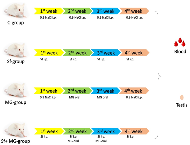
Figure 1: Experimental design. Control group (C-group) received 0.9 NaCl by intraperitoneal injection (i.p.) (for 4 weeks). Sulforaphane group (Sf-group) received sulforaphane by i.p. (for 4 weeks). Monosodium glutamate group (MG-group) received monosodium glutamate by oral gavage (at the 2nd and 3rd weeks). Sulforaphane + monosodium glutamate group (Sf+ MG- group) received sulforaphane (for 4 weeks) in addition to monosodium glutamate (at the 2nd and 3rd weeks).
Semen Analysis
Epididymis was cut into 4 pieces, and semen was mixed with four milliliters of physiological saline in a petri dish then collected using Eppendorf tube. Sperm counting was done using a cytometer. Sperm vitality and motility were assessed using SP/SFT/V-003 kit (Sperm Processor, India) as per manufacturer’s guides and CASA II Software (Hamilton ThorneTM, Spain) respectively [12].
Biochemical Analysis of Serum
Enzyme-linked immunosorbent assay (ELISA) examinations were used to estimate testosterone level (Tt) [ELISA Kit (ab108666) (Abcam, USA)] and luteinizing hormone (LH) [ELISA Kit (MBS764675) (Mybiosource, USA)] following manufacturer protocols. Lipid peroxides (LPO), nitric oxide (NO), and glutathione-S-transferase (GST) were estimated as mentioned by Ohkawa, et al. [13] and Ding, et al. [14] and Habig, et al. [15] respectively. Superoxide dismutase (SOD) and total antioxidant capacity (TAC) were estimated using commercial colorimetric kits.
Histological Examination of the Testis
Testis was cut into sections (5 μm) for hematoxylin and eosin (H&E) staining [16]. For each section, a total of ten fields were examined and scoring of the lesion was done per criteria enlisted in Table 1. Image J 1.24 software was used for field analysis. Immunohistochemistry staining was carried out in compliance with the protocol described by Goto, et al. [17]. Paraffin embedded tissue sections were deparaffinized and rehydrated. Hydrogen peroxide 3% was used to inhibit endogenous peroxidase. A two hundred microliter (200 μl) of the diluted primary antibodies [anti-caspase-3, anti- glutathione reductase (GR), and anti-superoxide dismutase 2 (SOD2)] were mounted to the tissue overnight then washed for 3 minutes by wash buffer. Slides were then stained with hematoxylin and examined by a histopathologists using the Image J 1.24 v. Software for image analysis.Ten fields per section were analyzed .
| Score | Severity |
|---|---|
| 0 | No lesion |
| 1 | Mild lesion |
| 2 | Moderate lesion |
| 3 | Sever lesion |
Table 1: Scoring criteria.
Statistical Analysis
Data analysis was done using the SPSS software, 20 V (Statistical Package for Social Sciences, SPSS Inc., USA). Group comparison was done by the the Post hoc Tukey-Kramer test. The one-way analysis of variance (ANOVA) test was used to evaluate the statistical significance of differences between groups. Data were presented in mean ± standard deviation. The probability (p) value was considered significant if <0.05.
Results
Effects of Sulforaphane and Monosodium Glutamate Supplements on Semen Parameters
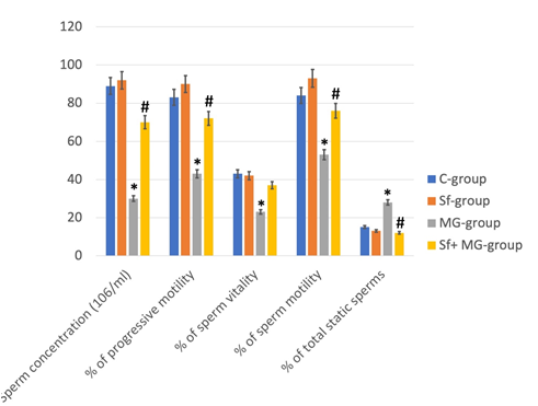
MG-group had a significant decrease of sperm concentration, percentage of sperm vitability, progressive motility, whole motility, and velocity beside a significant increase in the percentage of static sperms in comparison to C-group. The result of semen analysis of Sf-group and C-group was comparable. Ther was a significant improvement in these parameters in the Sf+MG-group compared to the MG- group (Figure 2).
Effects of Sulforaphane and Monosodium Glutamate Supplements on Serum Biochemical Analysis
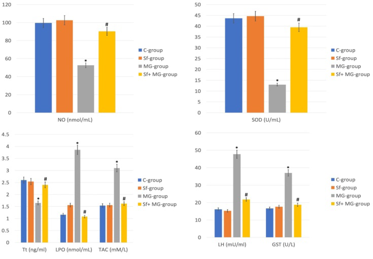
MG-group revealed a significant decrease of NO, SOD, and Tt levels beside a significant increase in the levels of LPO, TAC, LH, and GST levels in comaprison to C-group. The result of serum biochemical analysis of Sf-group and C-group was comparable. On the other hand, ther was a significant increase in NO, SOD, and Tt with a significant decrease in LPO, TAC, LH, and GST levels among the Sf+MG-group (Figure 3).
Effects of Sulforaphane and Monosodium Glutamate Supplements on the Histological Architecture of Testis
C-group showed normal testicular histological structure. Their seminiferous tubules appeared with a rounded or oval outlines with regular continuous basement membrane and lined with healthy spermatogenic cells and separated by interstitial cells of Leydig. Sf-group showed normal histological architecture of the testis as well. MG-group showed degeneration of seminiferous tubules which appeared associated with an irregular discontinuous basement membrane.
Sulforaphane Ameliorates Deleterious Effects of Monosodium Glutamate on Seminiferous Tubules
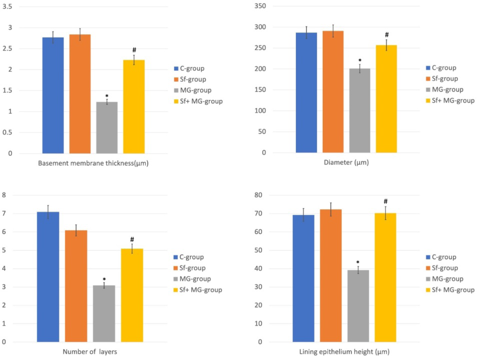
Several congested blood vessels appeared in the connective tissue separating between the tubules. In addition, seminiferous tubules parameters (basement membrane thickness, diameter, number of lining epithelium layers, lining epithelium height) showed a significant decrease relative to C-group. Sf+MG-group showed a regain of the normal histological structure of the testis and seminiferous tubules parameters comparable to that of C-group (Figures 4 & 5).
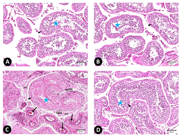
Figure 4: (A-D) photomicrograph of testis sections stained with H&E (X 400) (n = 10). (A) C group, (B) Sf-group, (C) MG- group, and (D) Sf+MG-group (Note: seminiferous tubules: star, spermatogenic cells: arrowhead, congested blood vessels: arrow, hyalinized interstitial tissue: hit, irregular and discontinuation of basement membrane: multiple arrow heads). (E) Histopathological soring. * Significant (p < 0.05) difference in comparison to C-group. # Significant (p < 0.05) difference in comparison to MG-group. Data are presented as mean ± SD, (n=10).
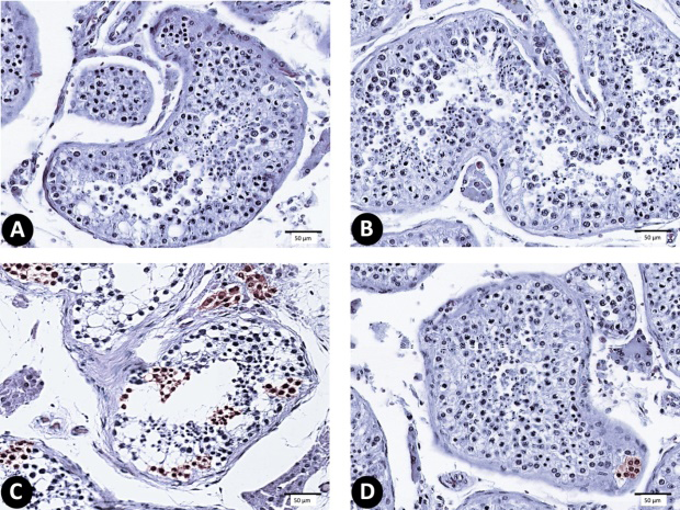
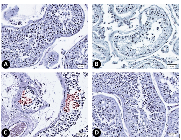
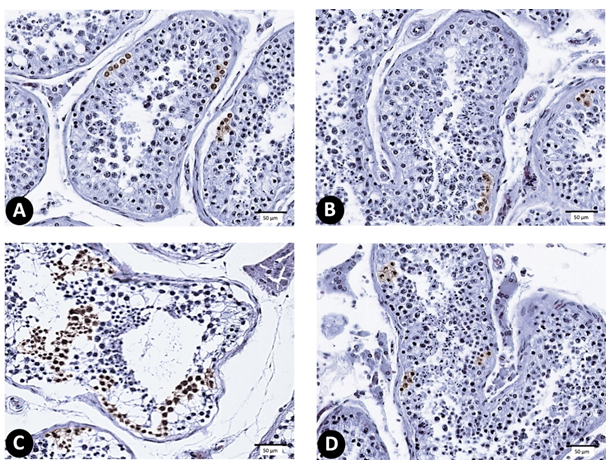
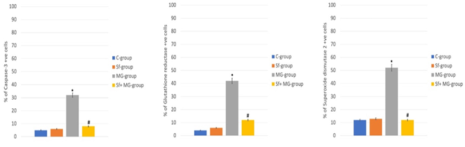
Effects of Sulforaphane and Monosodium Glutamate Supplements on the Immunoreactivity of Testis
MG-group showed a strong immunoreactivity after staining with anti-caspase-3, anti-glutathione reductase, and anti-superoxide dismutase 2 in comparison to C-group. It showed a significant increase of immuno +ve cells compared to C-group. Sf+MG-group showed a regain of immunoreactivity intensity relative to C-group and Sf-group. Moreover, there was a significant decrease of immuno +ve cells compared to MG-group (Figures 6-9).
Discussion
Sf is known for its potential preserving proteome homeostasis protecting the cell from degeneration. Several studies had reported the harmful effects of ingestion large amounts of MG such as neurotoxicity and hepatotoxicity in addition to its deleterious effects on the histological architecture and semen quality. Reproductive dysfunction may be caused by poor nutrition, drugs, or ingestion of toxic compounds such as MG. Therefore, this study was established to study the potential of Sf to protect the spermatic vitality and the histological architecture of seminiferous tubules from the deleterious effects after MG administration. MG- group revealed a significant decrease in sperm concentration, percentage of sperm viability, progressive motility, whole motility, and sperm velocity beside a significant increase in the percentage of total static sperms in comparison to C-group, this may be due to the decreased NO levels (shown in the results of biochemical analysis of MG-group’s serum), this is in accordance with Buzadzic, et al. [18]. The decrease Tt formation by Leydig cells could attribute to the spermatic parameters’ disruptions [19]. Sf+MG-group showed a significant increase of sperm concentration, percentage of sperm viability, progressive motility, total motility, and sperm velocity beside a significant decrease in the percentage of total static sperms relative to MG-group.
Similar findings were reported by Yang, et al. [20] due to increased Tt levels after Sf administration. In the current study, MG-group showed a significant reduction in Tt levels in addition to a significant increase of LH levels relative to C-group. In a similar study, Kianifard, et al. [21] reported the potential of MG to affect the factors regulating the pituitary- gonadal axis. Luo et al. [22] attributed the decreased Tt levels to the increased apoptotic activity of Leydig cells which was shown in the current study histological results. The decreased Tt levels may trigger a negative feedback mechanism causing the elevated LH levels [19]. Sf+MG-group showed a significant increase of Tt levels plus a significant decrease of LH levels compared to MG-group which denotes the potential of Sf to enhance testicular activities [20]. MG- group showed a significant decrease in NO and SOD levels relative to C-group. As reported by Bergh, et al. [23], the reduction in NO levels may cause histological disruption of the testis due to its effect on the blood supply of gonads. Fukai et al. reported that NO is essential for SOD upregulation [24] which may explain their concomitant decrease. Sf+MG-group revealed a significant increase of NO and SOD levels relative to MG-group which may improve mitochondrial functions [25].
MG administration was associated with a significant rise in LPO, TAC, and GST levels in comparison to control group. Similar findings were also reported by Kianifard, et al. [21]. Singh, et al. [26] reported the potential of MG to stimulate xanthine oxidase causing an extra formation of free radicles. Murphy, et al. [27] reported the potential of MG to reduce intracellular content of glutathione causing an extra formation of free radicles as well. The elevated TAC in serum denotes the compensation mechanisms of the tissue to overcome the oxidants/antioxidants disruption which was also mentioned by Ibegbulem, et al. [28]. Sf+MG-group revealed a significant improvement in LPO, TAC, LH, and GST levels relative to MG-group. As reported, the increased GST levels denote the enhancement of the body’s detoxification process against harmful compounds [27]. MG-group showed degeneration of seminiferous tubules due to accumulation of oxidants causing mitochondrial dysfunction [29] which appeared associated with an irregular discontinuous basement membrane. Several congested blood vessels appeared in the connective tissue separating between tubules which come in agreement with Sarhan [19].
In addition, seminiferous tubules parameters (Basement membrane thickness, diameter, number of lining epithelium layers, lining epithelium height) were significantlly decreased compared to C-group which is similar to previous findings by Sakr, et al. [30]. This may be due to the disruption of NO levels noticed in biochemical results. Sf+MG-group showed a regain of the normal histological structure of the testis and their seminiferous tubules which may be due to the antioxidant activity of Sf. In the current study, MG-group showed a strong immunoreactivity after staining with anti- caspase-3 which comes in agreement with Sarhan [19], anti-glutathione reductase, and anti-superoxide dismutase 2 compared to C-group. MG caused induction of oxidative stress pathway leading to the activation of apoptotic pathway [31]. The increase in glutathione reductase and superoxide dismutase 2 immuno +ve cells may be considered as the defensive mechanism of the tissue against free radicles.
Sf+MG-group showed a regain of immunoreactivity intensity relative to C-group and Sf-group beside a significant decrease of immuno +ve cells compared to MG-group. The current study used certain doses of supplements, further studies with different dosages are needed to determine the best sulforaphane dose that could protect against monosodium glutamate on seminiferous tubules. More studies are also needed to clarify the protective effect of sulforaphane against monosodium glutamate on different parts of male internal genital organs are also needed. Further studies to identify the exact molecular basis of the proven protection will enrich the scientific literature as well. In conclusion, the current study illustrated the possible protective effectl of sulforaphane against the deleterious effects of monosodium glutamate on spermatic vitality and motility. Sulforaphane showed its potential to restore the sex hormones and to enhance antioxidant markers, it also preserved the histological architecture of the testis after administration of monosodium glutamate.
Statement of Ethics
The current study protocol was approved by Research and Ethics Committee, College of Medicine, Tanta University, Egypt.
Conflict of Interest
The authors has no conflict of interst to be declared.
Funding
The authores declared that they didnot recieve any sort of fund for the current study.
Data Availability
The datasets generated in the current study are available upon a reasonable request.
References
-
Yanaka A, Fahey JW, Fukumoto A, Nakayama M, Inoue S, et al. (2009) Dietary sulforaphane-rich broccoli sprouts reduce colonization and attenuate gastritis in Helicobacter pylori infected mice and humans. Cancer prevention research 2(4): 353-360.
-
Schepici G, Bramanti P, Mazzon E (2020) Efficacy of Sulforaphane in Neurodegenerative Diseases. International journal of molecular sciences 21(22): 8637.
-
Wang Y, Mandal AK, Son YO, Pratheeshkumar P, Wise J, et al. (2018) Roles of ROS Nrf2 and autophagy in cadmium carcinogenesis and its prevention by sulforaphane. Toxicology and applied pharmacology 353: 23-30.
-
Tortorella SM, Royce SG, Licciardi PV, Karagiannis TC (2015) Dietary Sulforaphane in Cancer Chemoprevention The Role of Epigenetic Regulation and HDAC Inhibition. Antioxidants redox signaling 22(16): 1382-1424.
-
Rahimi AF, Baradaran R, Ghandy N, Jalali M, Reza NM, et al. (2019) Effects of monosodium glutamate on apoptosis of germ cells in testicular tissue of adult rat An experimental study. International journal of reproductive biomedicine 17(4): 261-270.
-
Zanfirescu A, Ungurianu A, Tsatsakis AM, Nițulescu GM, Kouretas D, et al. (2019) A review of the alleged health hazards of monosodium glutamate. Comprehensive reviews in food science and food safety 18(4): 1111- 1134.
-
Jubaidi FF, Mathialagan RD, Noor MM, Taib IS, Budin SB (2019) Monosodium glutamate daily oral supplementation: study of its effects on male reproductive system on rat model. Systems biology in reproductive medicine 65(3): 194-204.
-
Nosseir N, Ali M, Ebaid H (2012) A Histological and Morphometric Study of Monosodium Glutamate Toxic Effect on Testicular Structure and Potentiality of Recovery in Adult Albino Rats. Research Journal of Biology 2: 66-78.
-
Kayode OT, Rotimi DE, Kayode A, Olaolu TD, Adeyemi OS (2020) Monosodium Glutamate MSG Induced Male Reproductive Dysfunction A Mini Review. Toxics 8(1): 7.
-
Malaguti M, Angeloni C, Garatachea N, Baldini M, Leoncini E, et al. (2009) Sulforaphane treatment protects skeletal muscle against damage induced by exhaustive exercise in rats. Journal of applied physiology 107(4): 1028-1036.
-
El MEK, A., Osman HEH, Daghestani MH (2013) The effect of vitamin C administration on monosodium glutamate induced liver injury. An experimental study. Experimental and toxicologic pathology 65(5): 513-521.
-
Soleimanzadeh A, Saberivand A (2013) Effect of curcumin on rat sperm morphology after the freeze thawing process. Veterinary research forum 4(3): 185- 189.
-
Ohkawa H, Ohishi N, Yagi K (1979) Assay for lipid peroxides in animal tissues by thiobarbituric acid reaction. Analytical biochemistry 95(2): 351-358.
-
Ding AH, Nathan CF, Stuehr DJ (1988) Release of reactive nitrogen intermediates and reactive oxygen intermediates from mouse peritoneal macrophages Comparison of activating cytokines and evidence for independent production. Journal of immunology 141(7): 2407-2412.
-
Habig WH, Pabst MJ, Jakoby WB (1974) Glutathione S transferases The first enzymatic step in mercapturic acid formation. The Journal of biological chemistry 249(22): 7130-7139.
-
Ahmed AS, Hantash EM, Zakaria SS (2021) Potential of Amantadine to Ameliorate Glutamate Induced Pyramidal Cells Toxicity in Juvenile Rat’ Brain Cortex. Neurotoxicity research 39(4): 1203-1210.
-
Goto S, Morigaki R, Okita S, Nagahiro S, Kaji R (2015) Development of a highly sensitive immunohistochemical method to detect neurochemical molecules in formalin fixed and paraffin embedded tissues from autopsied human brains. Frontiers in neuroanatomy 9: 22.
-
Buzadzic B, Vucetic M, Jankovic A, Stancic A, Korac A, et al. (2015) New insights into male in fertility the importance of NO. British journal of pharmacology 172(6): 1455- 1467.
-
Sarhan NR (2018) The Ameliorating Effect of Sodium Selenite on the Histological Changes and Expression of Caspase-3 in the Testis of Monosodium Glutamate- Treated Rats Light and Electron Microscopic Study. Journal of microscopy and ultrastructure 6(2): 105-115.
-
Yang SH, Long M, Yu LH, Li L, Li P, et al. (2016) Sulforaphane Prevents Testicular Damage in Kunming Mice Exposed to Cadmium via Activation of Nrf2/ARE Signaling Pathways. International journal of molecular sciences 17(10): 1703.
-
Kianifard D, Ehsani A, Zeinolabedini DP, Akbari G, Maysam MS (2019). Effect of monosodium glutamate on testicular tissue of paclitaxel treated mice An experimental study. International journal of reproductive biomedicine 17(11): 819-830.
-
Luo DY, Yang G, Liu JJ, Yang YR, Dong Q (2011) Effects of varicocele on testosterone apoptosis and expression of StAR mRNA in rat Leydig cells. Asian journal of andrology 13(2): 287-291.
-
Bergh A, Collin O, Lissbrant E (2001) Effects of acute graded reductions in testicular blood flow on testicular morphology in the adult rat. Biology of reproduction 64(1): 13-20.
-
Fukai T, Siegfried MR, Ushio FM, Cheng Y, Kojda G, et al. (2000) Regulation of the vascular extracellular superoxide dismutase by nitric oxide and exercise training. The Journal of clinical investigation 105(11): 1631-1639.
-
Carrasco PC, Tan KN, Borges K (2015) Sulforaphane is anticonvulsant and improves mitochondrial function. Journal of neurochemistry 135(5): 932-942.
-
Singh K, Ahluwalia P (2012) Effect of monosodium glutamate on lipid peroxidation and certain antioxidant enzymes in cardiac tissue of alcoholic adult male mice. Journal of cardiovascular disease research 3(1): 12-18.
-
Murphy TH, Miyamoto M, Sastre A, Schnaar RL, Coyle JT (1989) Glutamate toxicity in a neuronal cell line involves inhibition of cystine transport leading to oxidative stress. Neuron 2(6): 1547-1558.
-
Ibegbulem CO, Chikezie PC, Ukoha AI, Opara CN (2016) Effects of diet containing monosodium glutamate on organ weights, acute blood steroidal sex hormone levels lipid profile and erythrocyte antioxidant enzymes activities of rats. J Acute Dis 5(5): 402-407.
-
Mekkawy AM, Ahmed YH, Khalaf A, El SMA (2020) Ameliorative effect of Nigella sativa oil and vitamin C on the thyroid gland and cerebellum of adult male albino rats exposed to Monosodium glutamate histological immunohistochemical and biochemical studies. Tissue cell 66: 101391.
-
Sakr SA, Badawy GM (2013) Protective effect of curcumin on monosodium glutamate induced reproductive toxicity in male albino rats. Glob J Pharmacol 7: 416-422.
-
Ryter SW, Kim HP, Hoetzel A, Park JW, Nakahira K, et al. (2007) Mechanisms of cell death in oxidative stress. Antioxidants redox signaling 9(1): 49-89.
- Pattern of Breast Lesions in Ovu Inland, Delta State, South Southern Nigeria
- Morphometric Analysis of the Human Femur: Exploring Platymetric and Robusticity Indices Among the Nigerian Population
- Anatomical Variation of Arteria Lusoria: Clinical Implications for Dysphagia Lusoria and Surgical Risk
- Morphometric Study of the Vertebral Body and Pedicle of Typical Cervical Vertebrae Using Radiological Image
- Epigenetic Mechanisms Driving Human Evolutionary Changes
- Neuroprotective Effects of Ginkgo Biloba Extract on Bilateral Common Carotid Artery Ischaemic Stroke Induced in Wistar Rat