Neural and Arterial Variations in the Right Upper Limb of a Single Cadaver- A Case Report
Background: During routine undergraduate dissection of a 43 years old male cadaver some important neural and arterial anatomic variations were seen in a single right upper limb in the form of double medial roots of median nerve and superficial brachial artery. Observation: During routine dissection of a 43-year-old male cadaver, some important neural and arterial anatomical variations were observed in a single right upper limb in the form of double medial roots of the median nerve and superficial brachial artery. The median nerve was formed in axilla by the union of one lateral and two medial roots on lateral side of axillary artery. The brachial artery which crossed the median nerve superficially from the medial to the lateral side, in the middle of the arm, continued as the superficial brachial artery. The variations were observed and photographed. Conclusion: The knowledge of such variations is however not only desirable, but also essential as these have important influences onBackground: During routine undergraduate dissection of a 43 years old male cadaver some important neural and arterial anatomic variations were seen in a single right upper limb in the form of double medial roots of median nerve and superficial brachial artery. Observation: During routine dissection of a 43-year-old male cadaver, some important neural and arterial anatomical variations were observed in a single right upper limb in the form of double medial roots of the median nerve and superficial brachial artery. The median nerve was formed in axilla by the union of one lateral and two medial roots on lateral side of axillary artery. The brachial artery which crossed the median nerve superficially from the medial to the lateral side, in the middle of the arm, continued as the superficial brachial artery. The variations were observed and photographed. Conclusion: The knowledge of such variations is however not only desirable, but also essential as these have important influences on
Introduction
Different types of variations pertaining to nerves and arteries of upper limb have been reported by several investigators earlier, so much so that is aptly said, variations are a rule rather than exception. The brachial plexus is formed by ventral primary rami of spinal nerves C5-C8 and T1, extends downwards and laterally, passes over the first rib behind the clavicle and results in formation of trunks, divisions, cords and terminal branches [1]. The median nerve is thus formed by union of lateral and medial roots from lateral (C5,6) and medial cords (C8,T1) of brachial plexus respectively [2]. In some cases, the median nerve may be formed by three roots or the brachial artery may course superficial to the median nerve called as superficial brachial artery. Usually, these variations are seen singly but a combination of neural and arterial variations in the same upper limb is extremely rare which is being reported here [3, 4]. Though all these variations have been described in the standard text books of Anatomy, and reported in literature but their occurrence, together in one limb, have never been encountered or reported earlier and that gave us an impetus to report this case. The knowledge of such variations is however not only desirable, but also essential to anatomists, radiologists, anaesthesiologists and surgeons who may contribute to the explanation of diagnosis and surgical treatment and can prevent any postoperative complications during surgery.
Neural-Double Medial Roots of Median Nerve
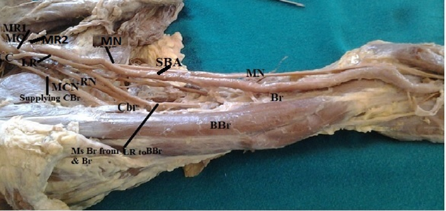
The Median nerve was formed by one lateral root (LR) of lateral cord and two medial roots (MR1 & MR2) of medial cord of brachial plexus. The lateral cord (LC) of the brachial plexus bifurcated into musculocutaneous nerve and lateral root of median nerve (MN) and the medial cord instead of bifurcation, trifurcated terminally into ulnar nerve and two medial roots of median nerve (MR1 & MR2). Thus, median nerve was formed by a lateral root coming from lateral cord and a double medial root (MR1 & MR2) of median nerve coming from the medial cord of brachial plexus. No such variation was seen in the contralateral limb. The coracobrachialis muscle of flexor compartment of the arm was being supplied by the musculocutaneous nerve and biceps brachii and brachialis were supplied by the muscular branches from the LR of median nerve (Figure 1 & Table 1).
| Case Report | |
|---|---|
| During routine undergraduate dissection conducted on the right upper limb of a 43 year old male cadaver two rare anatomic variations were encountered in the same arm in the form of: | |
| Neural-Double Medial Roots of Median Nerve | |
| Arterial-Superficial Brachial Artery | |
| Figure 1: Shows that median nerve was formed by a lateral root coming from lateral cord and two medial roots coming from the medial cord of brachial plexus in the right arm of a 43year old male cadaver. Muscles of flexor compartment of the arm are seen supplied by: cbrm- by mcn from lc of brachial plexus; bbrm & brm- by sba-superficial brachial artery. | |
| Neural – Double Medial Roots of Median Nerve | |
| Incidence | |
| Double medial root of median nerve is still rare though double lateral root has been reported in 14% of the subjects. | |
| This may be attributed to the following factors: | |
| Lack of coordination between the formation of limb muscles and their innervations [5]. | |
| Mis- expression of the signalling molecules which induce the differentiation of the dorsal and ventral motor horn cells, can lead to abnormalities in the formation and distribution of particular nerve fibres. | |
| Ontogeny | |
| Phylogeny | |
| In monotremes, marsupials, lemurs, dogs, monkeys, anthropoid apes & man brachialis longus inferior (a single nerve trunk seen in reptiles and amphibians) is divided into distinct median & ulnar nerves [6]. | |
| Evaluation of unexplained sensory/motor loss after trauma. | |
| Close proximity of the additional roots of median nerve with the axillary artery may result in its compression and reduced blood supply to the upper limb [7]. | |
| Clinical | |
| Significance | |
| Arterial- Superficial Brachial Artery | |
| Incidence | Incidence varies from 0.2-25% [8]. |
| Ontogeny | According to Baeza et al, 1995 the brachial artery has got two trunks of origin. One is passing deep to median nerve and other superficial to it. During later stages of development usually the deep artery gets hemodynamic preference leading to obliteration of superficial brachial artery. However if the artery passing superficial to median nerve gets the hemodynamic preference (as in the present case) the superficial brachial artery persists while the deep (Normal) one disappears [9]. |
| Phylogeny | Manners Smith, 1910 reported that Superficial Brachial Artery is a atavistic condition usually seen in primates [10]. |
| Clinical Significance | The superficial brachial artery as well as superficial position of ulnar and radial artery not only makes them more vulnerable to trauma and thus to bleeding but also makes them more accessible to cannulation, if needed. These arteries may also be mistaken for a vein. If certain drugs are injected into this vessel, the results may be disastrous like gangrene or loss of hand. Moreover, if the superficial brachial artery is retained, it is usually associated with a retarded development of the palmar arch. The presence of SBA may be a possible cause for idiopathic median nerve entrapment neuropathy [11]. The possibility of the superficial brachial artery’s presence should be considered during axillary cavity surgical exploration and anesthesiology procedures [12]. |
Table 1: Showing incidence, ontogeny, phylogeny and clinical significance of double medial root of median nerve.
Arterial-Superficial Brachial Artery
The Axillary artery continued as the brachial artery which crossed the median nerve superficially from the medial to the lateral side, in the middle of the arm, as the superficial brachial artery (Table 2).
Discussion
The variations related to the formation of median nerve by more than two roots are rare as revealed by survey of literatures. As depicted from (Figure 1), in the present case MN was seen formed by three roots i.e. 2 medial roots from MC of brachial plexus and one lateral root from LC of brachial plexus and along with this brachialis and biceps brachii were being supplied by the branches coming from LR of Median nerve. Sargon, et al. reported that the median nerve was formed of three branches, two originated from the lateral cord and one from the medial cord of brachial plexus [13]. Eglseder and Goldman also found that the median nerve was formed of two lateral roots in 14% of their specimens [14]. Similar findings have also been reported by Mohaptra, et al. [15]. Uzun, et al. found that the median nerve was formed by three branches coming from the lateral cord of the brachial plexus and one branch coming from the medial cord of the brachial plexus. Lalit and singla had also reported similar pattern of formation of median nerve where median nerve was formed by a lateral root coming from lateral cord and two medial roots coming from the medial cord but along with ulnar to median nerve communication christened as Piplani- Singla anastomosis [16]. No such anastomosis was observed in the present case instead brachial artery was observed as SBA Usually, the brachial artery passes deep to the median nerve from the medial to the lateral side in front of the arm. But when it crosses the median nerve superficially and replaces the main trunk, it is termed as the superficial brachial artery [17]. In the earlier literature which was related to this entity, the incidence is reported to vary between 0.2% to 22% [8]. No such variation was seen in the contra-lateral limb.
Conclusion
These findings may also be of importance in the evaluation of unexplained sensory and motor loss or vasomotor and trophic changes after trauma in that particular area. The median nerve with additional roots is more likely to be involved in entrapment syndromes and during surgical interventions. Thus a sound knowledge of such variations is essential for radiologists, surgeons, orthopedicians, interventional cardiologists and anaesthetists.
References
-
Williams PL, Bannister LH, Berry MM, Collins P, Dyson M, et al. (1995) Nervous system. Gray’s Anatomy: The anatomical basis of medicine and surgery. 38th(Edn.), Edinburgh, Churchill Livingstone, New York, pp: 1266- 1272.
-
Standring S, Borely NR, Collins P, Crossman AR, Gatzoulis MA, et al. (2008) Gray’s Anatomy: The Anatomical Basis of Clinical Practice. 40th(Edn.), Churchill Livingstone/ Elsevier, London, pp: 827-836.
-
Kerr AT (1918) The brachial plexus of nerves in man, the variations in its formation and branches. Am J Anat 23(2): 285-395.
-
Bergman RA (1988) Compendium of Human Anatomic Variation. Urban & Schwarzenberg, Baltimore, USA, pp: 139-143.
-
Sannes HD, Reh TA, Harris WA (2000) Axon growth and guidance. In: Sannes HD, et al. (Eds.), Development of nervous system. Academic Press, New York, pp: 189-197.
-
Miller RA (1934) Comparative studies on the morphology and the distribution of the brachial plexus. Am J Anat 54(1): 143-166.
-
Jurjus A, Sfeir R, Bezirdjian R (1986) Unusual variation of arterial pattern of the human upper limb. Anat Rec 215(1): 82-83.
-
Lalit M, Piplani S (2021) A Cadaveric study of Brachial Artery and its variations with its Ontogenic Basis: An Anatomical perspective. Int J Anat Res 9(1.1): 7844-7850
-
Baeza RA, Nebot J, Ferreira B, Reina F, Perez J, et al. (1995) An Anatomical study and ontogenic explanation of 23 cases with with variations in the main pattern of the human brachio-antebrachial arteries. J Anat 187 (Pt 2): 473-479
-
Manners-Smith T (1910) The limb arteries of primates. J Anat Physiol 45: 23-64.
-
Nkomozepi P, Xhakaza N, Swanepoel E (2017) Superficial brachial artery: A possible cause for idiopathic median nerve entrapment neuropathy. Folia Morphol 76(3): 527-531
-
Clarke E, Mazurek A, Radek M, Żytkowski A, Twardokęs W, et al. (2021) Superficial brachial artery – A case report with commentaries on the classification. Translational Research in Anatomy 23: 100112.
-
Sargon MF, Uslu SS, Celik HH, Aksit D (1995) A variation of median nerve at the level of brachial plexus. Bull Assoc Anat 79(246): 25-26.
-
Eglseder WA, Goldman M (1997) Anatomical variations of the musculocutaneous nerve in the arm. Am J Orthop 26(11): 777-780.
-
Mahapatra BB, Chinara PK, Dutta BK, Nayak AK (2004) Variations in the formation and branching pattern of median nerve. J Anat Soc Ind 53(1): 31-36.
-
Lalit M, Singla RK (2018) Double medial root of Median nerve and ulnar to median nerve communication in arm or Piplani Singla Communication - A Case Report. North States J anatomy 3(2): 30-34.
-
Adachi B (1928) The arterial system of the Japanese. Imperial University of Kyoto, Kyoto, Japan, pp: 205-210.
- Pattern of Breast Lesions in Ovu Inland, Delta State, South Southern Nigeria
- Morphometric Analysis of the Human Femur: Exploring Platymetric and Robusticity Indices Among the Nigerian Population
- Anatomical Variation of Arteria Lusoria: Clinical Implications for Dysphagia Lusoria and Surgical Risk
- Morphometric Study of the Vertebral Body and Pedicle of Typical Cervical Vertebrae Using Radiological Image
- Epigenetic Mechanisms Driving Human Evolutionary Changes
- Neuroprotective Effects of Ginkgo Biloba Extract on Bilateral Common Carotid Artery Ischaemic Stroke Induced in Wistar Rat