Innovative Preservation and Training Utility of Chemically Preserved Swine Viscera for Veterinary Surgical Education
The use of live animals during surgical practical classes exposes students to stressful situations that are often common in the operating room. However, this practice can generate a negative emotional state and hinder the student's cognitive mechanisms, causing different degrees of dissatisfaction and being detrimental to the learning process. Research involving alternative methods for diaeresis and synthesis training has already been carried out with promising outcomes, providing low-cost preserved anatomical pieces. Thus, the aim of the present study was to analyze the anatomical viability of chemically preserved swine viscera for operative techniques training and to evaluate the acceptance of students facing this educational approach, using viscera subjected to plastic bag vacuum conditioning in two different ways of storage: room temperature (20 to 25ºC) and refrigerated (0 to 6ºC). Thirty-six jejunum segments and 36 urinary bladders from swine were fixed in absolute ethanol (AE) for 30 days and subsequently preserved in sodium chloride hypersaturated solution (SCHS) for seven days. At the end of the fixation and preservation periods, the samples were placed in vacuum plastic bags and divided into two groups containing 18 urinary bladders and 18 jejunum segments each. After 60 days of storage, the samples were used for surgical technique classes. The results showed good acceptance by the participating students; the technique proved to be safe, has low cost, and has an easy implementation in terms of both preservation solutions and storage.
Abbreviations
AE: Absolute Ethanol; SCHS: Sodium Chloride Hypersaturated Solution; RT: Room Temperature; USP: University of São Paulo.
Introduction
Surgical practical classes with live animals aim to expose the student to stressful situations. They are in direct contact with the patient and exposed to organs, hemorrhages, and distinct technical procedure difficulties. This teaching method was used for a long time in many universities worldwide. However, this practice can generate a negative emotional state and hinder the student’s cognitive mechanisms, causing different degrees of dissatisfaction and being detrimental to the learning process [1]. These methods can also be considered unhealthy due to the risk of infections involving biological materials, such as secretions and/or blood. In addition, they can lead to accidents related to inexperience or even recklessness of the student regarding the inappropriate use of sharp objects like needles and scalpels [2, 3]. Currently, with the intellectual and technological advances in the development and application of alternative methods to vivisection, educational institutions have been able to adapt to the principles of ethics and animal welfare [4, 5, 6]. According to the Normative Resolution No. 17 of July 3, 2014, from CONCEA (National Council for Animal Experimentation Control, Brazil) [7], an alternative method is understood as any method that can be used to replace, reduce or refine the use of animals in research activities and by substitutive methods, educational resources or educational approaches that replace the use of animals or complement humanitarian teaching practices. In Veterinary Medicine, substitutive methods such as videos, anatomical models, computer programs, prototyping, and carcass preservation using formaldehyde, freezing, glycerin, and the Laskowski technique, among others, are often employed [8]. Teaching methods using cadavers for surgical practices can improve learning and provide exercise repetition, increasing students’ confidence and satisfaction when compared to the use of live animals [6]. Several authors have demonstrated the effectiveness of using chemically prepared cadavers for Surgery and Anatomy classes [9, 10, 11, 12, 13, 14, 15]. Research involving different animals fixed in absolute ethanol and preserved in a sodium chloride aqueous solution as an alternative method for diaeresis and synthesis training has already been carried out with promising outcomes, providing low- cost preserved anatomical pieces that keep satisfactory organoleptic morphological characteristics for surgical training, such as good malleability and resistance to incisions and sutures [16, 17, 18, 19]. Furthermore, Rocha [17] observed that 81.08% of the students are favorable to initial training in surgical practices using chemically preserved cadavers.
Currently, research using plastic bag vacuum packaging for preserving cadavers after fixation has shown good results in inhibiting microbial growth and maintaining the biomechanical properties of the anatomical parts [19, 20].
The preservative effect of vacuum packaging is due to the creation of an anaerobic environment, which inhibits the growth of microorganisms responsible for organic materials deterioration, especially those of animal origin [21, 22]. Thus, the aim of this study was to analyze the anatomical viability of chemically preserved swine viscera (urinary bladder and jejunum segments) subjected to plastic bag vacuum conditioning in two different ways of storage (room temperature and refrigerated) for operative techniques training and to evaluate the acceptance of students facing this educational approach.
Materials and Methods
This study was approved by the University of São Paulo Committee on Ethics in the Use of Animals (CEUA nº 9148181220) and by the Committee on Ethics in Research on Human Beings (CAAE 52551121.80000.5422) of the same institution. Thirty-six jejunum segments (20 cm in length) and 36 urinary bladders from swine were used. The anatomical pieces were obtained at the slaughterhouse school of the Faculty of Animal Science and Food Engineering (Faculdade de Zootecnia e Engenharia de Alimentos - FZEA) Campus “Fernando Costa” of the University of São Paulo. The viscera were fixed in absolute ethanol (AE, 99.8 ºGL) for 30 days and preserved in sodium chloride hypersaturated solution (SCHS, 30%) for 7 days. The parts were prepared as described by Guaraná [18], and the preservation time was based on the same study, which showed that swine viscera preserved using this methodology present favorable physical and histomorphological conditions and are efficient for training the operative technique with one week of conservation in SCHS.
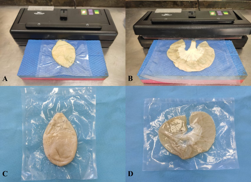
At the end of the fixation and preservation periods, the urinary bladders were placed in vacuum plastic bags measuring 15 x 20 cm and 12 microns thick, and the jejunum segments were accommodated in vacuum plastic bags measuring 20 x 25 cm with the same thickness. With the aid of an EVA plate for support, the viscera were positioned and subjected to packaging using a vacuum sealer (Commercial Vacuum Sealer with Reservoir - Cetro® - Bauru/SP - Brazil) and then had their end sealed for 3 seconds using the same device (Figure 1). The packaged anatomical pieces were separated into two groups containing 18 urinary bladders and 18 jejunum segments each. The room temperature group (RT) was placed on open shelves (20 to 25°C), and the refrigerated group (R) was placed in a refrigerator (0 to 6 ºC).
After the fixation periods in AE for 30 days and conservation in SHCS for seven days, before vacuum conditioning (D0), two urinary bladders and two jejunum segments were used to perform surgical techniques (cystotomy and cystotomy closure; enterotomy and enterotomy closure; intestinal resection and anastomosis) by the researchers (AFM and JBG) to analyze the samples physical characteristics. The same techniques were performed in two urinary bladders and two jejunum segments randomly chosen from the RT and R groups after 30 (D30) and 60 (D60) days of vacuum conditioning. The samples also had its physical characteristics examined and subjectively evaluated in each period (D0, D30, and D60). The results were recorded in a report following a scale of 1 to 5, with 1 being “unusable” - major physical characteristics changes, improper material; 2 “poor” - altered physical characteristics, impaired use; 3 “regular” - minor physical characteristics changes, use slightly impaired; 4 “good” - minor physical characteristics changes, use not impaired; 5 “excellent” - preserved physical characteristics.
At the same time as the execution of the techniques, urinary bladder and small intestine fragments were randomly collected for histological analysis just before the vacuum conditioning (D0), and at 30 and 60 days of vacuum conditioning (D30 and D60), from both groups (RT and R). Morphological characteristics (integrity/preservation of the mucosa, submucosa, muscular and serous layers) were compared with fresh samples of the urinary bladder and small intestine fixed in 10% formalin (microscopic evaluation control) and between the experimental groups to assess the differences of vacuum conditioning time and between the groups’ way of storage (RT and R).
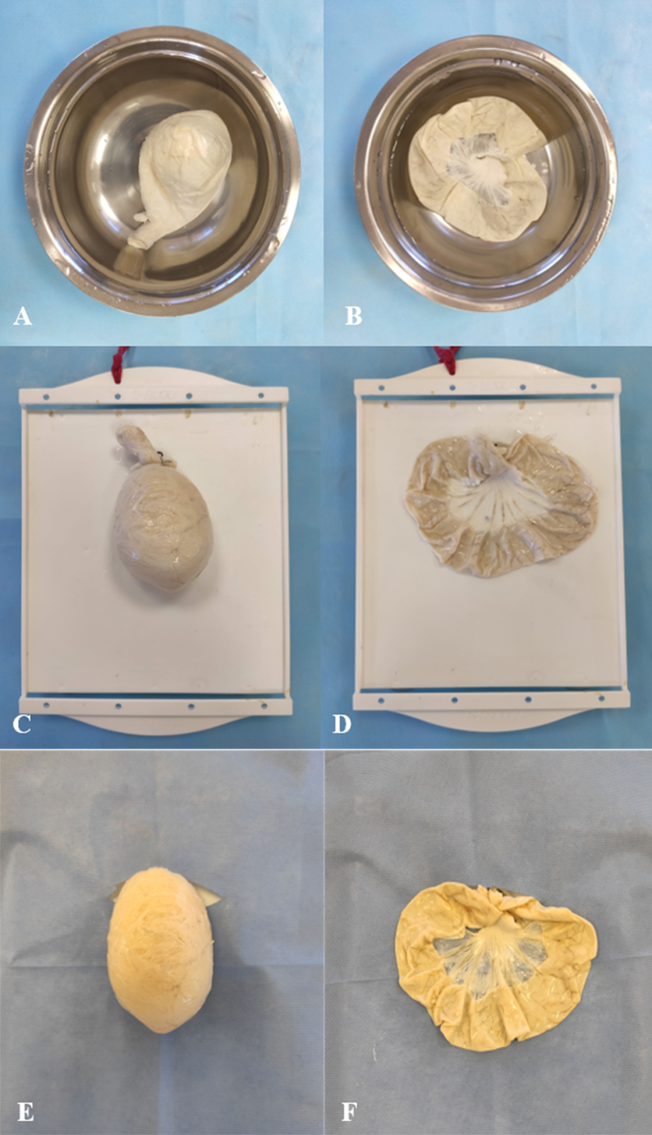
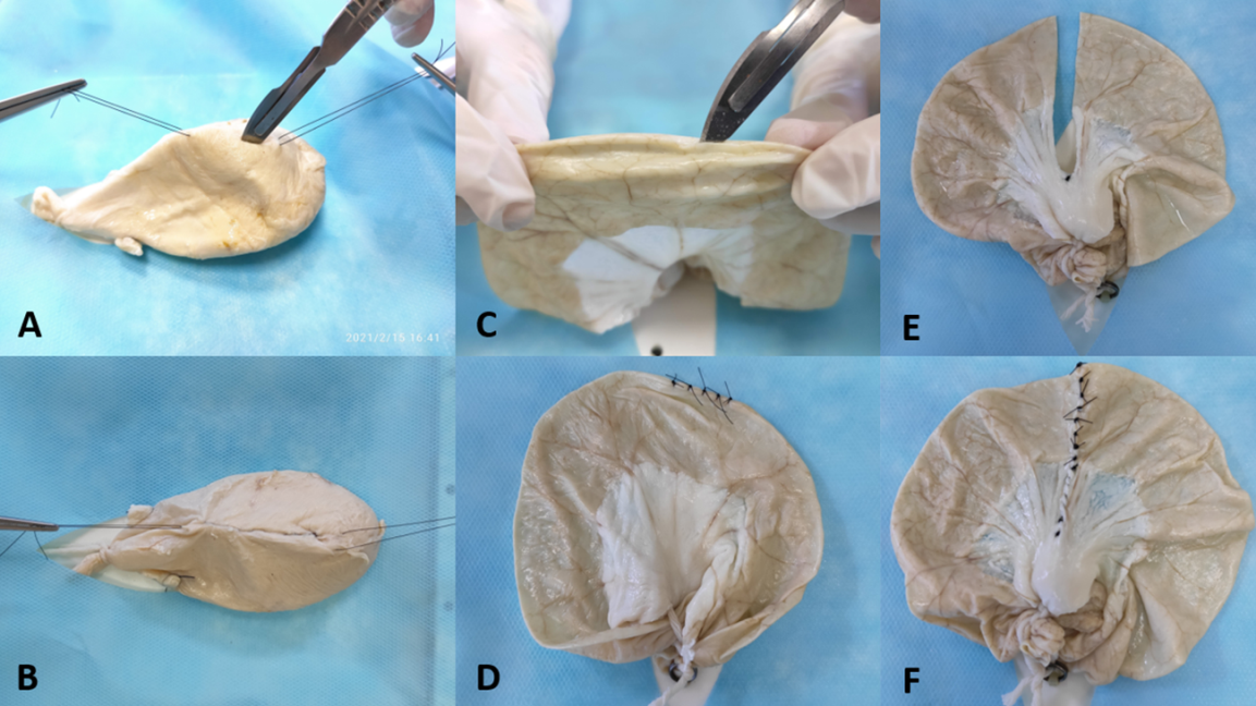
To survey the students’ impressions regarding the use of the samples (n=58), the swine’s viscera fixed for 30 days in EA, preserved in SCHS for seven days, and vacuum packed for 60 days, were removed from the packaging, placed in a container with potable water for rehydration for 20 minutes (Figures 2A and 2B), accommodated in a surgical model (Figures 2C and 2D), covered with unsterilized surgical drapes (Figures 2E and 2F) and provided to students enrolled in the Surgical Technique discipline of the Veterinary Medicine course at the Faculty of Animal Science and Food Engineering (FZEA) of the University of São Paulo (USP). The practice of the operative techniques performed (cystotomy and cystotomy closure; enterotomy and enterotomy closure; intestinal resection and anastomosis) (Figures 3A-3F) occurred simultaneously with their demonstration, through TV monitors in real-time, by the professor in charge of the discipline, as described by Fossum. Subsequently, the same students voluntarily filled out a form/questionnaire with objective and subjective questions regarding the fixed and preserved swine viscera. The first six questions were about the resistance of the viscera to the incision/suture and its malleability, in which the students should assign a score from 1 (unusable) to 10 (excellent) for each characteristic. The answers referred to the personal experience of each student and their experiences during the discipline using these and other educational approaches available: fresh pieces and synthetic suture models.
Results
The grades attributed by the evaluators (AFM and JBG) to the physical characteristics of the viscera (appearance, texture, flexibility, odor, and color) in each period in the two different ways of storage (RT and R) are shown in the following table (Table 1).
A - Urinary bladder physical evaluation (RT Group) on each preservation period.
| Features | D0 | D30 | D60 | General Evaluation |
|---|---|---|---|---|
| Aspect | good | good | good | good |
| Texture | good | good | good | good |
| Flexibility | good | good | good | good |
| Odour | excellent | excellent | excellent | excellent |
| Colour | good | good | good | good |
Table 1: Subjective evaluation report of the swine viscera physical characteristics (RT and R Groups). Note: Unusable: major phys
B - Jejunum physical evaluation (RT Group) on each preservation period.
| Features | D0 | D30 | D60 | General Evaluation |
|---|---|---|---|---|
| Aspect | good | good | good | good |
| Texture | good | regular | regular | regular |
| Flexibility | good | regular | regular | regular |
| Odour | excellent | excellent | excellent | excellent |
| Colour | good | good | good | good |
| 0.0556 in | D0 | D30 | D60 | General Evaluation |
| Aspect | good | good | good | good |
| Texture | good | good | good | good |
| Flexibility | good | good | good | good |
| Odour | excellent | excellent | excellent | excellent |
| Colour | good | good | good | good |
Table 2: Subjective evaluation report of the swine viscera physical characteristics (RT and R Groups). Note: Unusable: major phys
C - Urinary bladder physical evaluation (R Group) on each preservation period.
D - Jejunum physical evaluation (R Group) on each preservation period.
| Features | D0 | D30 | D60 | General Evaluation |
|---|---|---|---|---|
| Aspect | good | good | good | good |
| Texture | good | good | good | good |
| Flexibility | good | good | good | good |
| Odour | excellent | excellent | excellent | excellent |
| Colour | good | good | good | good |
Table 3: Subjective evaluation report of the swine viscera physical characteristics (RT and R Groups). Note: Unusable: major phys
Table 1: Subjective evaluation report of the swine viscera physical characteristics (RT and R Groups). Note: Unusable: major physical characteristics changes, improper material; Poor: altered physical characteristics, impaired use; Regular: minor physical characteristics changes, use slightly impaired; Good: minor physical characteristics changes, use not impaired; Excellent: preserved physical characteristics.
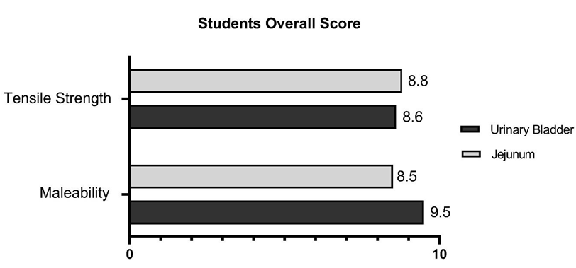
Histomorphological analysis revealed that the cell layers of the urinary bladder (serous, muscular, and submucosa) and intestine (muscular and submucosa) were preserved after vacuum conditioning for 30 and 60 days in both groups (RT and R) when compared to the control group (Figure 4). Mucosal degradation and damage to intestinal villi were similar to control in all experimental groups. The student’s responses to the form/questionnaire were collected, the arithmetic mean of these values was calculated, and the result is presented in the following figure (Figure 5). The mean grades attributed by undergraduate students about viscera malleability were 9.5 for the urinary bladder and 8.5 for the jejunum segment. Regarding the resistance of the viscera on the incision/suture, the average was 8.6 for the urinary bladder and 8.85 for the jejunum segment.
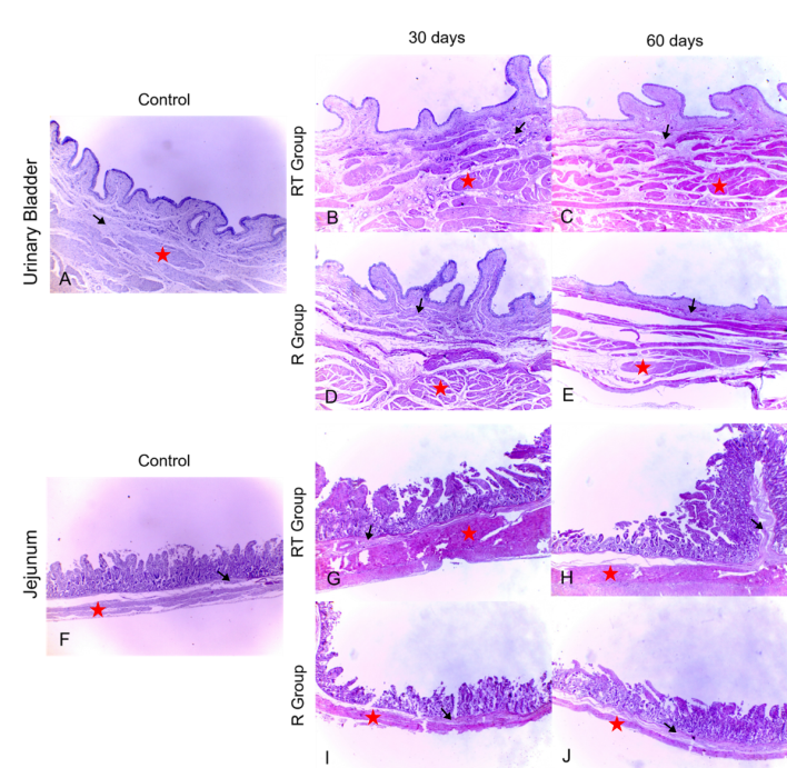
Figure 4: Histological photomicrographs of the analyzed viscera. (A) Urinary bladder and (F) jejunum control samples; Room Temperature urinary bladder experimental group after 30 (B) and 60 (C) days of storage; Refrigerated urinary bladder experimental group after 30 (D) and (E) 60 days of storage; Room Temperature jejunum experimental group after 30 (G) and (H) 60 days of storage; Refrigerated Temperature jejunum experimental group after 30 (I) and (J) 60 days of storage. Note that the cell layers (muscular – red stars and submucosa – black arrows) of the urinary bladder and intestine were preserved after conditioning for 30 and 60 days in both groups when compared to the control group.
The subsequent questions aimed to investigate the student’s acceptance (in favor of or against it) of the use of preserved viscera in surgical technique classes. It was also questioned whether the students had changed their opinion after their experience. The results showed that 100% of the students were in favor of using preserved viscera for teaching surgical techniques, and none of them changed their opinion after the study.
The last alternative questioned which kind of teaching method is preferred by the students for practicing surgical techniques among the options available throughout the discipline: training in synthetic models, training in small fresh anatomical parts, such as beef tongue and chicken thigh, or practices using chemically preserved viscera. The results showed that 3.45% (2/58) of the students prefer classes that provide training in synthetic models, such as plastic or silicone/foam models, 29.3% (17/58) prefer classes that provide training in small fresh anatomical parts, such as beef tongue and chicken thigh, and 67.25% (39/58) prefer classes that provide training with chemically preserved viscera (Figure 6).

Discussion
The desired characteristics for anatomical pieces used in surgical skills training include flexibility (elasticity) and malleability, which allow for a more reliable simulation of living tissues [18]. The numerous substances used for its chemical preservation can alter their properties, which may or may not make it unfeasible for operative techniques training. Furthermore, the different ways of packaging, depending on the choice of chemical substances used to preserve the tissues, influence its quality over time, the availability of space, and the cost of storing the pieces. All these factors make it possible or not to use these materials for specific purposes, such as surgical technique classes. Fresh anatomical parts, for example, require freezing, making it necessary to have available freezers for storage [23], besides presenting a potential biological risk. Freezers are also necessary for cadavers preserved in Larssen’s solution [24]. In literature, cadavers preserved in different compositions of saline solutions are kept refrigerated [25, 26, 27] or even in solution tanks [16, 25, 28] which requires abundant availability of material and room for storage.
Vacuum packaging of anatomical pieces after preservation in supersaturated saline solution, as described in this study, proved to be effective in maintaining physical characteristics favorable for operative techniques practices in swine viscera, allowing tissues division (diaeresis), identification and ligation of vessels, anastomosis, and different suture patterns application, among others, as shown in the swine viscera physical characteristics subjective evaluation report, in which the score attributed to nearly all parameters of the evaluated groups was between 4 and 5 in a scale from 1 to 5. Despite the regular evaluation of two parameters of the jejunum segment (RT) group, these pieces still allowed satisfactory execution of the proposed techniques. The histomorphological evaluation of the preserved viscera of different groups (RT and R) at different times (D0, D30, D60) demonstrated the integrity of the cell layers (except the mucosa), with few significant differences when compared to the control group. These results show that there are few significant differences in the material storage, making refrigeration/refrigerators unnecessary in this case.
The student’s acceptance of the use of preserved samples was promising. All declared themselves in favor of using this anatomical preservation technique for training surgical skills and kept their position at the end of the study. Furthermore, their form answers about the anatomical material show high value in the grades attributed to the quality of the viscera, all scoring above 8.0 on a scale of 0 to 10, in terms of both malleability and the sample’s resistance to suture/incision. Finally, the answers to the final question showed the students’ preference for chemically preserved viscera (67.25%) when compared to the other options for training operative techniques, such as synthetic models (3.45%) or fresh samples (29.3%). Thus, it is concluded that this approach had the students’ acceptance, and its implementation is feasible for surgical training classes.
Limitation of this Study
A limited sample group (n= 58 students) belonging to a single institution and monitored during only one semester did not allow a more accurate evaluation of the students’ surgical skills evolution. Furthermore, students in that stage of the program does not have clinical practice experience with surgery on live animals, which may influence the results obtained. Only two evaluators (AFM and JBG) with previous surgical skills rated the viscera for malleability and incision/ suture resistance.
Cost
The costs involved in the viscera’s preparation and storage are interesting factors to highlight. Considering that many places, mainly developing countries, do not have the financial resources to acquire advanced surgical models that faithfully simulate animal anatomy, a less expensive alternative that maintains the effective teaching and learning process is warranted. Preparation and storage of the viscera requires inexpensive, readily and widely available materials in most locations. The estimated amounts for preparing the anatomical pieces, already vaccum-packaged, are about US$1 per viscera (Table 2).
| Price in US$ | |
|---|---|
| Packages (un.) | 0.5 |
| Salt (kg) | 0.4 |
| Absolute ethanol 99,8% (L) | 2 |
| Visceras (un.) | 0.02 |
| Vacuum sealer (un.) | 172.8 |
Table 4: Average prices of the required materials for viscera preservation and storage in US dollars (US$). Abbreviation: un., un
Conclusion
Chemically preserved and vaccum-packed viscera using supersaturated sodium chloride solution are a viable option for storing anatomical material at room temperature, outside of solution tanks, exempting the use of refrigerators or freezers and occupying minor space in anatomy labs. Besides being well accepted by the participating students, the technique proved to be safe, low cost and easy to implement, in terms of both preservation solutions and storage. Vacuum packaging allows the preparation of didactic material in any period of the school year, depending only on the availability of materials and staff. Therefore, this may favor institutions that have fewer resources for the production and storage of anatomical pieces for practical classes
Acknowledgments
This study was financed in part by the Coordenação de Aperfeiçoamento de Pessoal de Nível Superior - Brasil (CAPES) -Finance Code 001. This study was funded by FAPESP 2019/26627-6. Authors thank Prof. Dr. Ricardo Strefezzi, Lindsay Baltel Paskosky, and the Pathology Laboratory of the Department of Veterinary Medicine at FZEA/USP for technical support, processing and analysis of histological samples.
References
-
Paixão RL (2008) What do we learn from physiology classes? In: TRÉZ T (Ed.), Instrumento Animal: the harmful use of animals in higher education. 1st(Edn.), Bauru, SP, Canal 6: 111-130.
-
Tudury EA, Potier GMA, Mesquita LS, Oliveira GK, Albuquerque VB, et al. (2004) Alternative methods for practical learning of the veterinary surgical technical discipline. Braz J vet Res Anim Sci 41(supl): 189.
-
Tudury EA, Potier GMA (2008) Alternative methods to the use of live animals in teaching: alternative methods for practical learning of the veterinary operative technique discipline. Vet Ci Trop 11(1): 92-95.
-
Singer P (2004) Libertação animal. 1st(Edn.), Porto Alegre, RS IN, Lugano, Switzerland, pp: 357.
-
Magalhães M, Filho HO (2006) Alternatives to using animals as teaching resources. UNIPAR Veterinary Sciences and Zoology Archives 9(2): 147-154.
-
Knight A (2007) The effectiveness of humane teaching methods in veterinary education. Altex 24(2): 91-109.
-
CONCEA (2014) Regulatory Resolution.
-
Silva RMGD, Matera JM, Ribeiro AACM (2004) Preservation of Cadavers for Surgical Technique Training. Vet Surg 33(6): 606-608.
-
Balta JY, Cronin M, Cryan JF, O’Mahony SM (2017) The utility of cadaver-based approaches for the teaching of human anatomy: A survey of British and Irish anatomy teachers. Anat Sci Educ 10(2): 137-143.
-
Homma H, Oda J, Sano H, Kawai K, Koizumi N, et al. (2019) Advanced cadaver-based educational seminar for trauma surgery using saturated salt solution-embalmed cadavers. Acute Med Surg 6(2): 123-130.
-
Hayashi S, Homma H, Naito M, Oda J, Nishiyama T, et al. (2014) Saturated salt solution method: a useful cadaver embalming for surgical skills training. Medicine (Baltimore) 93(27): e196.
-
Selçuk İ, Tatar İ, Huri E (2019) The effect of cadaveric hands-on training model on surgical skills and confidence for transobturator tape surgery. J Turk Ger Gynecol Assoc 20(4): 243-246.
-
Chai DQ, Naunton-Morgan R, Hamdorf J (2019) Fresh frozen cadaver workshops for general surgical training. ANZ J Surg 89(11): 1428-1431.
-
Reed AB, Crafton C, Giglia JS, Hutto JD (2009) Back to basics: use of fresh cadavers in vascular surgery training. Surgery 146(4): 757-762.
-
Braasch MC, Minchew HM, Riffel JDM, Berbel G (2022) Suture Education with Soft-Embalmed Cadavers: A Cut Above the Rest. Kans J Med 15: 78-81.
-
Oliveira FSD (2014) Assessing the effectiveness of 30% sodium chloride aqueous solution for the preservation of fixed anatomical specimens: a 5-year follow-up study. J Anat 225(1): 118-121.
-
TASS R, Yanagihara GR, Shimano AC, Rolim GS, Santos CCC, et al. (2018) Biomechanical analysis of the skin and jejunum of dog cadavers subjected to a new anatomical preservation technique for surgical teaching. J Plast 30(1): 16-23.
-
Guaraná JB, Müller AF, Strefezzi RFD, Oliveira FS, Machado LC, et al. (2021) Swine viscera preservation in hypersaturated salt solution after alcohol fixation as a preparation method for educational purposes. Anat Histol Embryol 50(6): 996-1006.
-
Queiroz ABPS, Rodrigues A, Cardozo MV, Costa NTB, Soares LG, et al. (2022) Biomechanical and microbiological analysis of embalmed cats - acute effect of conservation. An Acad Bras Cienc 94(1): e20201583.
-
Ferreira GC, Costa NTB, Cardozo MV, Queiroz ABPS, TASS R, et al. (2021) Canine corpses preserved with ethyl alcohol and curing salt and packaged in a vacuum for veterinary surgery training. Journal of Veterinary Research of Peru 32(4): e19075.
-
Gould GW (1996) Methods for preservation and extension of shelf life. Int J Food Microbiol 33(1): 51-64.
-
Wambui J, Stephan R (2019) Relevant aspects of Clostridium estertheticum as a specific spoilage organism of vacuum-packed meat. Microorganisms 7(5): 142.
-
Hayashi S, Naito M, Kawata S, Qu N, Hatayama N, et al. (2016) History and future of human cadaver preservation for surgical training: From formalin to saturated salt solution method. Anat Sci Int 91(1): 1-7.
-
Matera JM (2009) Level of learning in methods that replace the use of animals in teaching. Rev CFMV 15(46): 64-68.
-
Lombardero M, Yllera MM, Costa-e-Silva A, Oliveira MJ, Ferreira PG (2017) Saturated salt solution: A further step to a formaldehyde-free embalming method for veterinary gross anatomy. J Anat 231(2): 309-317.
-
Burns DM, Bell I, Katchky R, Dwyer T, Toor J, et al. (2018) Saturated salt solution cadaver-embalming method improves orthopaedic surgical skills training. J Bone Jt Surg Am 100(15): e104.
-
Gosomji IJ, Omirinde JO, Hena SA, Wanmi N, Azeez IA (2018) Saturated salt solution an alternative reagent in reducing formaldehyde concentration in embalming. MOJ anat physiol 5(3): 205-207.
-
Rocha TASS, Santos CCC, Iozzi MT, Dias RS, Zero RC, et al. (2019) Chemically prepared dog cadavers in teaching of surgical technique - evaluation by students of a veterinary medicine course. Acta Anat 1(2): 136-140.
- Pattern of Breast Lesions in Ovu Inland, Delta State, South Southern Nigeria
- Morphometric Analysis of the Human Femur: Exploring Platymetric and Robusticity Indices Among the Nigerian Population
- Anatomical Variation of Arteria Lusoria: Clinical Implications for Dysphagia Lusoria and Surgical Risk
- Morphometric Study of the Vertebral Body and Pedicle of Typical Cervical Vertebrae Using Radiological Image
- Epigenetic Mechanisms Driving Human Evolutionary Changes
- Neuroprotective Effects of Ginkgo Biloba Extract on Bilateral Common Carotid Artery Ischaemic Stroke Induced in Wistar Rat