Evaluation of Hepatoprotective Activity of Ethanolic Extract of Root of Prunus Persica on Paracetamol Induced Hepatotoxic in Rats
Objectives: The Objective of the present study is evaluation of hepatoprotective activity of root extract of Prunus persica on paracetamol induced hepatotoxic in rats. Methods: The roots of Prunus persica was dried under shade and then powdered and extracted with ethanol and the resultant extract was subjected for phytochemical analysis to identify different phytoconstituents. The effect of ethanolic extract of roots of Prunus persica on hepatotoxicity was evaluated by using experimental model of paracetamol induced liver necrosis. In this model the common parameter determined were physical parameter like wet liver weight and biochemical parameter like SGOT, SGPT, ALP and Total Bilirubin changes in liver. Histopathological changes in liver were assessed. Results: Phytochemical investigation of ethanolic extract of roots of P. persica revealed the presence of Carbohydrates, Tannins, Amino acids, Flavonoids, Steroids and Cardiac glycosides. Oral administration of Prunus persica extract (400 mg/kg) showed dose related hepatoprotective activity in Paracetamol induced liver necrosis. The parameters SGOT, SGPT, ALP and Total Bilirubin level were increased in paracetamol treated group whereas the treatment group like standard and extract decreased. The histopathological examination of the liver in the hepatotoxic animals in Paracetamol model shows liver parenchyma with distorted architecture. Some of the hepatocytes show degenerative changes, while some show apoptosis. Which were less in the treated group and regenerative hepatocytes show in treatment group? Conclusion: A significant hepatoprotective activity of ethanolic extract of Prunus persica observed in the present investigation may be due to synergistic potentiate action of its phytoconstituents since they contain a diverse array of active principles which are able to target multiple mechanisms involved in the pathophysiology of hepatotoxicity.
Introduction
Liver is the important organ of metabolism and excretion. Hepatotoxicity is a damage or injury to liver which is caused by various, chemicals, drugs and other agents. Amount of liver damage or injury depends on degree of exposure; mild liver damage reduced the function but extreme liver damage result in liver failure [1]. Damaged liver is not capable to perform all these functions properly and it may not able to secrete bile acid which is the primary way that liver dispose of waste product [2]. Plant drugs are known to play a vital role in the control of liver diseases. There are several plants and polyherbal formulations claimed to have hepatoprotective activities [3]. Prunus persica which belongs to family Rosaceae is a plant growing in temperate region used as laxative, sedative, anti-cancer, anti-inflammatory and also consists of glycosides, flavonoids, anthocyanins, vitamins etc [4]. It also possesses hepatoprotactive property.
Paracetamol (acetaminophen) is an extensively used analgesic and antipyretic, which produces acute liver damage if overdoses are used. Paracetamol is mainly metabolized in the liver to excretable glucuronide and sulphate conjugates [5, 6]. At toxic doses of Paracetamol, the normal metabolic pathways become saturated, causing both more N-acetyl- p-benzoquinone imine (NAPQI) to be formed and hepatic glutathione to be rapidly depleted. Paracetamol toxicity is caused by the reaction metabolite, N-acetyl-p-benzoquinoneimine (NAPQI), which is partly metabolized by cytochrome P- 450 [7]. This species causes severe oxidative damage and glutathione depletion leading to liver necrosis. Introduction of cytochrome or depletion of hepatic glutathione is a prerequisite for paracetamol-induced hepatotoxicity [8, 9]. To prove the activity scientifically the ethanolic extract of Prunus persica (roots) was studied against paracetamol induced Hepatotoxicity in albino wistar rats.
Materials and Methods
Chemicals
All chemicals used were of analytical grade. The kits for the estimation of serum glutamate oxaloacetate transaminase (SGOT), serum glutamate pyruvate transaminase (SGPT), alkaline phosphatase (ALP), and total bilirubin were purchased.
Plant Collection, Authentication and Extraction
The roots of Prunus persica were collected from the garden of Agricultural research center in Munnar, Kerala the month of September and were authenticated by Dr. R. Sridharan, Directorate of ISM & H, Govt. of Puducherry. The roots of Prunus persica were dried under shade and powdered with a mechanical grinder and passed through sieve no.40. The sieved powder was stored in airtight container and kept at room temperature. Coarsely powdered roots (350 g) were extracted with soxhlet apparatus using petroleum ether for about 24 hrs. After defatting, the marc was dried in hot air oven at 500C, packed in soxhlet apparatus, and further extracted with 95% ethanol. The solvent were removed from the extracts under reduced pressure by using rotary vacuum evaporator [10]. Then the yield was calculated. The percentage yield was 20.36 % w/w. The ethanol was selected to avoid the formation of prussic acid [11].
Animals
Wistar albino rats (200-250 g) were used for all other studies and they were housed at a temperature of 23±2 ºC and humidity (50-55 %) with 12 hrs. light and dark cycles. They were caged with a maximum of three animals in each polypropylene cage and were fed with standard diet and water adlibitum.
Hepatoprotective activity
Animals were randomly divided into five groups of six animals each. Group I served as normal control and received distilled water (5 ml/kg), Group II served as toxic control and received distilled water (5 ml/kg) for 7 days and 1:1(v/v) mixture of PCM in gum acacia (2 ml/ kg p.o.) on 8th day, Group III served as standard group and received silymarin (100 mg/kg) for 7 days and, while Group IV and V were treated with ethanolic extract of roots of Prunus persica at the dose of 200 and 400 mg/kg/day, p.o. for seven days, respectively [12, 13].
Analysis of liver function enzymes
After 24 hours of PCM administration (Day 9th) blood samples were collected by intra-cardiac puncture under mild chloroform. The collected blood was allowed to clot at room temperature and serum was separated by centrifugation at 2500 rpm for 15 min. Then serum was used for the estimation of biochemical parameters such as alkaline phosphatase (ALP), serum glutamate oxaloacetate transaminase (SGOT), serum glutamate pyruvate transaminase (SGPT) and total bilirubin (TB).The biochemical parameters were estimated as per the standard procedure prescribed by manufacturer’s instruction manual provided in the standard kit using autoanalyser [14, 15].
Statistical Analysis
All the data are expressed as Mean ± S.E.M. One way analysis of variance (ANOVA) was used for the statistical analysis of data. Tukey’s multiple comparison tests was used for determining the significance. A value of p<0.05 was considered as significant [16].
Results
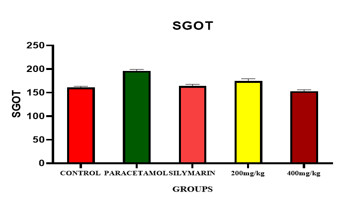
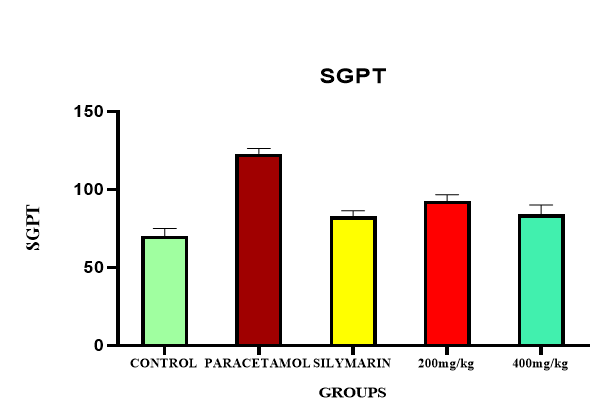
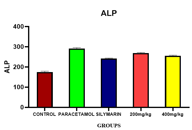
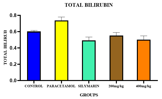
The result of current study showed that, the levels of SGOT [(196.3±2.77) IU/L], SGPT [(123.75 ±1.33) IU/L], ALP [(290.68 ±2.08) IU/L] and total bilirubin [(0.72±0.02)mg/dl] were significantly increased in toxic group (Group II) when compared with normal control group. Rats pre-treated with Prunus persica roots extract (200mg/kg) showed significant reduction in the levels of SGOT, SGPT, ALP, and total bilirubin compared with toxic control group. But maximum reduction of SGOT [(165.60±2.65) IU/L], SGPT [(84.03±2.53) IU/L], ALP [(256.25±3.26) IU/L] & bilirubin [(0.62±0.05)mg/dl] were observed in high dose group (400mg/kg) (Figures 1-5 & Tables 1-2).
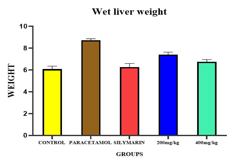
| Grou | p | Treatment | Dose | Wet liver weight(mg) | |||||||
|---|---|---|---|---|---|---|---|---|---|---|---|
| A | Control | 5 ml/kg, p.o. | 6.07±0.28 | ||||||||
| B | Negative control | Paracetamol 2g/kg, p.o. | 8.70±0.15 | ||||||||
| C | Standard or reference (Silymarin) | 100mg/kg, p.o. | 6.22±0.34*** | ||||||||
| D | Prunus persica roots extract (Low dose) | 200mg/kg, p.o. | 7.45±0.25* | ||||||||
| E | Prunus persica roots extract (High dose) | 400mg/kg, p.o. | 6.28±0.24** |
Table 1: Biochemical Parameters. Values are mean ± SEM (n=6) one way ANOVA. * represents significant at p<0.05, ** represents hig
Table1: WET liver weight.
| Total bilirubin | ||||||||||||||||||||
|---|---|---|---|---|---|---|---|---|---|---|---|---|---|---|---|---|---|---|---|---|
| G | roup | Treatment | Dose | S | GOT levels (U/L | ) | SGPT levels (U/L) | ALP levels (U/L | ) | |||||||||||
| (mg/dl) | ||||||||||||||||||||
| A | Control | 5ml/kg | 160.3±1.9 | 70.4±1.9 | 174.67±2.95 | 0.63±0.7 | ||||||||||||||
| B | Hepatotoxic PCM | 2g/kg | 196.3±2.7 | 123.7±1.3 | 290.68±2.08 | 0.72±0.02 | ||||||||||||||
| C | Standard (Silymarin) | 100 mg/kg | 171.2±1.6** | 82.80±1.5*** | 261.97±1.44** | 0.65±0.02** | ||||||||||||||
| D | Extract(P. p) | 200 mg/kg | 174.5±2.06* | 92.74±1.6* | 268.38±1.6* | 0.66±0.03* | ||||||||||||||
| E | Extract(P. p) | 400 mg/kg | 165.6±2.6*** | 84.03±2.5** | 256.2±3.2*** | 0.62±0.19*** |
Table 2: Biochemical Parameters. Values are mean ± SEM (n=6) one way ANOVA. * represents significant at p<0.05, ** represents hig
Histopathological Studies of the Liver in Paracetamol Induced Hepatotoxicity
The histopathological evaluation of paracetamol toxicity in all the groups was examined and shown in figures below (Figures 6-10). The description is as follows
Group 1 (Control group)
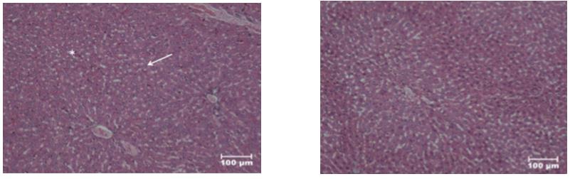
Liver showing normal architecture of hepatic cords with hepatocytes staining cytoplasm pink and vesicular nucleus blue in color (arrow) and red blood cells are seen in the sinusoidal spaces (asterisk). Haematoxylin and Eosin stain, scale bar=100μm.
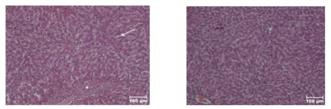
Group 2 (Hepatotoxic group)
Liver showing distortion of architecture, hepatic necrosis evident by condensation of nucleus, vacuolar degeneration and vesicular fatty change pushing the nucleus to one side (arrow). There was fibrosis formed by elongated fibroblast cells with oval shaped nucleus $$ \text{(asterisk). Haematoxylin and Eosin stain, scale bar=100 \mu m.} $$
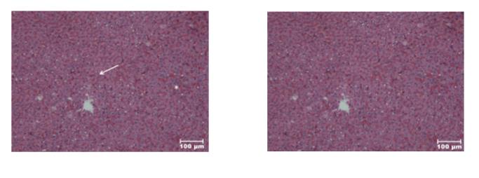
Group 3 (Standard/ Reference)
Liver showing normal architecture of hepatic cords with hepatocytes staining cytoplasm pink and vesicular nucleus blue in colour (arrow) along with portal triad. There was mild degenerative change in few areas with eosinophilic hepatic necrosis (Asterisk) indicating recovery from the hepatic injury. Haematoxylin and Eosin stain, scale bar=100μm.
Group 4 (Ethanolic Extract of Roots of Prunus persica)
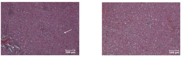
Liver showing normal architecture of hepatic cords with hepatocytes staining cytoplasm pink and vesicular nucleus blue in color (arrow). There were mild degenerative changes in few areas with vacuolar degeneration of hepatocytes (Asterisk) indicating recovery from the hepatic injury. Haematoxylin and Eosin stain, scale bar=100μm.
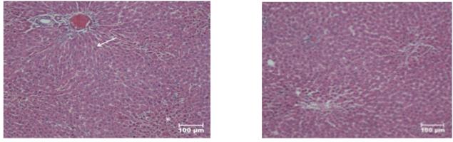
Group 5 (Ethanolic Extract of Roots of Prunus persica)
Liver showing normal architecture of hepatic cords with hepatocytes staining cytoplasm pink and vesicular nucleus blue in color (arrow) along with portal triad and congestion of blood vessels (asterisk) indicating recovery from the hepatic injury. Haematoxylin and Eosin stain, scale bar=100μm.
Discussion
Hepatotoxicity is the latent difficulty of paracetamol, which is normally, used in common medicine and an assessment of its relative toxicity is important. The primary toxicity of paracetamol is the result of drug metabolism in liver [17]. At therapeutic doses, paracetamol is metabolized through glucuronidation and sulphation reactions result in the water-soluble metabolites that are expelled through the kidney. The result of the metabolic transformation of paracetamol by the microsomal P-450 enzyme system is that, very reactive intermediate, namely, N-Acetyl-P Benzoquinone Imine (NAPQI) is produced. Glutathione (GSH) then decreases this metabolite. However, after Paracetamol high dose, capacity for glucuronidation and sulphation is exceed with the formation of additional NAPQI through cytochrome P450 2E1. This intern lead to depletion in glutathione, excess of NAPQI binds to hepatic cell protein and DNA resulting in mitochondrial dysfunction and the development of acute hepatic necrosis [18, 19].
Further NAPQI can also make covalent bound with cellular proteins, can modify the structure and functions of these proteins. This cellular damage causes a reduction in calcium ATPase activity and an increase in cytosolic calcium levels. As a result, cell permeability modifications and lead to the loss of cellular integrity [20]. In addition, it has been shown that NAPQI can avoid NADH and succinate dehydrogenase function. It was considered that damage of homeostasis was also responsible for high dose paracetamol induced hepatotoxicity. Because of destruction of homeostasis, intracellular Ca2+ accumulates and an increase is shown in catabolic enzymes, which causes cell death. Nitric oxide, reactive oxygen’s, lipid peroxidation and apoptosis are the major situations, which play vital roles in hepatotoxicity. In addition, peroxinitrite derivatives and protein nitrates also have roles in this condition [21].
So in the present study, paracetamol was employed as toxic agent and the protective effect of Prunus persica roots against the paracetamol induced hepatotoxicity was studied. Histopathological studies and biochemical enzyme markers like SGPT, SGOT, SALP and TB levels estimated the extent of toxicity. Strengthening the above stated mechanisms, the histopathological study also maintained the biochemical sign for the hepatoprotection shown by Prunus persica in the same group. The hepatic injury associated with Paracetamol is due to discharge of toxic metabolite N-acetyl-P-benzoquinone imine (NAPQ1) and its free radical generation. The protective effect exhibited by ethanolic extracts at a dose level of 200 mg/kg was comparable with the standard drug Silymarin. After the phytochemical study, it is reported that, the plant extracts contain carbohydrates, flavonoids, terpenoids, tannins and steroids. The presence of flavonoids in our extract may be responsible for its hepatoprotective activity [22, 23, 24, 25, 26]. In conclusion, the results of this study demonstrate that Prunus persica roots extract was effective for the prevention of Paracetamol-induced hepatic damage in rats. Our results show that the hepatoprotective effects of Prunus persica roots extract may be due to marked changes in various parameters of liver enzyme, some physical parameters which proved that it’s a powerful hepatoprotective in nature against the Paracetamol induced liver toxicity. The present study thus justifies the use of Prunus persica roots in the treatment of liver diseases and points out that Prunus persica roots deserves further detailed investigation as a promising hepatoprotective agent does. However, the exact mechanism(s) and the active compound(s) involved in these effects need to be clarified in future studies. The present study showed that ethanolic extract of Prunus persica roots possess hepatoprotective activity, as evidenced by the significant inhibition in the elevated levels of serum enzymes activities induced by PCM. There was an increase in SGOT (↑196.3.7%), SGPT (↑123.75%), ALP (↑290.68%) and bilirubin (↑0.72%) level of PCM+ distilled water treated group when compared to that of normal control group. Further, rats pretreated with standard drug silymarin (100 mg/kg) exhibited decrease in SGOT (↓171.25%), SGPT (↓82.80%), ALP (↓261.97%) and total bilirubin (↓0.65%) level as compared to that of toxic control group. Also, the rats pretreated with ethanolic extract of Prunus persica (200 mg/kg) showed decrease in SGOT (↓174.5%), SGPT (↓92.74%), ALP (↓268.38%), and total bilirubin (↓0.66%) level as compared to that of toxic control group. But rats’ pre administered with ethanolic extract 400 mg/kg (Group V) for one week; showed the more decrease in percentage of SGOT (↓165.60%), SGPT (↓84.032%), ALP (↓256.25%), and total bilirubin (↓0.62%) level when compared to that of toxic control group. The ethanolic extract of Prunus persica roots definitely possess hepatoprotective properties in the dose dependent manner, against PCM intoxication in rats, after one week pretreatment; at the dose level 200mg/kg and 400 mg/kg.
Conclusion
The present study reports the hepatoprotective activity of Prunus persica roots extract on PCM induced hepatotoxicity in rats. Phytochemical screening revealed the presence of carbohydrates, glycosides, flavonoids, phytosterols, saponins alkaloids and phenolic compounds in the extract. Several investigators have shown that plant extract containing flavonoids are responsible for hepatoprotective potential in various experimental animal models. Thus, it can be interpreted that the hepatoprotective effect may be due to the presence of flavonoids. The histopathological examination of the liver in the hepatotoxic animals in Paracetamol model shows liver parenchyma with distorted architecture. Some of the hepatocytes show degenerative changes, while some show apoptosis. Which were less in the treated group and regenerative hepatocytes show in treated group? On the basis of results obtained, it can be concluded that the ethanolic extract of Prunus persica roots seems to have hepatoprotective activity. The additional studies are needed to evaluate potential effectiveness of ethanolic extract in clinical condition related with liver damage.
References
-
Liver showing normal architecture of hepatic cords with hepatocytes staining cytoplasm pink and vesicular nucleus blue in colour (arrow) along with portal triad. There was mild degenerative change in few areas with eosinophilic hepatic necrosis (Asterisk) indicating recovery from the hepatic injury. Haematoxylin and Eosin stain, scale bar=100μm. [INLINE_FIGURE:4:3] Figure 8: H&E, X100.
- Management of Ear Keloid with Ksharsutra: A Case Study
- Yoga and Global Sustainability: A Holistic Path to One Earth, One Health
- Autoimmune Diseases in Ayurveda: A Narrative Review with Classical and Modern Perspectives
- Management of Cluster Headache Associated with Pituitary Apophysitis by CERT (Chakrasiddh Energy Release Technique): A Case Report on Energy Rebalancing
- Zygophyllum Geslini Coss : Biochemicals and Antioxidant Activity
- Observations of a Beginner Vaidya