In vitro Anti-Inflammatory Egg Albumin Denaturation Assay: An Enhanced Approach
As a physical reaction to injury, infection, or destruction, inflammation is characterized by heat, redness, discomfort, swelling, and abnormal physiological processes. The inflammatory process is a complicated series of relationships among soluble substances and cellular elements that frequently include connective tissue damage. An uncontrolled inflammatory response is the primary cause of a wide range of disorders, including allergies, cardiovascular dysfunctions, metabolic syndrome, cancer, and autoimmune diseases, placing a significant financial burden on individuals and, in turn, on society. Steroids, nonsteroid anti-inflammatory drugs, and immunosuppressants are some examples of common medications for controlling and suppressing inflammatory crises; but they have several side effects, particularly stomach irritation that can cause gastric ulcers. Using natural goods has had a significant positive impact on the development of modern medicine. On such background, plants have historically been an excellent source of medication, and there are several records on the use of herbs in traditional medicine to treat a variety of inflammatory illnesses. However, there has been no scientific attempt to verify these spontaneous uses reported in the literature. Therefore, to conduct more studies and gather more data, scientists need to develop low-cost, straightforward in vitro methodologies to assess the efficacy of natural anti-inflammatory compounds. With this context in mind, this methodology focuses on in vitro anti-inflammatory methodology that can be used to evaluate natural anti-inflammatory substances. They are cost-effective, easy to operate, and dependable with minor alterations.
Introduction
A physical reaction to injury, infection, or destruction known as inflammation is characterized by heat, redness, discomfort, swelling, and disrupted physiological processes [1]. Inflammation is a typical protective response when tissues are harmed by physical trauma, toxic chemicals, or microbial infections. The body’s response is to get rid of the irritants, neutralize the invasive organisms, and get ready for tissue healing [1]. It happens as a result of the release of chemical mediators from migrating cells and injured tissue. Non-steroidal anti-inflammatory medicines are the most widely used medication for treating inflammatory diseases, but they have several side effects, particularly stomach irritation that can cause gastric ulcers [2]. Inflammation is a complicated biological reaction of vascular tissues to potentially damaging stimuli. It is also the organism’s protective attempt to eliminate harmful stimuli and start the healing process [3]. At the beginning of inflammation, cells are stimulated and release inflammatory mediators. Some of these mediators include histamine, serotonin, SRS-A, prostaglandins, and several plasma enzyme systems, such as the complement system, the clotting system, the fibrinolytic system, and the kinin system [4]. These mediator molecules work together to increase vasodilation and blood vessel permeability. Consequently, this causes the blood to flow more freely, plasma proteins and fluids to exude from the wounded tissues, and leukocytes—primarily neutrophils— to go outside the blood vessels. Acute inflammation and chronic inflammation are two different types of inflammation. The body’s initial reaction to harmful stimuli is acute inflammation, which is brought on by an increase in the flow of plasma and leukocytes from the blood into the wounded tissues the process of acute inflammation is initiated by pre-existing cells in the tissues. Vasodilatation and enhanced capillary permeability, which are brought on by the actions of the numerous inflammatory mediators, are two prominent vascular changes that characterize this [5]. Chronic inflammation is characterized by the simultaneous destruction and healing of the tissues damaged by the inflammatory process and is a prolonged inflammatory response that results in a progressive change in the kind of cells at the inflammatory site [6]. The usage of natural products has significantly helped modern medicine’s evolution. The world has begun to reconsider conventional medicine as a result of recent extensive research on numerous plant species and their active medicinal principles. The analysis of anti-inflammatory natural plants offers a chance to find and use natural chemicals as an alternative to synthetic medications [7]. Studying natural plants’ anti-inflammatory potential enables us to draw on this huge body of knowledge and consider long-lasting therapy alternatives [8]. Natural plants have been utilized for millennia in traditional medicine for their medicinal capabilities. Natural anti-inflammatory substances derived from plants are frequently used by people for a long time and are usually regarded as harmless can discover chemicals that, when compared to synthetic medications, may have lower toxicity profiles and fewer adverse effects by analyzing these plants [9]. This is crucial in chronic illnesses when long-term anti-inflammatory drug use may have negative health implications. Polyphenols, flavonoids, terpenoids, and alkaloids are only a few examples of the wide variety of bioactive substances that are known to be present in natural plants. These substances can target numerous inflammatory pathways and have promising anti- inflammatory activities [10]. Discovery of novel bioactive substances that have the potential to have strong anti- inflammatory effects by investigating and analyzing natural plants. The creation of novel medicines has benefited greatly from the inspiration provided by natural flora. Aspirin and other nonsteroidal anti-inflammatory medicines (NSAIDs), among other treatments, are made from natural substances that are found in plants. By analyzing naturally occurring anti-inflammatory plants, can find novel chemicals or chemical structures that could be used as the basis for drug discovery and development, resulting in the development of more potent and precisely targeted anti-inflammatory drugs [8]. Therefore, to perform more research and collect more data, scientists need to develop simple, low-cost in vitro approaches to evaluate the efficacy of natural anti- inflammatory substances. This methodology concentrates on in vitro anti-inflammatory methodologies that can be used to assess natural anti-inflammatory chemicals in light of the surrounding circumstances. They require little maintenance and are reliable, affordable, and simple to use. In such a context, the egg albumin denaturation approach might be beneficial for researchers working in this field. Thus, the enhanced egg albumin denaturation approach is as follows.
Principle of In vitro Egg Albumin Denaturation Method
The principal objective behind the egg albumin denaturation assay is to determine whether agents or compounds can stop or hinder egg albumin from becoming denatured under particular circumstances. Denaturation is the term used to describe how a protein changes in structure and loses its biological activity [11]. Egg albumin is employed as a model protein in the experiment, and denaturation is brought about by exposing it to extremes of heat, pH, or other denaturing agents. Egg albumin’s original conformation is disrupted during denaturation, changing its physical characteristics, and causing it to lose its functional activity. The egg albumin denaturation assay measures a drug or compound’s capacity to prevent or lessen egg albumin denaturation to evaluate its anti-inflammatory effects [12]. The egg albumin denaturation assay is based on the idea that substances with anti-inflammatory qualities may be able to stabilize protein structures and prevent denaturation, which is frequently linked to inflammation and tissue damage. As a result, agents or chemicals that significantly decrease the denaturation of egg albumin in this assay may have potential anti-inflammatory properties [13]. One of the causes of inflammation is assumed to be protein denaturation. NSAIDs prevent protein denaturation and inhibit the COX enzyme at the same time [14]. The different concentrations of the test sample can be incubated with egg albumin solution in controlled experimental conditions and let the reactions happen and then the determination of absorbance to calculate the percentage inhibition. And then IC50 values can be calculated using GraphPad Prism software. Diclofenac sodium can be used as a reference drug [15].
Materials
Chemical and Reagents
- Egg albumin solution
- Phosphate buffered saline
- Distilled water/DMSO
Equipment
- Clean pipettes and puppet tips
- Khan tubes or test tubes
- Incubator
- Spectrophotometer
- Water bath
Egg Albumin Denaturation Assay: Methodology
Method
Preparation of plant extract: To initiate the dilution series, in our previous studies there were several herbal plants used. Namely Evolvulus Alsinoides (L.) L., Terminalia Arjuna (Roxb. ex DC.) Wight & Arn. and Hemidesmus indicus R. Br. In our previous studies maceration technique was utilized to prepare the plant extract. Maceration was a technique used in homemade preparations of tonic for a long time. It became a popular and economical way to obtain essential oils and bioactive compounds. For small-scale extraction, maceration usually consists of several steps. First, the plant materials, either leaves or stem bark, or root bark, are ground into small particles which increases the surface area for proper mixing with solvent. Then, the suitable solvent named menstruum is added in a closed vessel. Here we used the most polar solvent as water. Next, the liquid is strained off but the march which is the solid residue is pressed to recover many occluded solutions. The obtained strained and the press-out liquid are mixed and separated from impurities by filtration. Occasional shaking in maceration facilitates extraction in two ways; (a) increase diffusion, and (b) remove the concentrated solution from the sample surface for bringing new solvent to the menstruum for more extraction yield [16]. Fresh plant components were collected and cleaned under running water to remove any visible contaminants. The cleaned sample was allowed to dry for one to two days in a shed away from direct sunlight. The plant components were then ground using a mechanical blender. The powdered areal components were placed in a separate, airtight container. Then, distilled water was added to the powdered plant material in a 3 to 1 (volume to weight) ratio. The container was shaken and placed in a lid-tight container for 48 hours. Afterward, by employing three layers of muslin cloth filtering took place. Samples were maintained in a fume hood for 48 hours while the filtration was divided into a tray and allowed to evaporate to eliminate all solvents. A spatula was used to scratch the dried powder once the water had evaporated. Finally, amber- colored vials were used to keep the aqueous plant powder dry until future use. One gram of the aqueous plant extract powder was dissolved in 1 mL of distilled water. This initial concentration represents an undiluted extract that served as the highest concentration point in the dilution series, and from this 1mL was taken and two-fold dilution series was made. The dilution series was established using a two-fold dilution method, where each subsequent concentration was half of the previous concentration. This process was repeated for a total of 10 points, resulting in a range of different concentrations of the aqueous plant extract concentration ranging from 1g mL-1 to 2×10⁻³ g mL-1 concentration. Then, 2mL of the prepared solution was taken into the respective test tubes, and conducted the assay.
Preparation of 1% of egg albumin solution: Fresh hen’s eggs or egg albumin powder that is readily accessible in stores can be used to make a 1% egg albumin solution. Making egg-albumin solution using a fresh hen’s egg properly involves carefully cracking an egg, transferring 1 mL of the translucent portion to 100 mL of w/V distilled water, and stirring thoroughly. The clear component of the egg is called egg albumin. The water should be cold when making the solution. Water will coagulate if it is heated to a boil.
Egg Albumin Assay
The anti-inflammatory activity of unknown crude extracts can be determined in vitro for inhibition of the denaturation of egg albumin (protein).
- 0.2 mL of 1-2% egg albumin solution (from fresh hen’s egg/ or commercially available egg albumin powder), 2 mL of sample extract or standard (Diclofenac sodium) at varying concentrations, and 2.8 mL of phosphate- buffered saline (pH 7.4) were mixed to form a reaction mixture of a total volume of 5 mL.
- A total volume of 5 mL of the control was created by combining 2 mL of triple-distilled water, 0.2 mL of 1-2% egg albumin solution, and 2.8 mL of phosphate-buffered saline.
- The reaction mixtures were then incubated at 37±2°C for 30 min and will be heated in a water bath at 70±2°C for 15 min.
- After cooling, the absorbance was measured at 280 nm by a suitable UV/Vis spectrophotometer using triple distilled water as the blank [1].
- The following equation was used to determine the % inhibition of protein denaturation.
100 Absorbanceof control Absorbanceof test sample Percentageinhibition Absorbanceof control − = ×
Then plant extract/positive control concentration for 50% inhibition (IC50) was determined by plotting percentage inhibition concerning control against concentration [1, 15, 17].
Statistical Analysis
As a statistical analysis tool, GraphPad Prism software for Windows versions up to 9 (GraphPad Software, San Diego, CA, USA) can be used. To calculate the mean and the standard error of the mean, all results (absorbance) must be triplicated. Nonlinear regression was applied using the GraphPad Prism software to determine the half-maximal inhibitory concentration (IC50) value and concentration relationships [18]. Or student’s t-test was used to assess the results for statistical significance. Then p-value was used for the statistical significance evaluation and/or one-way analysis of variance (ANOVA) followed by Turkey post-test comparisons carried out [19, 20, 21]. As well as some literature evidence of the use of the SPSS version for Windows (IBM, Somers, NY, USA) for statistical data analysis. Data were expressed as mean ± standard error of the mean. An Independent sample t-test was used to examine the difference in inhibition rate percentage between the various groups. Then p-value was used for the statistical significance evaluation [13].
Observations
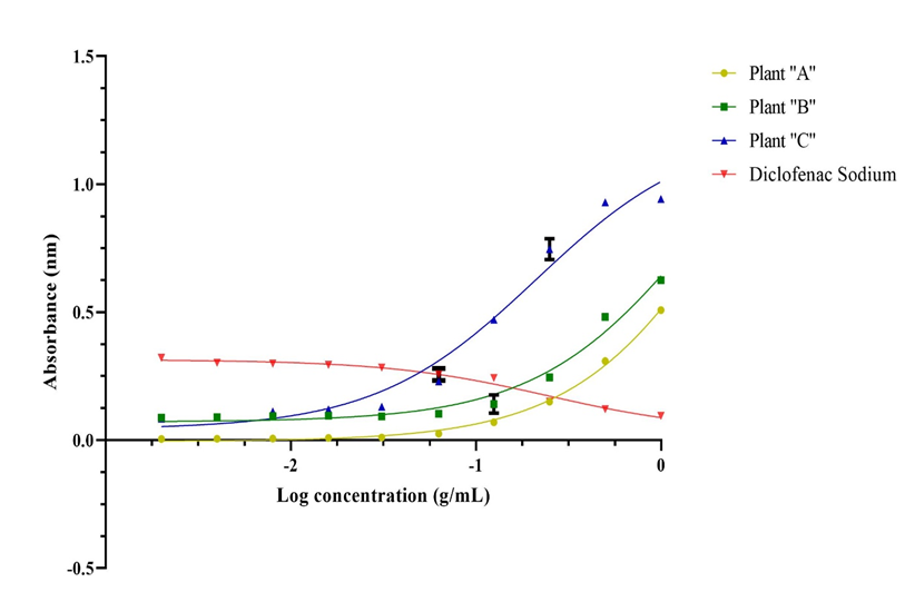
Several plant extracts were previously tested for anti- inflammatory efficacy utilizing an egg albumin denaturation assay with diclofenac sodium as the positive control. Figure 1 depicted the activity of various plant extracts in comparison to the positive control diclofenac sodium. Plants “A”, “B”, and “C” were all included. We can get an idea of the anti- inflammatory potential of aqueous plant extracts of plants A, B, and C by looking at the diagram below. This graph depicts data transfer in plants A, B, and C with the positive control of diclofenac sodium. By comparing these data transformations, one can quickly get an understanding of anti-inflammatory activity by comparing them to the standard.
Conclusion
Increased vascular permeability, increased protein denaturation, and membrane rearrangement are only a few of the complicated processes that occur during inflammation, which is frequently accompanied by discomfort. Thus, investigating and examining natural plants can lead to the discovery of novel bioactive compounds that have the potential to have substantial anti-inflammatory effects. The inspiration offered by natural flora has greatly aided the development of innovative medications. Therefore, to do more studies and gather more data, scientists must require straightforward, affordable in vitro methods to assess the effectiveness of natural anti-inflammatory compounds. Here considered, one of the causes of inflammation is assumed to be protein denaturation. NSAIDs, which also inhibit the COX enzyme, reduce protein denaturation. In controlled experimental settings, the various test sample concentrations can be incubated with egg albumin solution to allow the reactions to occur, and then the absorbance can be measured to determine the % inhibition. Following that, IC50 values can be computed using GraphPad Prism software. Diclofenac
sodium may be utilized as a model medication, and then using statistical analysis methods, researchers can be able to conclude the findings regarding their studies.
Authors’ Contributions
H.D.T. Madhuranga conceptualized and drafted the manuscript and conducted the experimental methodology and obtained the assay results and statistically validated the protocol. D.N.A.W. Samarakoon substantively reviewed the draft. The final manuscript was read by all authors and got their approval.
Acknowledgments
Not applicable
Disclosure
The author reports no conflicts of interest in this work.
References
-
Chandra S, Chatterjee P, Dey P, Bhattacharya S (2012) Evaluation of In Vitro Anti-Inflammatory Activity of Coffee against the Denaturation of Protein. Asian Pac J Trop Biomed 2(1): 178-180.
-
Vonkeman HE, Van de Laar MAF (2010) Nonsteroidal Anti-Inflammatory Drugs: Adverse Effects and Their Prevention. Semin Arthritis Rheum 39(4): 294-312.
-
Ferrero Miliani L, Nielsen OH, Andersen PS, Girardin SE (2007) Chronic Inflammation: Importance of NOD2 and NALP3 in Interleukin-1β Generation. Clin Exp Immunol 147(2): 227-235.
-
Perianayagam JB, Sharma SK, Pillai KK (2006) Anti- inflammatory activity of Trichodesma indicum root extract in experimental animals. J Ethnopharmacol 104(3): 410-414.
-
Okoli CO, Akah PA, Nwafor SV, Anisiobi AI, Ibegbunam IN, et al. (2007) Anti-inflammatory activity of hexane leaf extract of Aspilia africana C.D. Adams. J Ethnopharmacol 109(2): 219-225.
-
Eming SA, Krieg T, Davidson JM (2007) Inflammation in wound repair: Molecular and cellular mechanisms. J Invest Dermatol 127(3): 514-525.
-
Yuan H, Ma Q, Ye L, Piao G (2016) The traditional medicine and modern medicine from natural products. Molecules 21(5): 559.
-
Furst R, Zundorf I (2014) Plant-derived anti- inflammatory compounds: Hopes and disappointments regarding the translation of preclinical knowledge into clinical progress. Mediators Inflamm 2014: 146832.
-
Ekor M (2014) The growing use of herbal medicines: Issues relating to adverse reactions and challenges in monitoring safety. Front Pharmacol 10(4): 177.
-
Riaz M, Khalid R, Afzal M, Anjum F, Fatima H, et al. (2023) Phytobioactive compounds as therapeutic agents for human diseases: A review. Food Sci Nutr 11(6): 2427- 3617.
-
Clark JH (1943) Denaturation changes in egg albumin with urea, radiation, and heat. J Gen Physiol 27(2): 101- 111.
-
Goryanin I, Ovchinnikov L, Vesnin S, Ivanov Y (2022) Monitoring Protein Denaturation of Egg White Using Passive Microwave Radiometry (MWR). Diagnostics 12.
-
Dharmadeva S, Galgamuwa L, Prasadinie C, Kumarasinghe N (2018) In vitro anti-inflammatory activity of Ficus racemosa L. bark using albumin denaturation method. AYU 39(4): 239-242.
-
Ahmadi M, Bekeschus S, Weltmann KD, Von Woedtke T, Wende K (2022) Non-steroidal anti-inflammatory drugs: recent advances in the use of synthetic COX-2 inhibitors. RSC Med Chem 13(5): 471-496.
-
Sen S, Chakraborty R, Maramsa N, Basak M, Deka S, et al. (2015) In vitro anti-inflammatory activity of amaranthus caudatus L. Leaves. Indian J Nat Prod Resour 6(4): 326- 329.
-
Azmir J, Zaidul ISM, Rahman MM, Sharif KM, Mohamed A, et al. (2013) Techniques for extraction of bioactive compounds from plant materials: A review. J Food Eng 117(2013): 426-436.
-
Burn JH (1961) Medical Pharmacology. BMJ 1961(2): 1131.
-
Ileperuma KG, Senanayaka RU, De Silva HPD, Samanmali BLC, Jayasuriya WJABN, et al. (2022) Evaluation of the in vitro anti-inflammatory activity of different fractions of the aqueous extract of Curcuma zedoaria Roscoe rhizome and formulation of a cream with anti-inflammatory potential. Pharm J Sri Lanka 12(1): 20-32.
-
Anokwah D, Kwatia EA, Amponsah IK, Jibira Y, Harley BK, et al. (2022) Evaluation of the anti-inflammatory and antioxidant potential of the stem bark extract and some constituents of Aidia genipiflora (DC.) dandy (rubiaceae). Heliyon 8(8): e10082.
-
Banerjee S, Biswas S, Chanda A, Das A, Adhikari A (2014) Evaluation of phytochemical screening and anti- inflammatory activity of leaves and stem of Mikania scandens (l.) wild. Ann Med Health Sci Res 4(4): 532- 536.
-
Shunmugaperumal T, Kaur V (2016) In Vitro Anti- inflammatory and Antimicrobial Activities of Azithromycin After Loaded in Chitosan- and Tween 20-Based Oil-in-Water Macroemulsion for Acne Management. AAPS PharmSciTech 17(3): 700-709.
- Management of Ear Keloid with Ksharsutra: A Case Study
- Yoga and Global Sustainability: A Holistic Path to One Earth, One Health
- Autoimmune Diseases in Ayurveda: A Narrative Review with Classical and Modern Perspectives
- Management of Cluster Headache Associated with Pituitary Apophysitis by CERT (Chakrasiddh Energy Release Technique): A Case Report on Energy Rebalancing
- Zygophyllum Geslini Coss : Biochemicals and Antioxidant Activity
- Observations of a Beginner Vaidya