Primary Brain Calcification in a Case and Review of the Literature
Primary brain calcifications are most often described in a family context (PFBC). It is a rare neurodegenerative disease characterized by calcifications of the basal ganglia and other brain regions. the diagnostic criteria for this disease are defined by: bilateral calcifications of the nuclei of the base; progressive neurological disorders; the absence of abnormalities suggestive of another metabolic or mitochondrial disorder, the absence of an infectious, traumatic or toxic cause and an evocative family context. His pathophysiology is unclear. We report an observation of sporadic primary cerebral calcification in a Congolese subject.
Introduction
Primary family calcification (PFBC), also known as Fahr's disease or familial calcification of idiopathic basal ganglia, is a rare neurodegenerative disease with characteristic calcium deposits characterized by basal ganglia calcifications and other areas of the body. brain [1, 2]. Calcification of basal ganglia may be asymptomatic or associated with neuropsychiatric and motor symptoms [1, 2]. Currently, the diagnostic criteria for this disease are defined by: bilateral calcifications of the nuclei of the base; progressive neurologic disorders generally including abnormal movements and / or psychiatric disorders; the absence of abnormalities suggestive of another metabolic or mitochondrial disorder, the absence of an infectious, traumatic or toxic cause and an evocative family context [3, 4]. The clinical manifestations of the disease occur at very young ages or later and do not correspond to any specific table. Its pathophysiology is unclear [2]. A few sporadic cases have been reported under the name of Fahr's disease only, in these cases, the involvement preferentially affects the basal ganglia and the cerebellum [5]. We report an observation of sporadic primary cerebral calcification in a Congolese subject.
Cases Report
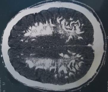
It is Mr. CY, 49 years old, with no particular antecedents, who present progressively over 12 months a gait disorder, with type of fatigability of the lower limbs, instability when standing and walking. , associated with dysarthria-like speech disorders and almost constant memory disorders. Clinical examination revealed good hemodynamic status, spatial disorientation, concentration disorder, attention deficit, pyramidal lower limb syndrome, resting tremor with extrapyramidal hypertonia. The brain scan performed revealed voluminous bilateral and symmetrical spontaneous calcium hyper-densities of the serrated nuclei, all the basal ganglia, the semi-oval center and the juxtacortical white matter, with no mass effect on the adjacent structures and without of contrast (Figures 1-3). The electroencephalogram was normal.
The biological assessment i.e, calcemia, phosphoremia and parathyroid hormone (PTH) are normal; as well as the blood ionogram, renal, hepatic function, inflammatory assessment, blood count, and the search for anti-HIV1 and anti-HIV2 antibodies). Ophthalmological examination was normal. The patient received symptomatic levodopa treatment at 250 mg / day for 6 months. The evolution is marked by the persistence of the clinical picture. The patient was lost to view. The association of neurological abnormalities, images of symmetrical brain calcification and the absence of phosphocalcic balance and PTH abnormalities argue for familial primary brain calcification, but the absence of other cases in the family makes us consider one case sporadic.
Discussion
Primary family calcification (PFBC), also known as Fahr's disease or idiopathic calcification of the basal ganglia, is a rare neuropsychiatric disorder [2, 4]. In this disease, most symmetrical calcifications occur in the basal ganglia and other brain regions, including the dentate nucleus, thalami, brainstem, supratentorial white matter, and cerebral cortex [2]. The clinical presentation is characterized by psychiatric signs, cognitive impairment and movement disorders, including chorea, dystonia, athetosis and Parkinsonism. Cerebellar signs, pyramidal signs, seizures and headaches are also associated with this condition. Age at diagnosis is usually around 40-50 years and is therefore rarely worn in the geriatric population, although several cases have been described in geriatric patients [3]. The age of onset of our patient's symptoms corresponds to that described in the literature. Doumbia finds a similar age. Pathophysiologically, calcium deposits and other minerals are found in the vascular walls as well as in the perivascular spaces at the cerebral level, mainly in the basal ganglia. These calcifications are thought to be the cause of neuronal and glial involvement [2, 6, 7]. However, the exact pathophysiological mechanisms are not yet clearly understood. Genetically, this disease is classically described as autosomal dominant, but recessive and sporadic autosomal forms have also been described. The role of chromosome 14q is the most frequently mentioned, but abnormalities of chromosomes 8 and 2 are some times reported [8, 9]. To date, mutations in SLC20A2, PDGFRB, PDGFB and XPR1 have been identified. Reported as PFBC responsible and detected in familial and sporadic cases [2]. Nevertheless, these four genes responsible for the disease do not represent all PFBC cases, indicating additional genetic heterogeneity. Although true sporadic cases resulting from de novo mutations have been reported, the majority of cases apparently sporadic presentations are due to inadequate analysis of asymptomatic limbs in the family [1, 2, 10].
Global Exome Sequencing (WES) is increasingly being adopted as an effective strategy for identifying mutations in genetically heterogeneous diseases [2]. Our case did not benefit from genetic analysis in search of a mutation, by insufficiency of the technical plateau. The most common neurological signs are abnormal movements (including choreic movements, tremor, dystonia, athetotic movements and dyskinesias), cognitive disorders, cerebellar signs, and language disorders. The analysis of the literature also allows finding epilepsy, gait disorders, pyramidal signs and psychiatric disorders. [1, 3, 7]. On the paraclinical level, the imaging shows calcifications of the nuclei of the base, easily visualizable by a brain scanner. These calcifications are symmetrical and mainly affect the serrated nuclei, basal ganglia, thalami, and semi-oval centers [10, 11]. The systematic examination of the cerebral scans makes it possible to highlight brain calcifications in 0.3 to 1.5% of the patients outside of any context of Fahr's disease, and this all the more in the elderly subjects. But the localization of the latter, their symmetrical character and the association with a neurological or psychiatric array whose age is between 40 and 50 and a lack of biochemical abnormalities or other known causes (infection, toxic exposure, trauma ) makes the diagnosis very likely [10, 12].
In addition, genetic screening for SLC20A2, PDGFB, PDGFRB, XPR1, and MYORG mutations that cause CBFP should be performed as it is currently the best way to determine with certainty whether an individual has CBFP. This screening requires molecular diagnostic tests on DNA and can usually be performed by private diagnostic laboratories or by CBFP research groups. Molecular assays can be simple DNA sequencing or any high throughput DNA sequencing technology, such as Whole Exome Sequencing (WES) or Whole Genome Sequencing (WGS). Genetic counseling is recommended for those affected and their loved ones, especially for those with mutations in the genes associated with PFBC [2]. These aspects need to discuss other causes of intracerebral calcification [6, 9]. The search for hypoparathyroidism is classic [13, 14]. Calcifications of hyperparathyroidism are accompanied by disturbances of the calcium phosphate balance. In our context, parasitic calcifications are systematically mentioned, but they have different seats and aspects. Toxoplasmic calcifications are bilateral, disseminated throughout the cerebral substance and grouped in the periventricular and cortical regions; those of cysticercosis are multiple, punctate, disseminated and asymmetrical; in hydatidosis they tend to look cystic and in trichinosis they appear as multiple small, scattered ovoid spots [6, 7]. In the case of our patient, no specific therapy has been implemented. To date, no therapy has shown real effectiveness in the management of these patients. Quetiapine has already been tried in the literature on choreic movements occurring in the context of Fahr's diseases [2]. The treatment is most often symptomatic. Parkinson's syndrome generally responds poorly to dopa therapy. Psychiatric signs usually require treatment with antipsychotics, but in this case atypical antipsychotics will be preferred given the frequency of Parkinson's syndromes and the gait disorders most often associated [1, 2].
Conclusion
Primary family cerebral calcification remains a rare entity and should not be confused with the commonplace calcifications commonly found in imaging. The presence of neuro-cognitive and or psychiatric disorders associated with characteristic brain images must be reminiscent of the diagnosis. Further research is needed to bridge the gap in our current knowledge of prevalence, etiology, symptoms and treatment. The management so far is symptomatic.
Conflict of Interest
None
References
-
Ramos EM, Oliveira J, Sobrido MJ (2004) Primary Familial Brain Calcification. In: Adam MP, (Eds.), GeneReviews® University of Washington, Seattle, pp: 1993-2019.
-
Mufaddel AA, Al-Hassani GA (2014) Familial idiopathic basal ganglia calcification (Fahr’s disease). Neurosciences 19(3): 171-177.
-
Calabro RS, Spadaro L, Marra A, Bramanti P (2014) Fahr’s disease presenting with dementia at onset: a case report and literature review. Behav Neurol 2014: 750975.
-
Manyam BV (2005) What is and what is not ‘Fahr's disease’. Parkinsonism Relat Disord 11(2): 73-80.
-
Shahidi GA, Safdarian M (2017) Fahr disease: Idiopathic basal ganglia calcification. Iran J Neurol 16(1): 53-54
-
Doumbia M, Kouassi L, Kouamé-Assouan AE, Douayoua-Sonan TH, Boa-Yapo F (2006) Maladie de fahr revelée par des troubles de la marche et de la parole. Rev Int Sc Med 8(2): 32-35.
-
Saleem S, Aslam HM, Anwar M, Anwar S, Saleem M, et al. (2013) Fahr’s syndrome: literature review of current evidence. Orphanet J Rare dis 8: 156.
-
Kobari M, Nogawa S, Sugimoto Y, Fukuuchi Y (1997) Familial idiopathic brain calcification with autosomal dominant inheritance. Neurology 48(3): 645-649.
-
Geschwind DH, Loginov M, Stern JM (1999) Identification of a locus on chromosome 14q for idiopathic basal ganglia calcification (Fahr disease). Am J Hum Genet 65(3): 764-772.
-
D’Anglejean Chatillon J, de Billy A, Dormont D (1988) Calcifications idiopathiques des noyaux gris centraux (maladie de Fahr). Apport de l’IRM. Presse Med 17(34): 1760.
-
Rafaia MA, Boulaajaj FZ, Oumaria S, Moutawakkil BE, Slassi I, et al. (2011) Syndrome de Fahr. Feuillets de radiologie 51(2): 105-107.
-
Doghmi N, Elkoundi A, Belghiti A, Baite A, Haimeur C (2018) Accident vasculaire cérébral ischémique révélant un syndrome de Fahr. Pan Afr Med J 30: 259.
-
Nicolas G, Hannequin G (2013) Idiopathic basal ganglia calcification (Fahr's disease) Pratique. Neurologique-FMC 4(3): 143-150.
-
Rharrabti S, Darouich I, Alouane R (2013) Unsyndrome confusionnel révélant un syndrome de Fahr avec hyperparathyroïdie. Pan Afr Med J 14: 123.
- A Review of Gene Therapy for Parkinson's Disease to Control Dopaminergic Neurons
- Late-Onset Myasthenia Gravis in a Patient with Recurrent Breast Cancer: A Case Report
- Covid-Induced Dystonia and Opsoclonus: A Case Report
- Generalized Tonic-Clonic Seizure in a Pediatric Patient with Sunflower Syndrome: A Case Report
- Comparison of Doppler Guided Seldinger Technique Versus Classic Palpatory Seldinger Technique for Radial Artery Cannulation-an Open Label Randomized Controlled Trial
- Brown Sequard Syndrome: Understanding the Complexities of Spinal Cord Injury