Pleomorphic Xanthoastrocytoma Report of Four Cases and Review of Literature
We present a review of four patients who developed pleomorphic xanthoastrocytomas, presenting the clinical features, pathology findings and therapeutic approach of these rare tumours which almost always occur in children and young adults. The pleomorphic xanthoastrocytomas (P.X.A.), a low grade leptomeningeal glioma are reported. Prominent histological features used for diagnosis were a cellular pleomorphism of G.F.A.P. positive cells, with intracytoplasmic lipidic vacuols. Our cases were observed during the surgical management of young patients with resistant epilepsy. Neuroradiological examinations showed a tumor superficially located within the temporal or the parietal lobe. This tumor could be calcified and/or cystic. Operative aspects showed a firm and non-encapsulated leptomeningeal tumor with possible various colors. Our patients were seizuresfree after surgery even during the follow-up. From the currently reported cases clinical follow-up ranging for 1.5 to 7 years is not sufficient to predict a favorable carcinologic prognosis. P.X.A. is an uncommon tumor and less than 50 cases are reported throughout the literature. This tumor affecting young subjects mainly during the second decade is revealed in the majority of cases (3/4) by epileptic seizures or by an intracranial hypertension. Optimal management of P.X.A. seems to be primary surgical resection with later surgery for residual or recurrent tumor. The role of radiotherapy in the management of P.X.A. is at this time uncertain.
Introduction
Pleomorphic Xanthoastrocytomas are rare tumors, accounting for less than 1% of all central nervous system tumors, they are often confused with Glioblastomas, but have a better prognosis. In this presentation we will report to you the experience of our service over 07 years, with a review of the literature.
Materials and Methods
In our department of neurosurgery of Ait IDDIR Health
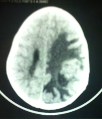
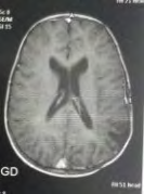
Hospital Establishment in Algiers, we grouped together 04 cases of cerebral Xanthoastrocytomas over a period of 07 years from January 2010 to December 2017. For all the patients, the diagnosis was made after surgery. All patients received the adjuvant treatment. All patients underwent routine physical examination; they also received a thorough neurological evaluation. A special neuro-surgical sheet was applied in all cases included in the study to cover all suspects needed. CT as well as MRI, with and without contrast enhancement, was done for all cases before surgery (Figure 1).
Results
Between January 2010 and 2017, 04 patients with pleomorphic xanthoastrocytomas were operated, 03 (75%) patients were males and 01 (25%) were females. The mean age was 14 years (ranged 07 –23 years). A male and pediatric predominance were observed in our cases. We had 75% child and 25% adults. The most frequent clinical manifestations
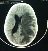
Here is the imagery of the 07-year-old little KA, with no particular history who presented a HIC syndrome of headache, vomiting for 02 months, with clinical examination stage II papillary edema in the funds without an associated neurological deficit. Preoperative brain CT as well as MRI
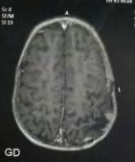
found were intracranial hypertension syndrome without neurological deficit. All our patients underwent radiological exploration such as CT and MRI of the brain (Figures 1 and 2). For all our patients, the diagnosis of glioma was made after imaging. All our patients were operated, the diagnosis of pleomorphic Xanthoastrocytoma was made after the pathological study. 75% experienced tumor recurrence after one year despite postoperative adjuvant treatment
supported a glial malignant process. The patient was operated on with favorable post-operative consequences. Referred for radiotherapy after an anatomical pathological study of the operative specimen which returned in favor of a Pleomorphic Xanthoastrocytoma.
Review of Literatures
Pleomorphic Xanthoastrocytoma is a low grade glial tumor (grade II, according to the WHO classification), it represents 0.5 to 1% of all tumors of the central nervous system [1]. It is mainly found in adolescents and young adults, with an average age at diagnosis of 12 years, with a sex ratio of 1. It develops from the supportive glial cells of the central nervous system (astrocytes). Its most frequent location is hemispherical cerebral, but it can be found at the spinal level [2]. Usually has two parts: cystic and fleshy. The fleshy part presents a leptomeningeal attachment. It is most often manifested by HIC syndrome, neurological deficit and epileptic seizures [3]. Generally, on CT, it is hypo or isodense, enhancing after injection of contrast product, more or less well limited, with peri-lesional edema, can be the cause of scalloping of the covering bone. On MRI it is hypo or iso intense in T1, iso or hyperintense in T2, on angiography the vascularization appears to come from the branches of the meningeal arteries. Treatment is based on surgery (total excision) and postoperative radiotherapy. Xanthoastrocytoma is often confused with Glioblastoma, but due to its solid constitution and its often superficial location, it has a better prognosis with a 5-year survival rate of 70- 80%. Nevertheless, a malignant transformation is described in 20% of cases [4].
Conclusion
Pleomorphic Xanthoastrocytoma is a low grade glioma. It is interesting the big child and the young adult. Often confused with Glioblastoma, but has a better prognosis with a 5-year survival rate of 70-80%, nevertheless a malignant transformation is possible, hence the interest of early diagnosis and treatment, in particular excision complete.
References
-
Michael Yang MH, Ash Singhal, Shahrad Rod Rassekh, Stephen Yip, Patrice Eydoux, et al. (2012) Possible differentiation of cerebral glioblastoma into pleomorphic xanthoastrocytoma: an unusual case in an infant, Case report. J Neurosurg Pediatrics 9(5): 517-523.
-
Bucciero A, De Caro M, De Stefano V, Tedeschi E, Monticelli A, et al. (1997) Pleomorphic xanthoatrocytoma: clinical, imaging and pathological features of four cases. Clin Neurol Neurosurg 99(1): 40-45.
-
Giannini C, Scheithauer BW, Burger PC, Brat DJ, Wollan PC, et al. (1999) Pleomorphic xanthoastrocytoma: what do we really know about-it? Cancer 85(9): 2033-2045.
-
Tonn JC, Paulus W, Warmuth-Metz M, Schachenmayr W, Sörensen N, et al. (1997) Pleomorphic xanthoastrocytoma: report of six cases with special consideration of diagnostic and therapeutic pitfalls. Surg Neurol 47(2): 162-169.
- A Review of Gene Therapy for Parkinson's Disease to Control Dopaminergic Neurons
- Late-Onset Myasthenia Gravis in a Patient with Recurrent Breast Cancer: A Case Report
- Covid-Induced Dystonia and Opsoclonus: A Case Report
- Generalized Tonic-Clonic Seizure in a Pediatric Patient with Sunflower Syndrome: A Case Report
- Comparison of Doppler Guided Seldinger Technique Versus Classic Palpatory Seldinger Technique for Radial Artery Cannulation-an Open Label Randomized Controlled Trial
- Brown Sequard Syndrome: Understanding the Complexities of Spinal Cord Injury