Morphoanatomical Characterization of an Ectomycorrhizal Species of Tricholoma (Tricholomataceae) from Kashmir Himalaya, India
The present study includes the comparative study of characteristic field ectomycorrhizae between Tricholoma aurantium associated with Cedrus deodara and Tricholoma pardinum with Pinus wallichiana. The carpophores and their ectomycorrhizal morphotypes were collected under their respective host plants. These species were taxonomically identified as Tricholoma aurantium and Tricholoma pardinum. These species are unique in their characters such as Tricholoma aurantium is characterized by growing carpophore in ectomycorrhizal association with Cedrus deodara. Its pileus is yellow orange (4A8) with non-striate, involute margin. The lamellae changes from white to rusty brown, the stipe consists fluid like substance at surface with mycelium. The cystidial elements are frequently present in infected roots while rhizomorphs are not found. Tricholoma pardinum is characterized by tiger spotted scales covering the entire pileus, with unchanging flesh, interveined notched lamellae. It has been found in ectomycorrhizal association with Pinus wallichiana with frequently present rhizomorphs and absence of cystidia on mantle of root. Morpho-anatomical description of ectomycorrhizae and carpophores are illustrated in this paper. The Tricholoma aurantium associated with Cedrus deodara is first time morpho-anatomically described from Kashmir Himalaya while Tricholoma pardinum is first time recorded from India.
Introduction
The Kashmir Himalaya being located at the bio- geographically pivotal position, representing a unique biospheric unit in the North -Western Himalayas Rodgers WA, et al. [1], lies between 33°20’ and 34°54’N latitude and 73°55’ and 75°35’E, longitudes covering an area of 15,948km2 with forest area about 8,123Km2. The forests of Kashmir Himalaya are dominated with conifers with dominant members as Cedrus deodara, Pinus wallichiana and Pinus gerardiana with pockets of Picea smithiana, Abies pindrow, Abies spectabilis, Betula utilis, Taxus wallichiana, and Rhododendron anthopogan. The best season for the collection of ectomycorrhizal agarics starts in mid April and ends in November. The Kashmir Himalaya is a hot spot for the diversity of ectomycorrhizal agarics particularly tricholomas. Tricholoma aurantium and T. pardinum have been reported from pure coniferous forest under Cedrus deodara and Pinus wallichina. The coniferous forests are dominant in the Kashmir province. The ectomycorrhizal hosts belong to Pinaceae, especially with Cedrus deodara , Pinus wallichiana , Picea smithiana, Abies pindrow and Betulaceae. The Kashmir Himalaya harbors a hub of diversity of agarics and their ectomycorrhizas. The present study was undertaken to document the ectomycorrhizal diversity of Kashmir forests associated with Cedrus deodara and Pinus wallichiana in the Kashmir Himalaya, India. Watling R, et al. [2] had published a comprehensive list of 119taxa of mushroom from Jammu and Kashmir and the list was further extended to 145species by Beig MA, et al. (2008) [3] with the addition of 26taxa [4] added more taxa to this list thereby extending the list to 250 from the state of Jammu and Kashmir.
Tricholoma (Fr.) Quel is an ectomycorrhizal genus that is characterized by fairly fleshy, white spored agaric which is growing in forests and distributed worldwide. They are ectomycorrhizal, associated with various species of coniferous and broad-leaved forests. These ectomycorrhizal agarics are more common under Pinus wallichiana and Cedrus deodara, thus these have been targeted for the collection. Kirk PM, et al. [5] recognized 200 species of genus Tricholoma world over. The recent Mycobank (http://www. mycobank.org/) shows 984species of this genus. From India this genus is reported by 13species [6, 7].
On the basis of morphotyping, 5800 fungal species belonging to 184 genera form ectomycorrhizal associations. As per Alexander I, et al. [8], 7000-10000 fungal species form ectomycorrhizal association worldwide. Recently Rinaldi AC, et al. [9] documented 343 genera including 11, 950 species, of which 252 genera belong to Basidiomycota, 84 to Ascomycota and 5 to Zygomycota that form ectomycorrhizal association. The members of Betulaceae, Pinaceae, Fagaceae, Salicaceae and Dipterocarpaceae families form ectomycorrhizal association with fungal species Buscot F, et al. [10] described mycorrhizae formation of Tricholoma sulfureum and T. vaccinum with spruce. Tricholoma aurantium is widely distributed in North America Kuo M, et al. [11]; Niazi AR, et al. [12] described putative ectomycorrhizae of T. aurantium with Pinus wallichiana and Abies pindrow from Pakistan Comandini O, et al. [13] described mycorrhizae of T. aurantium with Abies alba in Italy. About 5400 species of fungi form ectomycorrhizal associations with members of families Betulaceae, Fagaceae, Pinaceae and Myrtaceae. The morphological and anatomical characterization of ectomycorhizae and identification of fungal partner are pre- requirement for recognizing mycorrhizal diversity in an forest ectosystem. In the present paper, ectomycorhhizae of Tricholoma aurantium with Cedrus deodara and T. pardinum associated with Pinus wallichiana are documented in detail.
Materials and Methods
Study Area
The Kashmir Himalaya was taken as investigation area. The present collections were examined from Kulgam at an altitude of 2130m with coordinates 330 31.554´ N -0750. 269E and village Langate of district Kupwara at an altitude of 1791m during summer season. The agarics were collected, examined and identified in the laboratory.
Methodology for Systematics
The classification, terminology and generic concepts as given in the “Dictionary of Fungi” by Kirk PM, et al. [5] will be followed. The macroscopic & microscopic details will be worked out as per methodology given by Atri et al.. The characters pertaining to the gross morphology like, shape, colour, size of the pileus, stipe, lamellae, presence or absence of annulus and volval types, etc will be noted. The macroscopic features of the collected material will be documented on the “Field Key to Mushroom Collector”. The colour terminology of Kornerup A, et al. [14] will be used. A small portion of the collection will be preserved in a liquid preservative Hawksworth DL, et al. [15]. The major portion of the same collection was hot dried as per the standard protocol. The dried material will be packed in the cellophone sheets along with few crystals of 1,4 paradicholorobenzene or naphthalene balls. The microscopic details will be studied by cutting free hand sections of the wet preserved material. In case of dried material it is revived in 10% KOH solution. The sections will be stained with 1% Cotton Blue. Camera Lucida drawings of the sections will be done. The observations for basidia, cystidia, epicutis, hymenophoral trama, root sections, etc. will be made with the help of Camera Lucida at accurate magnification. The research work will consist of systematics including the description and illustrations of the families with its genera, section, subsections, species and varieties. A full synonymy and author citations will be quoted for each species and collection data of all specimens will be mentioned. All the collections have been deposited in the Herbarium of the Department of Botany, Punjabi University, and Patiala under (PUN) for further references.
Methodology for ECM preservation and Morpho- Anatomical Studies
The fruiting bodies of putative ectomycorrhizal agarics growing under Cedrus deodara and Pinus wallichiana have been collected. The ECM roots just beneath the fruit bodies growing on the fine feeder roots of host plant have been accessed. The ECM roots were preserved in FAA (Formalin acetic acid alcohol) for morpho anatomical studies. Morpho anatomical details of ectomycorrhizal roots were worked out as per methodology given by Agerer and Rambold, et al. (2004-2007).
Results
Key to the Investigated Taxa of Genus Tricholoma
• Carpophores growing in scattered condition in a pure coniferous forest under Cedrus deodara on humus soil;
Pileus yellow orange (4A8); Odour farinaceous; Lamellae changes to rusty brown; Rhizomorphs not observed. Cystidia present on outer mantle of root..Tricholoma aurantium. • Carpophores growing in groups in ECM association with Pinus wallichiana in coniferous forest; Pileus reddish grey (9B2) with pinkish to violet shade; Odor mild; Lamellae unchanging; Rhizomorphs frequently present; Cystidia not found on outer mantle of root... Tricholoma pardinum.
Tricholoma aurantium (Schaeff.) Ricken, Die Blätterpilze 1: 332 (1915)
[MycoBank No. 356852; Legitimate] Figures 1&2 Carpophores 4.5-11.0cm in height Pileus 2.8-7cm broad, convex to flattened depressed; surface brownish (6D7) towards margin, yellow orange (4A8) in center, irregular, splitting at maturity, non-straite, sticky when young; involute, areolate, dry, brown colour appressed fibrillose scales cover entire pileus surface; cuticle half peeling; flesh offwhite, unchanging, upto 0.8cm thick; odour farinaceous. Lamellae adnexed, subdistant, unequal, not in series, moderately broad (upto 0.3cm broad), yellowish white (3A2), changing; changes to rusty brown, tinges of yellow colour on gill edges, white fragments on gill surface, lamellulae present; forking absent; gill edges smooth, fragile. Spore print not found. Stipe excentric, 4.0-7.0 cm long and upto 1.2cm broad near apex, 1.5cm at middle, and 1.0cm broad at base, tapering at both ends and broad in the middle; brownish orange (6C8), brownish (7D7) colour appressed fibrillose scales on surface except at apex; surface areolate and flaring, base offwhite in color, fluid like substance at surface, basal mycelium white to off-white.
Basidiospores 3.7-6.0×3.0-4.5µm (Q=1.23), subglobose, single walled, smooth; inamyloid; apiculate, apiculus 0.5-0.7µm long. Basidia 16.0-25.0×4.5-6.0µm, clavate, tetrasterigmate, granular, basal clamps absent; sterigmata 3.0-4.5 µm long; acute type. Cheilocystidia 22.0–29.0 × 4.5–6.5 µm, clavate, clavate to cylindrical having inflated apices. Pleurocystidia absent. Gill edge heteromorphous. Hymenophoral trama regular. Pileus surface hyphal, gelatinized (ixocutis) made up of horizontally placed 0.9- 18.0 µm, interwoven, broad septate hypha. Pileus context hyphal made up of sub-horizontally placed interwoven, 0.9-19.0µm broad septate hyphae interspersed with celluar cells. Stipe cuticle hyphal, made up of longitudinally placed loosely interwoven 6.0-16.0 µm broad septate hyphae. Context hyphal made up of parallarly running, tightly woven, 9.0-16.0µm broad septate hyhae. Clamp connections absent throughout.
Collection Examined: Jammu and Kashmir: Kupwara,village
Langate (1791 m) 34022’42‶N– 74018’32E, growing in scattered condition in a pure coniferous forest in ECM association with Cedrus deodara on humus soil during summer season, Hilal Ahmad Rather, PUN ( 10778), June 19th, 2017.
Distribution and Ecology: Tricholoma aurantium growing scattered or gregariously and in clusters in mycorrhizal symbiosis with conifers, quite extensively dispersed in northern and montane North America includes the Rocky mountains and the Appalachians Kuo M, et al. [11]. This species is widely distributed in North America Kuo M, et al. [11]. The ectomycorrhizal characterization of T. aurantium has been documented with Pinus wallichiana and Abies pindrow from Pakistan Niazi AR, et al. [12]. This species also form ectomycorrhizal association with silver fir (Abies alba) in Italy Comandini O, et al. [13]. From India, it has been reported from Kashmir Abraham, et al. 1993. The presently worked out collection have been found growing in scattered condition in a pure coniferous forest under Cedrus deodara on humicolous soil during summer season at an altitude of (1791m). Edibility: The mushroom should be considered to be toxic Boa E, et al. [16].
Description of putative Ectomycorrhizae: Tricholoma aurantium (Schaeff.) Ricken, Die Blätterpilze with Cedrus deodara (Roxb. ex D. Don.) G. Don.
Morphological characters: Mycorrhizal system dichotomously branched to irregularly pinnate with 0-1 order of ramification, upto 13.0-20mm long; main axes 3.0-5.0mm in diam. Unramified ends sinous to constricted between older and young parts and tapering to enlarged at tip, 0.5-0.9mm in length and 0.1-0.3mm in diam; tips rounded to slightly pointed. Surface of unramified ends not smooth, loosely hairy and covered with soil particles, younger mycorrhizae grayish brown to redish gray and older dark gray to deep black, mycorrhizae color changes to redish brown on bruising or injury, no latex or any other fluid exuded when injured; mantle not transparent, cortical cells not visible; mantle hydrophobicity absent, root tip rounded, straight to tapering, mostly swollen, dark brown to deep black, mantle dots present, reddish brown, carbonisation absent. Emanating hyphae present, not specifically distributed, cystidia present. Rhizomorphs not observed. Sclerotia not found.
Anatomical characters in cross section: Mantle thickness 56.6–65.5µm, differentiated into outer mantle layer and inner mantle layer. Outer mantle layer 20.9.–35.7µm thick, almost plectenchymatous, with patches of rounded cells representing type F Agerer and Rambold, et al. (2004-2007).; giving rise to prominent emanating hyphae and cystidia, tightly arranged, ampullate, not gelatinous; hyphal cells 2.0–4.5µm thick in outer mantle layer, smooth, no contents present inside, septate, thick walled (0.7µm), not constricted at septa,septa clamps absent; septa as thick as hyphal wall. Inner mantle layer 28–46µm, pseudoparenchymatous representing type K Agerer and Rambold, et al. (2004- 2007).; hyphal cells hyaline, thin walled, variable in shape measuring 4.5–13.5µm, tangentially and 3–5µm radially. Emanating hyphae 3.0–6.0µm in diam; longitudinally placed, woven, septate with normal ends, slightly swellings, thick walled upto (0.8 µm), ramified, not constricted, clamps absent throughout. Cystidia 14.9–26.8 × 3.7–6.0µm, arising from outer mantle, the most distinct and often frequent with type 1(Agerer and Rambold, et al. (2004-2007), unramified, clavate to fusiform representing type F Agerer and Rambold, et al. (2004-2007), agranulated, smooth, thick walled upto (0.2µm), septate to aseptate without clamps. Rhizomorphal hyphae not observed.
Longitudinal section: Mantle thickness 56.6–65.5µm, differentiated into outer mantle layer and inner mantle layer. Outer mantle layer 20.9–35.7µm thick, almost plectenchymatous with patches of rounded cells representing type F Agerer and Rambold, et al. (2004-2007), giving rise to emanating hyphae and cystidia, hyphal cells 2-5µm in diam., smooth, no contents inside as no contents present in case of sporophores hyphal cells. Inner mantle layer 28-46µm, pseudoparenchymatous possessing angular to rounded cells representing type K Agerer and Rambold, et al. (2004-2007), hyphal cells thick, hyaline, variable having rounded, angular, elliptic to oval in shape measuring 4.5- 13.5µm tangentially and 3-5µm radially. Labyrinthine Hartig net hyphae protruding towards endodermis around cortical cells, measuring 19–38.7µm tangentially and 13.5–31.2µm radially, rounded, oval to elliptic in shape. Epidermal cells measuring 3–6µm tangentially and 3-5µm radially, angular to elliptic in shape. Tannin cells in 1-2 rows, measuring 17.9–47.7µm tangentially and 3–6µm radially, oval, elliptic to cylindrical in shape and oriented parallarly. Root tip mantle upto 75µm thick, different from rest of the mantle, plectenchymatous with prominent emanating hyphae and cystidia representing type D Agerer and Rambold, et al. (2004-2007).
Colour reaction with different reagents: FeSO4 (no reaction); Ethanol (70%): brownish orange, KOH (10%): brownish green; Melzer’s reagent: brownish gray, Cotton blue: bluish green, Acetic acid: (no reaction).
Remarks: All the morphological and internal details of the present worked out taxa are similar to the description given for Tricholoma aurantium (Schaeff.) by Kuo M, et al. [11]. Putative ectomycorrhizae of T. aurantium are reported as new record from Kashmir Himalaya, India. The ectomycorrhizae T. aurantium have already been described Niazi AR, et al. [12] with Abies pindrow from Pakistan. The putative ECM of this species have been described by (Uhl, 1988) with Picea sp. Reshi ZA, et al. [17] also collected this species under Cedrus deodara from Kashmir Himalaya but did not described field ectomycorrhizae and morphological and anatomical characterization of ECM of Tricholoma aurantium with Cedrus deodara in detail. Presently, the ectomycorrhizae of this species have been illustrated morphologically and anatomically in detail. The ECM of Tricholoma aurantium are reported as a new record from Kashmir Himalaya. Here C. deodara is reported as a new host for this species.
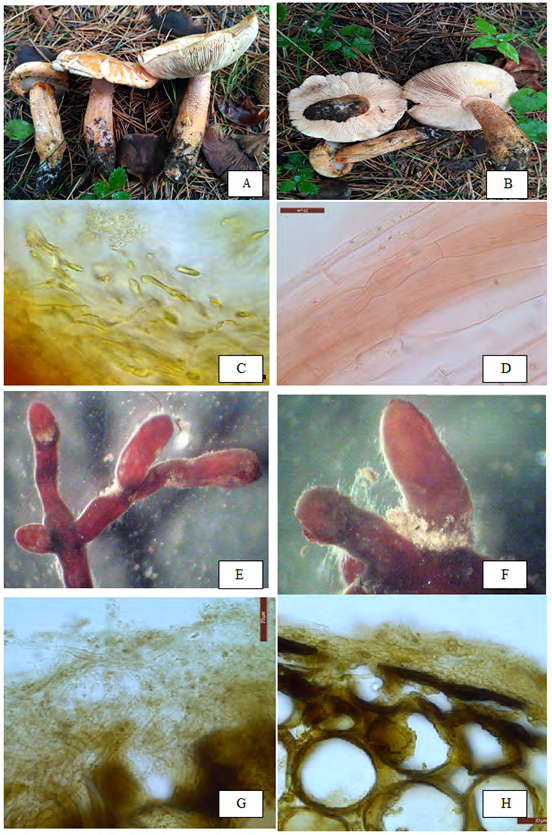
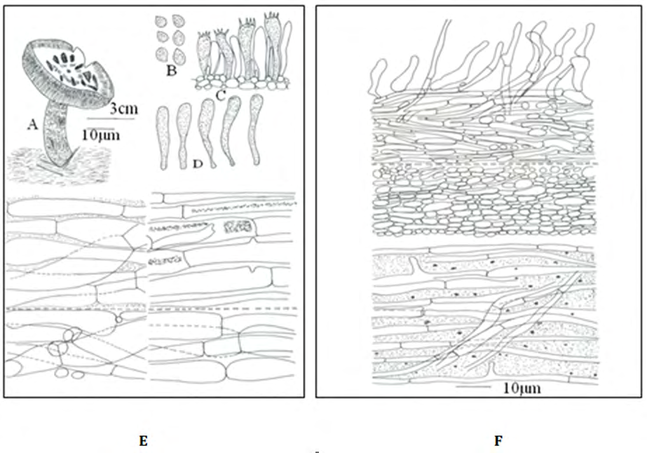
Figure 1: Tricholoma aurantium: (A) Carpophores in groups. (B) Underview of cap with adnexed to notched lamellae. (C) Pileus cuticle an ixocutis. (D) Stipe cuticle hyphae closely septate. ECM (Tricholoma aurantium+ Cedrus deodara): (E) Dichotomously branched ecto- mycorrhizal system. (F) Emanating hyphae proximally positioned. (G) Plectenchymatous mantle type ‘F’. (H) Hartig net (hn) between cortex cells of root and parallarly oriented layers of tannin cells.
Tricholoma pardinum (Pers.) Quél., Mém. de la Soci. d’Émul. de Montb.5: 339, 1873. [Mycobank No.356853; Legitimate] Figures 3 & 4 Synonymy: =Agaricus myomyces var. pardinus Pers., Syn. meth. fungor.: 346, 1801.=Tricholoma pardalotum Herink & Kotl., Ceská Mykologie 21 (1): 5, 1967.
Carpophores 3.0-4.5cm in height. Pileus 2.0-3.0cm broad, convex, incurved margin when young; umbo absent; surface reddish grey (9B2) with pinkish to violet shade; scaly, scales, appressed fibirillose, tiger spotted, cover the entire pileus, reddish brown (9E4); velvety; dry; margin regular; cuticle half peeling; flesh up to 0.1cm thick, white, unchanging; odor and taste mild. Pileal veil appendiculate. Lamellae up to 0.4 cm broad, adnexed to notched, distant, and unequal, interveined, white (2A1) with pinkish tinge, not grey spotted, furcate, unchanging; lamellulae present. Gill edges smooth, fimbriate to wavy near margin. Stipe excentric, 3.0-3.5cm long, up to 1.0cm broad in middle, up to 0.3cm broad near base; clavate, slightly tapering downward; surface white (15A1), unchanging; scaly, scales fibrillose, off white; solid; exannulate.
Basidiospores 5.81-8.3×4.15-5.81µm, Q=1.4, ellipsoidal, granular, double walled, thick, non–guttulate, inamyloid; apiculate, apiculus up to 0.83µm long, eccentric. Basidia 25.0-36.52×5.0- 8.3µm, clavate, granular, thick walled, bi to tetrasterigmate, commonly tetrasterigmate; sterigmata 2.49- 3.32µm long, hyaline. Pleurocystidia and cheilocystidia absent. Hymenophoral trama regular. Gill edge sterile.
Pileus cuticle cellular, ixocutis, made up of hyaline, spherical cells intermixed with septate, hyaline hyphae, giving rise to the regular turf of 8.3–13.28µm broad, closely septate, hyaline, encrustated hyphae; context hyphal, made up of 9.96–18.26µm broad, septate, gelatinized, irregularly placed, hyaline, inflated hyphae intermixed with broad, hyaline spherical cells. Stipe cuticle hyphal, made up of 3.2–6.4µm broad, septate, granular, longitudinally placed hyphae; context hyphal made up of 3.2–9.6µm broad, hyaline, longitudinally placed hyphae. Clamp connections absent.
Collection Examined: Kulgam, Banimulla (2130m), 330 31.554´ N–075000.269E, growing in groups in ECM association with Pinus wallichiana in coniferous forest, 18th May, 2013, Nazir Ahmad Malik, PUN 9079.
Edibility: It is a poisonous species (Alder, 1960).
Distribution and Ecology: Zeitlmayr L, et al. [18] found Tricholoma pardinum in ectomycorrhizal (ECM) association with fir, beech and conifers during summer and autumn in South Europe while Lamaison JL, et al. [19] have found this species growing solitary or in groups or fairy rings from France, Belgium Germany and North America. In India, presently this species have been found growing in groups in putative ECM association with Pinus wallichiana during starting summer in coniferous forest of Jammu and Kashmir at an altitude of 2130m.
Description of Putative Ectomycorrhizal root system of Pinus wallichiana with Tricholoma pardinum (Pers.) Quél
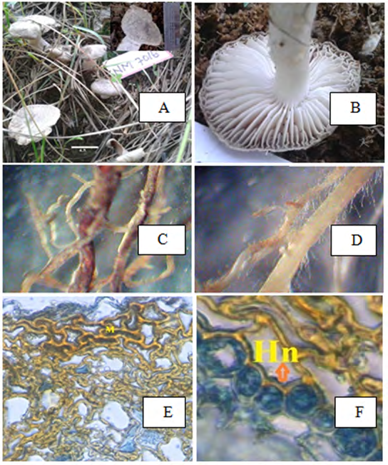
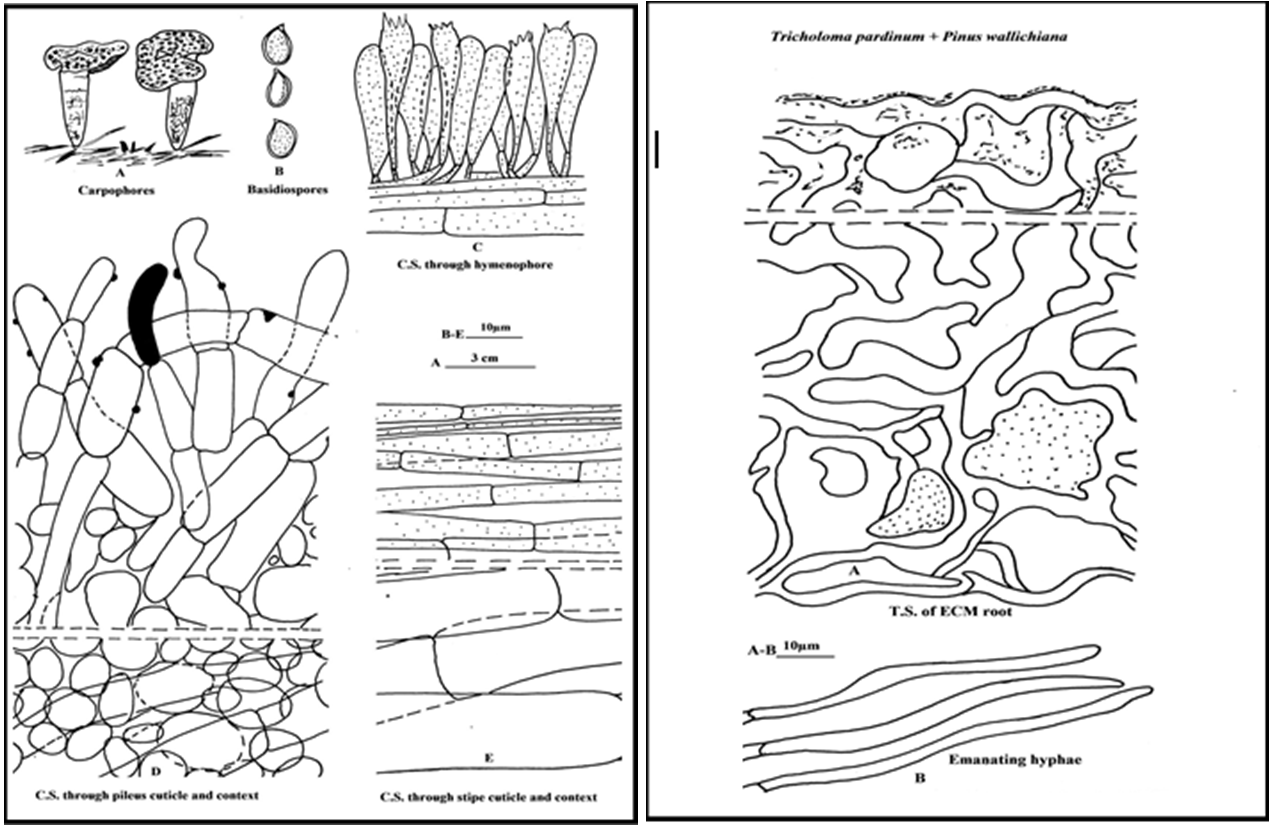
Morphological characters: Ectomycorrhizal system hydrophobic, dichotomously branched up to 3.0mm long, ectomycorrhizea small to large in numbers, clusters, coralloid; main axis 2.0mm diameter; Unramified ends 1.0-1.5mm diameter, bent, sineous with pointed tips, reddish brown with silvery appearance, glabrous, mantle transparency opaque; cortex cells are not visible; appearance dense cottony. It does not consist debris of soil particles. Emanating hyphae present. Rhizomorphs frequently present. Sclerotia absent. Anatomical characters: Mantle is plectenchymatous, Type ‘H’. Mantle 155.42–204.5µm thick, non-gelatinized, Pseudoplectenchymatous net of coarse and irregularly shaped hyphae at an angle of 1200. Hartig net extending deep up to many layers of cortex. Cystidia not seen. Emanating hyphae, 3.2–4.8µm broad, septate, narrow, thick walled, rarely present. Chemical reactions: Potassium hydroxide: The mantle becomes a dull brown. Melzer’s Reagent: There is a little reactivity with Melzer’s Reagent. Sulfovanillin: ECM becomes reddish-brown with sulfovanillin expect the oldest roots. Remarks: All the morphological and internal details of the present worked out collection are similar to the description given for Tricholoma pardinum (Pers.) Quél. By Bas et al. (1999); Arora (1986). This collection is characterized by reddish grey (9B2) with pinkish to violet shade cap having reddish brown (9E4) tiger spotted scales covering the entire pileus, white unchanging flesh, notched lamellae, interveined and are not spotted grey. Although present specimen shows variation in the basidispore size which is 7-9.5(10)×5-6.5(7) µm in the literature while it is 5.81- 8.3 × 4.15-5.81µm in presently examined specimen but its all other characters matches and fits well with the T. pardinum as given by Bas et al. (1999); Arora (1986) in their description. It has also been founded from southwestern Turkey Solak MH, et al. [20], China Deng H, et al. [21] and Japan Miyauchi S, et al. [22]. This species is first time recorded from India.
Discussion
Carpophores and their ECM root surveys carried out during the present study resulted in identification of Tricholoma aurantium associated with Cedrus deodara and Tricholoma pardinum with Pinus wallichiana. Previously, the putative ECM of Tricholoma aurantium have been described by Uhl M, et al. [23] with Picea species. Hence Cedrus deodara is reported as a new host for this species. Itoo ZA, et al. [17] collected this species under Cedrus deodara from Kashmir Himalaya but they did not described field ectomycorrhizae and morphological and anatomical characterization of ECM of Tricholoma aurantium with Cedrus deodara in detail. There is no an earlier record of Tricholoma pardinum from India, hence it constitutes a new addition to the tricholomas of India. Earlier, Zeitlmayr L, et al. [18] reported Tricholoma pardinum in ECM association with fir and beech but presently it has been found in having putative ECM association with Pinus wallichiana. Abraham and Kaul (1985) listed 175 taxa of agarics from the Kashmir of which 53 were found to form ectomycorrhizal associations with tree species. Watling and Abraham (1992) reported 77 ECM taxa from Kashmir forests. Beig MA, et al. [3] reported 24 ectomycorrhizal species during describing mycorrhizal biodiversity of Kashmir Himalayas. Pande (2004) described 98 ectomycorrhizal species from western Himalaya out of which 55 species were found in ectomycorrhizal association with Pinus wallichiana and Cedrus deodara.
Conclusion
The present study disclosed that coniferous forests of Kashmir Himalaya, constitutes several ectomycorrhizal agaric taxa. The diverse forests of Kashmir Himalaya need to be intensively surveyed for agaric taxa springing during different seasons. The present study stresses the need for the vast and specialized exploration of these forests which could clearly lead to the detection of many new and unnoticed ectomycorrhizal agaric taxa from this diverse region.
Acknowledgements
The author’s are highly thankful to the Head Department of Botany, Punjabi University Patiala for help in identification of ECM species. The Ministry of Social Justice and Empowerment, Govt of India for providing financial assistance under the scheme of “National Fellowship for Persons with Disabilities” (NFPwD) as SRF fellow.
References
-
Rodgers WA, Panwar SH (1988) Biogeographical classification of India. New Forest, Dehra Dun, India.
-
Watling R, Gregory NM (1980) Larger fungi of Kashmir. Nova Hedwigia Z Kryptogamenkd 32: 473-563.
-
Beig MA, Dar GH, Ganai NA, Khan NA (2008) Mycorrhizal biodiversity in Kashmir forests and some new records of macrofungi from J&K State. Applied Biol Res 10(1-2): 26-30.
-
Dar GH, Beig MA, Ganai NA (2009) Biodiversity of macrofungi in Kashmir forests-1. Indian J 32: 119-121.
-
Kirk PM, Ainsworth GC, Cannon PF, Minter DW (2008) Ainsworth Bisby’s Dictionary of Fungi 10th (Edn.), CAB International, UK.
-
Mohanan C (2011) Macrofungi of Kerala. KFRI Handbook No. 27, Kerala Forest Research Institute, India.
-
Henry LDC, Rajakumar RS (2014) Nutritional status of certain wild edible mushrooms from the eastern ghats of Tamil Nadu. Mysore Journal of Agricultural Sciences 23(2): 131-136.
-
Taylor AFS, Alexander I (2005) The ectomycorrhizal symbiosis: Life in the real world. Mycologist 19(3): 102- 112.
-
Rinaldi AC, Comandini O, Kuper TW (2008) Ectomycorrhizal fungal diversity separating wheat from the chaff. Fungal Diversity 33: 1-45.
-
Buscot F, Munch JC, Charcosset JY Gardes M, Nehls U, et al. (2000) Recent advances in exploring physiology and biodiversity of ectomycorrhizas highlight the functioning of these symbioses in ecosystems. FEMS Microbiol Rev 24(5): 601-614.
-
Kuo M, Methven A (2010) 100 Cool Mushrooms. University of Michigan Press, pp: 191-192.
-
Niazi AR, Khalid AN, Iqbal SH (2010) New records of ectomycorrhiza from Pakistan. Pakistan Journal of Botany 42(6): 4335-4343.
-
Comandini O, Pacioni G, Rinaldi AC (1998) “Fungi in ectomycorrhizal associations of silver fir (Abies alba Miller) in Central Italy”. Mycorrhiza 7(6): 323-328.
-
Kornerup A, Wanscher JH (1978) Methuen Handbook of Colour, 3rd (Edn.), Eyre Methuen, London.
-
Hawksworth DL, Sutton BC, Ainsworth GC (1983) Ainsworth and Bisby’ Dictionary of the Fungi. International Books and Periodicals Supply Service, New Delhi.
-
Boa E (2004) Wild Edible Fungi: A Global Overview of their Use and Importance to People. Non-Wood Forest Products Series, No. 17, FAO, Rome.
-
Itoo ZA, Reshi ZA (2014) Ectomycorrhizal Diversity Associated with Cedrus deodara and Pinus wallichiana in the Kashmir Himalaya, India. Pak J Biol Sci 17(1): 32-40.
-
Zeitlmayr L (1976) Wild Mushrooms: An Illustrated Handbook. Hertfordshire, UK: Garden City Press, pp: 72- 73
-
Lamaison JL (2005) The Great Encyclopedia of Mushrooms. Cologne, Germany Könemann, pp: 89.
-
Solak MH, Mustafa I, Fahrettin G, Isa G (1999) Macrofungi of İzmir Province. Turkish Journal of Botany 23: 383- 390.
-
Deng H, Yao YJ, Pegler DN (2004) An annotated checklist of Tricholoma from China. Journal of Fungal Research (in Chinese and English) 2(1): 1-18.
-
Miyauchi S (1997) A poisonous species, Tricholoma pardinum newly recorded in Japan. Nippon Kingakukai Kaiho 38(2): 85-86.
-
Uhl M (1988) Identification and Characterization on Ectomycorrhizae of Pinus sylvestris and ectomycorrhizae Gattung Tricholoma. Dissertation Munchen.
-
Pande V, Palni UT, Singh SP (2004) Species diversity of ectomycorrhizal fungi associated with temperate forest of Western Himalaya: A preliminary assessment. Curr Sci 86(12): 1619-1623.
-
Champion G, Harry S, Seth K (1965) Forest types of Pakistan. Pakistan Forest Institute, Peshawar, pp: 233.
-
Farjon A (1990) Pinaceae. Drawings Descriptions of the Genera. Koeltz Scientific Books.
-
Frank AB (1885) On the nutrient providing root- symbiosis between underground fungi and certain trees. Journal of the American Botanical Society 3: 128-145.
-
Hibbett DS, Matheny PB (2009) The relative ages of ectomycorrhizal mushrooms their plant hosts estimated using Bayesian relaxed molecular clock analyses. BMC Biology 7: 13.
-
Siddiqui MF, Shaukat SS, Ahmed M, Khan N, Khan IA (2013) Vegetation-Environment relationship of conifer dominating forests of moist temperate belt of Himalayan and Hindukush regions of Pakistan. Pakistan Journal of Botany 45(2): 577-592.
-
http://www.mycobank.org/.
- Enhancement of Vegetative Growth and Fruit Yield in Cucumber (Cucumis sativus L.) via Spiritual Blessing (Biofield) Energy Intervention
- Production of Açaí (Euterpe oleracea Mart.) under Different Agroforestry System Management Intensities in Amazonian Floodplain (Varzea) Forests
- Coffee and the Production Region: What is the Secret to the Expression "Quality"?
- Experiential Agripreneurship Training in Sub-Saharan Africa: Integrating a Business Incubator into Postgraduate Livestock Education at the University of Buea
- Advances in Agricultural High-Quality Development
- Linking Compost Residue to ABAGE in Plants - a Short Note