Isolation and Characterization of Dermatophytes from Students Bathing Places
Background: Dermatophyte comprises of a vast range of filamentous pathogenic fungi that infect keratinous tissues of skin (the stratum corneum layer), hair, and nail in humans and animals via their keratinase enzymes which are capable of causing superficial infections. Aim: This study aimed to isolate and characterized dermatophytes isolated from students bathing places. Methodology: Four sampling sites were chosen from both male and female students bathing places; the floor, walls, sinks and door handles. Three hundred samples were collected and cultured on sterile plates containing Potato dextrose agar using spread plate method. The plates were incubated at 350C for 72 hours. Results: Out of three hundred samples collected from the various sampling sites, a total of four hundred and ten fungi were isolated, 286 (70%) were dermatophytes belonging to three genera (Trichophyton, Microsporum and Epidermorphyton) while124 (30%) were non dermatophytes. The female bathing places yielded 167(58%) and 48(39%) dermatophytes and Candida species respectively higher than male counterparts which yielded 119 (42%) dermatophytes without any species of Candida. There was no significant difference between the isolates from female and male bathrooms P ≥ 0.05. Conclusion: Hence the students bathing places were found to be rich in pathogenic and potentially pathogenic fungi and need thorough disinfectants to avoid dermatophytes infections’ like Tinea pedis, Tinea corporis Tinea capitis and Tinea cruris.
Introduction
Dermatophytes are group of filamentous fungi that cause infections of the skin. Diseases caused by dermatophytes include athlete’s foot, ringworm, jock itch, and nail infections (onychomycosis). The medical terminology for dermatophyte infections is to use the word tinea (to denote a fungal infection of the skin) followed by a word that describes the site of infection [1]. For example, Tinea pedis refers to athlete’s foot and Tinea capitis is scalp ringworm. In general, dermatophytes remain localized to keratinized surfaces such as skin, hair, and nails and do not invade deeper tissues [1]. That said, dermatophyte infections in immune compromised patients can be quite severe. Dermatophytes are grouped into three general categories based on their natural environment: anthropophilic (live exclusively on humans), zoophilic (live on an animal host), and geophilic (live in the soil) [1]. The majority of human infections are caused by anthropophilic species; however, species from all three groups have been associated with human disease. Fungal infections of the skin, hair, and nails due to dermatophytes are common problem across the globe. Dermatophyte comprises of a vast range of filamentous pathogenic fungi including three important genera of Epidermophyton, Microsporum, and Trichophyton which may lead to superficial infections in both humans and animals-zoonosis [2]. However, Pityriasis versicolor, Saccharomyces cerevisiae, and Candida spp. as opportunistic pathogenic fungi are capable of causing superficial mycotic infections in human beings [3, 4]. Dermatophytes as keratinophilic fungi are able to infect keratinous tissues of skin (the stratum corneum layer), hair, and nail in humans via their keratinase enzymes. They also degrade claws, feathers, hooves, horns, wools in animals. Dermatophytes are fungal agents of dermatophytoses.
Superficial mycoses of dermatophytoses are named after anatomic localization of the lesions. Dermatophytosis (tinea) is a general name for acute to mild and chronic lesions of the outer layers of keratinized tissues caused by dermatophytes. Dermatophytoses include Tinea barae, Tinea faciei, Tinea capitis, Tinea favosa, Tinea corporis, Tinea cruris, Tinea manuum, Tinea pedis, and Tinea unguium [4]. In recent years, there are a vast range of traditional and modern diagnostic approaches and treatments for managing superficial infections of dermatophytes. Although dermatophytes are not life threatening microbial agents but they are distributed around the world and cause mycotic infections with high morbidity. The prevalence of dermatophytosis can vary from one climatic condition or lifestyle to another [5]. Such life styles include poor hygienic conditions, communal sharing of hair-care tools, close contact with animals, playing with sand among others. Dermatophytes are found in the different environment with variable distribution patterns depending on various factors the most important is human association. Bathing places contribute to spread of dermatophytes infections like Tinea pedis, Tinea barbae, Tinea faciei, and Tinea capitis because of common use of them. Thus the environmental sanitation of various bathing places is important.
Materials and Methods
Study Area
The study was carried out in Novena university Ogume, Delta state Nigeria. Novena university is one of the private universities in Nigeria located in Ukwuani local government, Ndokwa west, Delta state Nigeria.
Collection of Samples
Three hundred samples were collected from the floors, walls, door handles and sinks of bathrooms in students’ hostels (one hundred and fifty samples each from both male and female hostels) of Novena University Ogume, Delta state Nigeria using sterile swab sticks. A total of one hundred and twenty samples were collected from the male and female bathroom walls while one hundred samples were from bathroom floors. Total of 60 and 20 samples were collected from door handles and sinks respectively.
Fungal Isolation
Each sample was cultured on sterile plates containing Potato dextrose agar using spread plate method. The plates were incubated at 35oC for 72 hours. Pure colonies were sub-cultured and slanted in potato dextrose agar for further studies.
Characterization of the Isolates
Macroscopic Examination of Fungal Isolates: The different characteristics of the colonies were examined. These included the colour and texture of the colony on the surface of the medium used, nature and colour on the reverse side, rate of growth, consistency, presence of pigments nature and shape of fruiting bodies and other peculiar features of the colony. These were noted as guide to final identification of isolates.
Microscopic Examination of Fungal Isolates: A drop of lactophenol cotton blue solution was placed on the center of clean grease free slide. The mycelia mats of the isolates were collected with sterile needle and placed on the lactophenol blue drop and teased. Sterile cover slip was placed on the material and passed through a flame. The slide was examined under the microscope using ×10 and ×40 objective lens.
Slide Culture Technique
Slide culture was prepared to examine and identify the fungi colonies in-situ with as little disturbance as possible. Sterile filter paper was aseptically placed in petri dish with a pair of forceps and sterile V-shaped glass rod was placed on the filter paper. About 3ml of sterile water was poured on the filter paper to completely moisten it. With forceps, sterile slide was placed on the V-shaped rod. The scalpel was sterilize by flaming and gently used to cut a 5mm square block from the plate of already prepared sabouraud dextrose agar fortified with 0.5µg/ml chloramphenicol. The agar block was aseptically transferred to the center of the slide. Four sides of the agar square were inoculated with mycelia fragments of the fungus to be examined using sterile needle. Sterile cover slip was aseptically place on the upper surface of the agar cube. Then the petri dish was covered and incubated for 48 to 72 hours. After incubation a drop of lactophenol blue stain was placed on a clean microscopic slide. The cover slip was removed from the slide culture and placed side down on the drop of lactophenol blue stain on the slide. The block of agar was discarded and lactophenol blue was drop on the slide and covered with sterile cover slip. The two slides preparations were sealed with nail polish and were examined under low power ×10 and high power ×40 objective lens.
Statistical Analysis
The statistical analysis was done using SPSS (statistical package for social sciences) version 21.0. The data collected were analyzed using chi- square test, t- test and one way anova. The results were tested for significant levels of 0.05.
Results
Out of three hundred (300) samples collected from students’ bathrooms floors, walls, door handles and sinks. A total of four hundred and ten fungi were isolated. Out of these fungi 286 (70%) were dermatophytes belonging to three genera (Trichophyton, Microsporum and Epidermorphyton) while124 (30%) were non dermatophytes. The dermatophytes consist of Trichophyton mentagrophytes 142 (50%), Trichophyton rubrum 61(21%), Microsporum gypseum 32(11%), Microsporum canis 5(2%), Trichophyton verrucosum 19(7%), Epidermorphyton floccosum 7(2%), Trichophyton concentrichum 3(1%), Trichophyton erinacei 12(4%) and Trichophyton schoenlainii 5(2%) as shown in Figure 1.
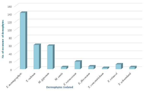
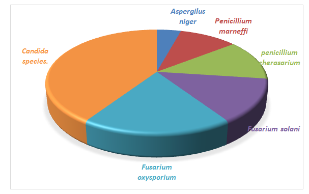
Figure 2 shows the frequency of non-dermatophytes isolated, which include Aspergillus niger 6(5%), Penicillium marneffi 17(14%), Penicillium cherasanum 15 (12%), Fusarium solani 16(13%), Fusarium oxysporium 22 (18%) and Candida species 48 (39%).
Tables 1 & 2 show the morphological characteristics of dermatophytes and non-dermatophytes respectively, with respect to the mould texture, colour on the surface, colour on reverse and the pigment production.
| Texture of growth | Rate of growth | Colour on surface | Colour Reverse | Pigmentation | |
|---|---|---|---|---|---|
| Trichophyton concentrichum | raised and folded | slow | white / cream | yellow/ brown | - |
| Trichophyton Schoenlaini | waxy, folded | slow | white/ cream | no colour | - |
| Trichophyton erinacei | flat, powdery downy to fluffy | slow | white | lemon yellow | - |
| Trichophyton Verrucosum | velvety, raised centre | slow | white/ cream | yellow | + |
| Trichophyton Mentagrophytes | powdery, granular, central folding | slow | white/ cream | reddish- brown | - |
| Trichophyton tonsuran | powdery, radially or irregular furrowed | slow | dark brown | reddish brown | - |
| Trichophyton rubrum | downy, heaped and folded | slow | white | yellow | + |
Table 1: Characteristics of Dermatophytes Isolated.
| Fungi species | Texture of growth | Rate of growth | Colour on surface | Colour reverse | Pigmentation |
|---|---|---|---|---|---|
| Aspergillus niger | powdery | rapid | black | black | - |
| Fusarium oxysporum | powdery | rapid | white | dark purple | + |
| Penicillium cherasanum | downy | rapid | rapid | green with white edges | + |
| Penicillium marneffi | downy Suede-like | rapid | white with yellowish green | wine red | + |
| Fusarium solani | powdery | rapid | white | Brown | + |
Table 2: Characteristics of non-Dermatophytes Isolated.
| Dermatophytes | Walls | Floors | Sink | Door handle | Total |
|---|---|---|---|---|---|
| T. mentagrophytes | 38 | 18 | 30 | 56 | 142 (50%) |
| T. rubrum | 20 | 26 | 4 | 11 | 61(21%) |
| M. gypseum | 6 | 5 | 5 | 2 | 18(6%) |
| M. canis | 3 | 3 | 2 | 1 | 9(3%) |
| T. verrucosum | 2 | 6 | 3 | 1 | 12 (4%) |
| E. floccusum | 3 | 8 | 1 | 2 | 14(5%) |
| T. concentrichum | 8 | 2 | 0 | 0 | 8(3%) |
| T. erinacei | 5 | 4 | 2 | 3 | 14(5%) |
| T. schoenlainii | 2 | 0 | 4 | 2 | 8(3%) |
| Total | 85(30%) | 72 (25%) | 51(18%) | 78(27%) | 286(70%) |
Table 3: Distribution of Dermatophytes according to samples.
Table 3 shows the distribution of dermatophytes according to samples, 85 (30%) dermatophytes were isolated from bathroom walls, 72 (25%) floors, 51(18%) from sink and 78(27%) from door handles. The dermatophyte with highest frequency was Trichophyton mentagrophytes 142 (50%), followed by T. rubrum 61(21%).Bathroom sinks produced the lowest number of dermatophytes while the bathroom walls had the highest number; Trichophyton concentrichum was isolated only from the bathroom walls and floors while T. schoenlainii were not isolated from bathroom floors. Only one M. canis and T. verrucossum were isolated from door handles.
Table 4 shows the distribution of dermatophytes isolated from different bathrooms according to gender. The female bathroom has higher frequency of 167(58%) dermatophytes isolated while 119 (42%) dermatophytes were isolated from male students’ bathrooms. M. canis and Trichophyton concentrichum were not isolated from male bathrooms. The only dermatophyte that was not present in female bathing places was T. schoenlainii. The most abundant and frequently isolated species in the bathing places were Trichophyton mentagrophytes 142 (50%), Trichophyton rubrum 61(21%).
| Females | Males | Total | |
|---|---|---|---|
| T. mentagrophytes | 82 | 60 | 142 |
| T. rubrum | 43 | 18 | 61 |
| M. gypseum | 15 | 17 | 32 |
| M. canis | 5 | 0 | 5 |
| T. verrucosum | 6 | 13 | 19 |
| E. floccusum | 5 | 2 | 7 |
| T. concentrichum | 3 | 0 | 3 |
| T. erinacei | 8 | 4 | 12 |
| T. schoenlainii | 0 | 5 | 5 |
| Total | 167 | 119 | 286 |
Table 4: Distribution of dermatophytes isolated according to gender.
Discussion
Dermatophytes are among the most common causes of skin diseases in the world causing mostly superficial and cutaneous infections involving the skin, hair and nails. The occurrence of dermatophytes and some fungal species which are known to be opportunistic pathogens might be related to low concentration of disinfectants and attendance of bathers with fungal infections. The present study investigated the distribution of dermatophytes and opportunistic fungi in students bathing places as human risk factors. The results of this research were in agreement with those of previous investigations that indicated a possible relationship between this important group of fungi and the environment [6, 7]. These fungi species are pathogenic and potential pathogens to humans, animals and plants.
T. mentagrophyte and T. rubrum were two dermatophytes species that have been isolated from humans infected with cutaneous mycoses, other species such as Microsporum gypseum , Microsporum canis ,Trichophyton verrucosum , Epidermorphyton floccosum, Trichophyton concentrichum, Trichophyton erinacei and Trichophyton schoenlainii are considered to be etiological agents of opportunistic mycoses, the results are in agreement with those of similar surveys which showed that dermatophytes are especially frequent in bathing places where keratin occurs [8]. These fungi are common zoophilic dermatophytes widely distributed in both indoor and outdoor bathing places and are common causes of inflammatory or chronic non inflammatory finely scaling lesions of skin, nails and scalp. They are also common causes of tinea capitis in human and animals [9]. T. mentagrophytes has been reported as the causal agent of tinea pedis, tinea corporis, tinea curis and onychomycoses [9].
Also higher number of dermatophytes recovered in the current study 286 (70%) can be attributed to the fact that these fungi survive and grow best in moist places which is in agreement with the study of (10) who isolated more than 60% of dermatophytes from communal bathing paces. These dermatophytes are also successors on human hairs, nails and skin, as some of the students will prefer to shave and cut their nails when they enter the bathing places. Maghzay, et al. isolated dermatophytes from two communal bathing places in Egypt. The isolation of the dermatophytes might be a continuous contamination of the communal bathing places by fungi through air, soil and human bodies. The dermatophytes are transmitted by contact with infected hair, formites or from the environments (spores in soil). Most of the saprophytic organisms isolated represent transient contamination by fungi from moist environment, air borne fungi and potential pathogens causing mycoses. These fungi which are human pathogen could be considered as bio indicators of environmental pollution with animal faces, hair and plant debris and can pose risk of human and animal mycoses.
Furthermore, the current study revealed differences in species biodiversity, among the four sampling sites, the highest number of dermatophytes was encountered in the walls followed by the floors. Higher contamination of the floors and walls by dermatophytes species especially T. mentagrophytes, T. rubrum, Epidermophyton floccussum and Microsporium mainly comes from the shed skin scales or fragments of bathers who usually walk bare footed on the floors. This is in accordance with the study of which isolated higher number of these organisms from the floors and walls of communal bathing places [9]. Bathers are therefore apt to contract fungal skin infections including Tinea pedis, Tinea cruris, Tinea coporis, and Tinea barbae etc. to avoid such infections people are advised not to walk into the bathing places bare footed and users should clean the floors and walls of the various bathing places frequently with disinfectants. This is in agreement with the studies of who indicated that bathroom users with dermatophytoses could spread debris containing dermatophytes on the floors and walls of bathing places [10, 11].
Moreover, this study also shows that female bathing places produced the highest number of dermatophytes, which is in accordance with the work of who isolated the highest number of dermatophytes from female communal bathing places [12]. This may be as a result of enough breeding sites like long hairs and nails harbor by women, as this organism grows effectively in keratinized tissues. The genus Trichophyton was represented by Trichophyton mentagrophytes, Trichophyton verrucosum, Trichophyton concentrichum, Trichophyton erinacei and Trichophyton schoenlainii species. These organisms are known to be major causative agents of human dermatophytosis. This is in agreement with the results that reported these species from swimming pools. Trichophyton concentrichum was only isolated from the floors of the bathing places in the present study. It has been commonly isolated by several researchers from their bathing places and swimming pools [12]. It is pathogenic dermatophytes and has been isolated from several mycotic infections. Microsporum canis, a zoophiliic species can be transmitted to humans via direct contact with infected animals and cause severely inflammatory infections, Tinea capitis and Tinea corporis [13, 14, 15]. Infections in humans, mostly in young adults after contact with the pet, guinea pigs and cats.
In this study Aspergillus niger was the second most commonly isolated species 40(9.83%). It was previously reported as one of the most dominant species in soil by Moallaei, et al. [16]. This fungal species is present in different soil samples and it is distributed worldwide. The species also occurs in external ears and involved in otitis [17]. On the other hand this species is a potential mycotoxin producer [18]. A. Niger causes skin, eye and ear infections [19]. Also in this present study no dermatophytes were isolated from female doors and male sinks, the reason may be related to the different techniques used in this research. The findings in this research showed a relationship between the number of bathrooms users and the number of isolated fungal cases. The higher number of isolated fungal cases may be related to the higher number of students using the bathing places. Therefore sanitary quality of bathing places is concerned due to spread of fungi and other organisms because of common usage of them. Thus the environmental sanitation of the bathing places is very important.
Conclusion
The existence of pathogenic dermatophytes in the students bathing places is plausible. This high degree of contamination is due to continuous use of the bathing places by many people within a restricted time period. Thus the isolation of dermatophytes in the bathing places reveals the important of these bathrooms in skin disease transmission. Therefore the environmental sanitation and disinfection of the bathing places is very important.
Acknowledgements
My profound gratitude goes to all those who helped in the preparation of this research work especially Dr. Valentine Unegbu of Department of microbiology, Spiritan University Umunneochi, Abia State, Nigeria and the entire staff of Department of Biological sciences, Novena University Ogume Delta state, Nigeria.
References
-
Achterman RR, White TC (2012) A foot in the door for dermatophyte research. PLoS Pathog 8(3): e1002564.
-
Annual Prevalence Report (2007) Birmingham Research Unit. Royal College of General Practitioners 57(537): 7-14.
-
Behzadi P, Najafi A, Behzadi E (2013) Detection and Identification of Clinical Pathogenic Fungi by DNA Microarray_._ Infectioro 35(3): 6-10.
-
Chaya AK, Pande S (2007) Methods of specimen collection for diagnosis of superficial and subcutaneous fungal infections. Indian J of Dermato Vene and Lep 73(3): 202-205.
-
Garg J, Tilak R, Garg A, Prakash P, Gulati AK, et al. (2009) Rapid detection of dermatophytes from skin and hair. BMC Res Notes 2: 60.
-
Hainer BL (2003) Dermatophyte infections. Am family physician 67(1): 101-108.
-
Sahin I, Oksuz S, Kaya D, Sencan I, Cetinkaya R (2004) Dermatophytes in the rural area of Duzce, Turkey. Mycoses 47(11-12): 470-474.
-
Zahra LV, Gatt P, Boffa MJ, Borg E, Mifsud E, et al. (2003) Characteristics of superficial mycoses in Malta. Int J Dermatol 42(4): 265-271.
-
Barnes T, Vale M (2005) Isolation and identification of fungi. Mycoses 31(10): 495-500
-
Attye A, Auger P, Joly J (1990) Incidence of occult athletes foot in swimmings. Eur J Epidemiol 6(3): 244-247.
-
Maghazy SMN, Mallek AYA, Bagy MMK (1989) Fungi in two swimming pools in Assiut town Egypt. Zientrabl Microb 144(3): 213-216.
-
Bolanos B (1991) Dermatophyte feet infection among students enrolled in swimming courses at a university pool. Bol ASO Med PR 83(5): 181-184.
-
Weitzman I, Summerbell RC (1995) The dermatophytes. Clin Microbiol Rev 8(2): 240-259.
-
Seebacher C, Bouchara JP, Mignon B (2008) Updates on the epidemiology of dermatophyte infections. Mycopathologia 166(5-6): 335-352.
-
Moallaei H, Zaini F, Pihet M, Mahmoudi M, Hashemi J (2006) Isolation of Keratinophilic Fungi from Soil Samples of Forests and Farm Yards. Iranian Journal of Public Health 35(4): 62-69.
-
Al-Musallam AA, Al-Zarban SS, Al-Sane NA, Ahmed TM (1995) A report on the predominant occurrence of dermatophytes species in cultivated soil from Kuwait. Mycopathol 130: 159-161.
-
Irshad HS, Yasmeen FK, Miandad Z, Abdul HS (2007) Isolation of Keratinophilic Fungi from Soil in Khairpur City, Sindh, Pakistan. Bangladesh J Microbiol 24(1): 79- 80.
-
Fozia I, Suhail M, Abro H (2007) Keratinophilic fungi from the soil of district, Jamshoro, Sindh (Pakistan). Pakistan Journal of Botany 39(4): 1377-1382.
-
Ali ZM, Majid Z (2008) Isolation of dermatophytes and related keratinophilic fungi from the two public parks in Ahvaz. Jundishapur Journal of Microbiology 1(1): 20-23.
- Antifungal Activity of New Acetophenone Derivatives
- Interconnected Microbiomes Human Health Within an Environmental Framework
- Silkworm-Based Vaccine Production for H5N1: A One Health Approach to Pandemic Preparedness
- Microbial Diversity and Lipolytic Activity of Bacteria and Fungi from Oil-Contaminated Sites in Makurdi Metroplois
- Antibiotic Resistance Profile of Bacteria Isolated at the Central Laboratory of the National Hospital Center of Nouakchott
- Epidemiology and Sensitivity to Antibiotics of Germs Isolated from Blood Cultures in the Laboratory of the National Hospital Center of Nouakchott-Mauritania