Isolation, Identification and Antimicrobial Resistance Profile of Pathogenic Staphylococcus Aureus from Intensive Dairy Farms in Modjo and Adama, Central Ethiopia
Staphylococcus aureus plays its most significant animal pathogenic role as cause of intra mammary infections in cattle and small ruminants leading to considerable economic losses in cattle farming. A cross sectional study was conducted from February to June 2019 in Adama and Modjo town, to isolate and identify S. aureus and their susceptible to different antimicrobials. Out of a total of 160 samples examined,20.6 % (33) were positive for S. aureus. Of this, 13/86 (15.1%), 20/74(27%) were positive for S. aureus atModjo and Adama, respectively. All 33 isolates were tested for antimicrobial resistance with 12 selected antibiotics. The isolates were highly susceptible to gentamycin (100%), streptomycin 24(72.7%), ciprofloxacin21 (63.6)), and kanamycine20 (60.6%), however, they were highly resistant to methicillin 31(93.9%), penicillin G 30(90.9%), tetracycline 21 (63.6%) and erythromycin (60.6%) respectively. Out of the resistance S. aureus isolates, 31 (93.9%) were developed multidrug resistant against 8 antibiotics. The S. aureus isolates from the dairy farms in the study area were multi drug resistant. Hence, public awareness about good management in dairy farms and milk handling practices, rational use of drugs and periodic assessment of drug sensitivity test should be done to prevent the current global issue which is antimicrobial resistance.
Introduction
Ethiopia has the largest cattle population in Africa with an estimated population of 49.3 million. Cows represent the biggest portion of cattle population of the country, around 42% of the total cattle heads are milking cows [1]. However, milk production often does not satisfy the country’s requirements due to a multitude of factors. Mastitis is among the various factors contributing to reduced milk production [2]. Bovine mastitis is an infectious inflammation or irritation of the mammary glands that interferes with the normal flow and quality of milk [3]. Among mastitis causing pathogens, the S. aureus bacterium is a major pathogen of intramammary infections in dairy cattle [4]. Staphylococci are Gram positive bacteria, with diameters of 0.5 -1.5 μm and characterized by individual cocci, which divide in more than one plane to form grape like clusters. The staphylococci are non motile, non spore forming facultative anaerobes that grow by aerobic respiration or by fermentation. Staphylococci are tolerant to high concentrations of salt and show resistance to heat. Pathogenic staphylococci are commonly identified by their ability to produce coagulase, and thus clot blood .This distinguishes the coagulase positive strains, S. aureus, from the other staphylococcal species such as Staphylococcus epidermidis that are coagulase negative [5]. It is both commensal and pathogen. It is found as a commensal associated with skin, skin glands and mucous membranes. S. aureus is a versatile pathogen of humans and animals that causes a wide variety of the disease [6]. The bacterium is a colonizer of the skin and mucosae from which it can invade multiple organ, skin, soft tissue and to lesser extent infections of the locomotory system. Surgical site infections (SSI) in which S. aureus is isolated have been increasingly reported in small companion animals and horses [7]. S. aureus plays its most significant animal pathogenic role as cause of intra mammary infections in cattle and small ruminants leading to considerable economic losses in cattle farming [8]. It also causes the subclinical mastitis and contaminate the udder and milk; acting as the main source of contaminants. Contaminated milking equipment’s and the milker’s hands also may be the source of infection [9]. Presence of S. aureus on the skin and mucosae of food animals and their frequent association with mastitis, often leads to contamination of milk which may result in food poisoning in human beings [10]. Contamination of milk can also occur from environmental sources during handling and processing [11]. Milk is agood substrate for S. Aureus growth and dairy products are common sources of staphylococcal food poisoning [12].
Preformed enterotoxins of S. aureus are responsible for causing food poisoning and these ranks third among reported food borne illnesses in the world [13]. Enterotoxins are proteins produced by some strains of staphylococci [14] which, if allowed to grow in foods, may produce enough enterotoxin to cause illness when the contaminated food is consumed. These structurally related, toxicologically similar proteins are produced primarily by S. aureus, although Staphylococcus intermedius and Staphylococcus hyicus have also been shown to be enterotoxigenic [15].
Prevention of staphylococcal food poisoning from the infected food handlers may be difficult as carriers are asymptomatic [16]. Staphylococcal food poisoning (SFP) is usually self limiting and typically resolves within 24- 48hr after onset. Symptoms like vomiting, abdominal pain and diarrhea usually occur approximately 2-6 hr after the consumption of food containing enterotoxins [17]. It grows as smooth, circular, convex colonies of 0.5-1.5 μm in diameter with lustrous growth. The colony pigmentation may vary from grey, grey-white, grey white with yellowish to orange shades and in blood agar typical β-hemolysis may be produced depending on the growth condition [18]. In recent years, there has been increased concern about antibiotic resistant strains of S. aureus. Development of resistance has been attributed to the extensive therapeutic use of antimicrobials or to their administration as growth promoters in food animal production [19].
Isolates of S. aureus are frequently resistant to methicillin and essentially all other β-lactam antibiotics. The resistance to methicillin in staphylococci is mediated by the chromosomal gene mecA that encodes a modified penicillin binding protein (PBP), the PBP2a or 2’, which shows reduced affinity to penicillins, such as methicillin and oxacillin and for all other beta lactam antibiotics. Therefore, the present study was carried out to address the following objectives; to isolate and identify S. aureus from the collected samples of dairy cow and determine the antimicrobial resistance patterns of the S. aureus isolates.
Materials and Methods
Study Area
The study was conducted in Modjo and Adama Town from February to June 2019. Adama is district located in the Rift Valley, about 95 Km southeast of Addis Ababa (8.33°N and 39.17°E) with an altitude of 1622m above sea level. It receives an annual Rainfall ranging from 400 to 800mm. The temperature range is 13.9 to 27.7°C [20]. Adama is one of the most populous townships in the country with a significant Number of households engaged in smallholder dairying [21]. Modjo is the administrative center of Lome district, located in the East Shewa Zone of the Oromia Region, Ethiopia. It is located at 66 Km Southeast of Addis Ababa and lies at latitude 8°35’N and longitude 39°7’E at an altitude of 1790 meters above sea level. The area gain rainfall twice a year those known as long and short rainy season. The main rainy season extends from June to September. The average annual rainfall, temperature, and mean relative Humidity is: 776mm, 19.4 ºC and 59.9% respectively [22].
Study Population
The study population was lactating dairy cows from Modjo and Adama town dairy farms. All lactating cows were kept under intensive management system.
Study Design
A cross-sectional study design was conducted to generate the required data from February to June 2019 to isolate, identify and antimicrobial resistant profile of the S. aureus from dairy farms. Sample types were milk from lactating dairy cows, bulk milk from containers, swabs of milkers‘hands, nasal swab from the cow and environmental contamination from the floor.
Sample Size and Sampling Technique
Simple random sampling technique was applied on all available dairy cows in the study area. A total of 160 samples were collected from Modjo and Adama town.
Collection and Transportation of Sample
Strict aseptic procedure was followed when collecting milk samples in order to prevent contamination with microorganisms present on the skin udder and teats, on the hands of samplers and on the farm environment. Teat ends were cleaned and disinfected with ethanol (70%) before sampling. Strict foremilk (first jets) were discharged to reduce the number of contamination of teat canal [23]. Sterile test tubes with tight fitting cups were used. The test tube was labelled with permanent marker before sampling. To reduce contamination of teat ends during sample collection, the near teats were sampled first and then followed by the far ones. Swabs from nasal, floor sample, hands of the milking personnel and milking containers were collected using sterile, cotton-tipped swabs. After agitating the bulk tank milk, sample was taken from the top of bulk milk using a sanitized dipper. The collected samples were transported in an ice box to Addis Ababa University College of veterinary medicine and agriculture microbiology laboratory for microbiological examination if immediate inoculation was not convenient; samples were kept at 4°c until cultured for isolation.
Isolation and Identification of Staphylococcus aureus
The selective medium used for isolation of S. aureus was Mannitol salt Agar (Mannitol fermentation) and each sample from milk, nasal swab, bulk thank milk , floor sample, and hand swab samples were directly inoculated onto Mannitol salt agar (MSA) and incubated at 37 ºC for 24 hrs and examined after 24-48 hrs for growth. The presence of growth and change of PH in the medium (red to yellow) was regarded as presumptive identification of S. aureus or coagulase positive S. aureus. Phenol red pH indicator detected the acidic metabolic product of Mannitol. Fermentation of Mannitol by S. aureus causes yellow Discoloration of the medium within 24 hrs of incubation [24]. The coagulase tests used were slide coaglase tests . the presumptively identified S. aureus from Mannitol salt Agar were sub cultured to nutrient agar plate and after 24 hours culture colonies of S. aureus was picked by bacteriological loop and placed on clean slide with a small drop of saline water and emulsified. The test suspension was treated with a drop of rabbit plasma and mixed well with a needle for 5-10 seconds. Those forming Clumping of cocci were taken as positive.
Antimicrobial Resistant Testing
The 33 S. aureus isolates were tested for anti microbial resistant by disc diffusion method. Drugs like Ciprofloxacin, Gentamycine, erythromycin, streptomycin, tetracycline, penicillin G, Methicillin, Cefoxitin, nalidixic acid, Kanamycin, Nitrofurantoin, and Cefuroxime were used for Antimicrobial resistant testing.Well isolated colonies of the same morphological type were selected from a non-selective agar plate and suspension was made in sterile saline. The turbidity of the suspension was adjusted by comparison with a 0.5 McFarland turbidity standard.
Muller Hinton Agar plate was prepared and a sterile swab was dipped into the standardized suspension of bacteria and excess fluid was expressed by pressing and rotating the swab firmly against the inside of the tube. The swab was streaked in three directions and continuously brushed over the Mueller Hinton agar and inoculated plates were allowed to stand for 3-5 minutes. Then, the antibiotic discs were placed on the agar plate using disc dispenser. The plates were read after 24hrs of incubation at 37oc under aerobic condition. The isolates were classified in accordance with the guideline of the National Committee for Clinical Laboratory Standards [25] as susceptible, intermediate or resistance for each antibiotic tested according to the manufacturer’s instructions by measuring the zone of inhibition around the antibiotic disc [26] (Table 1).
| Antimicrobials | Disc code | Potency | Zone diameter nearest whole mm | ||
|---|---|---|---|---|---|
| R | I | S | |||
| Ciprofloxacin | CIP | 5µg | ≤15 | 16-20 | ≥21 |
| Methicillin | Met | 5µg | ≤11 | 14-Dec | ≥15 |
| Gentamycine | Gm | 10µg | ≤12 | 13-14 | ≥15 |
| Cefoxitin | Cxt | 30µg | ≤14 | 15-17 | ≥18 |
| Tetracycline | T | 10µg | ≤14 | 15-18 | ≥19 |
| nalidixic acid | NAl | 30µg | ≤13 | 14-18 | ≥19 |
| Streptomycin | S | 10µg | ≤11 | 14-Dec | ≥15 |
| Erythromycin | Ery | 5µg | ≤13 | 14-22 | ≥23 |
| Kanamycin | K | 30µg | ≤13 | 14-17 | ≥18 |
| Nitrofurantoin | Nit | 300µg | ≤14 | 15-16 | ≥17 |
| Penicillin G | P | 10µg | ≤14 | - | ≥15 |
| Cefuroxime | Crx | 30µg | ≤14 | 15-17 | ≥18 |
Table 1: Zone diameter interpretive standards for Staphylococcus aureus. Key R: Resistant, I: Intermediate, S: Susceptible
Data Management and Analysis
Collected data was coded and entered to MS Excel spreadsheet and checked for accuracy. After validation, it was transferred and processed using computer software SPSS version 20 for analysis. Pearson’s chi square tests were used when appropriate to analyze the proportions of categorical data. The results were considered significant at P < 0.05
Results
In the study area from the total 160 samples 33/160 (20.6%) of Staphylococcus aureus was isolated. Out of the 33 S. aureus isolates, 12/56 (21.4%), 14/78 (17.9%), 2/9(22.2%), 4/9(44.4), and 1/8(12.5%) were from lactating cow’s milk, nasal swab, hand swab, bulk milk and Floor sample, respectively. There is no statistically significant difference between isolates derived from different types of samples of studied dairy cows (χ2=3.819, p=0. 431) (Table 2).
| Sample types | Number of samples Examined | Positive (%) |
|---|---|---|
| Milk | 56 | 21.4 |
| Nasal swab | 78 | 17.9 |
| Hand swab | 9 | 22.2 |
| Bulk milk | 9 | 44.4 |
| Floor sample | 8 | 12.5 |
| Total | 160 | 20.6 |
Table 2: Distribution of Staphylococcus aureus isolates from different type of sample in dairy farms. χ2=3.819, p=0. 431 In Adama
Table 2: Distribution of Staphylococcus aureus isolates from different type of sample in dairy farms. χ2=3.819, p=0. 431 In Adama Out of the total 74 samples, 20/74(27%) were positive for S. aureus. There by, the proportion of positive samples ranged from milk sample7/24 (29.2%), nasal swab sample 7/36(19.4%), Hand swab1/5(20%), Bulk tank milk 4/5(80%), and Floor sample 1/4(25%).On the other hand the number and percentage of S. aureus isolates In Modjo Out of the 86 total samples, 13/86(15%) were positive for S.
aureus. The proportion of positive samples ranged from milk sample5/32 (15.6%), nasal swab sample7/42(16.6%), Hand swab 1/4(25%), Bulk tank milk (0%), and Floor sample (0%). Hence the result Showed that There is no statistically significant difference between isolates derived from Adama and Modjo site of studied dairy cows (χ2=3.447, p=0. 63) (Table 3).
| Sites | Type of Sample | No of Examined | No of Positive | Positive (%) |
|---|---|---|---|---|
| Modjo | Milk from cow | 32 | 5 | 15.63 |
| Nasal swab | 42 | 7 | 16.6 | |
| Hand swab | 4 | 1 | 25 | |
| Bulky tank milk | 4 | 0 | 0 | |
| Flour sample | 4 | 0 | 0 | |
| Total in Modjo | 86 | 13 | 15 | |
| Milk from cow | 24 | 7 | 29.2 | |
| Nasal swab | 36 | 7 | 19.4 | |
| Hand swab | 5 | 1 | 20 | |
| Bulky tank milk | 5 | 4 | 80 | |
| Flour sample | 4 | 1 | 25 | |
| Total in Adama | 74 | 20 | 27 | |
| Total | Grand total | 160 | 33 | 20.6 |
Table 3: Identification of Staphylococcus aureus from lactating cow in Adama and Modjo town. χ2=3.447, p=0. 63 All the 33 isolate
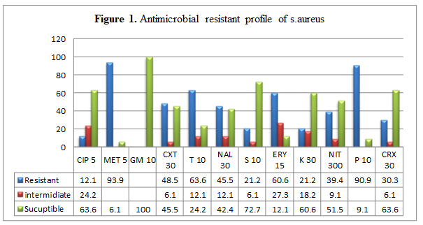
Table 3: Identification of Staphylococcus aureus from lactating cow in Adama and Modjo town. χ2=3.447, p=0. 63 All the 33 isolates of S. aureus were tested for antimicrobial resistant by 12 selected antibiotics. Of the 33 isolates all are resistant to one or more antimicrobials. The antibiotic resistant profiles of the isolates showed that the isolates were 31(93.9), 30(90.9), 21(63.6) and 20(60.6) resistant to Methicillin, Penicillin G, Tetracycline and Erythromycin, respectively. On the other hand, all isolates were 100%, 24(72.7), 21(63.6) and 20(60.6) Sensitive to Gentamycin, Streptomycin, Ciprofloxacin and Kanamycin respectively (Table 4, Figure 1).
| Antimicrobials | Status Resistance | Intermediate | Susceptible |
|---|---|---|---|
| Ciprofloxacin | 4(12.1) | 8(24.2) | 21(63.6) |
| Methicillin | 31(93.9%) | - | 2(6.1%) |
| Gentamycine | - | - | 33(100%) |
| Cefoxitin | 16(48.5%) | 2(6.1%) | 15(45.5%) |
| Tetracycline | 21(63.6%) | 4(12.1%) | 8(24.2%) |
| nalidixic acid | 15(45.5%) | 4(12.1%) | 14(42.4%) |
| Streptomycin | 7(21.2%) | 2(6.1%) | 24(72.7%) |
| Erythromycin | 20(60.6%) | 9(27.3%) | 4(12.1%) |
| Kanamycin | 7(21.2%) | 6(18.2%) | 20(60.6%) |
| Nitroforton | 13(39.4%) | 3(9.1%) | 17(51.5%) |
| Penicillin G | 30(90.9%) | - | 3(9.1%) |
| Cefuroxime | 10(30.3%) | 2(6.1%) | 21(63.6%) |
Table 4: Antibiotic Resistant Profiles of staphylococcus aureus isolates in dairy farm.
Among 33 resistant isolates, 93.9% (31/33) of the isolates were resistant to two or more of the antimicrobials tested (Multiple antimicrobial resistances (MDR)). The total of eight different antimicrobial resistance patterns were observed and the large proportions of the MDR isolates 29 (93.5%) were resistant to four to eight different antimicrobials. Whereas the others four isolates were resistant to one to three antimicrobials (Table 5).
| No antimicrobial resistance | Antimicrobial resistant patterns (no of isolates) | Total no of isolates | No of isolates (%) |
|---|---|---|---|
| Three | BM(1),NS(1) | -2 | 6.5 |
| Four | FS(1),HS(1),milk(3),NS(2) | -7 | 22.6 |
| Five | BM(1),HS(1),milk(1),NS(3) | -6 | 19.4 |
| Six | BM(1),milk(3),NS(5), | -9 | 29 |
| Seven | milk(4),NS(2) | -6 | 19.4 |
| Eight | NS(1) | -1 | 3.2 |
Table 5: Multi Drug Resistant. KEY: BM: Bulk milk, NS: Nasal swab, FS: Floor sample, HS: Hand swab
Discussion
The present study was carried out to isolate and identify Staphylococcus aureus in the sample collected from dairy cows and determine the antimicrobial resistance patterns of the S. aureus isolates. Following this study S. aureus strains (20.6 %) were isolated from lactating cows following culturing and biochemical tests of the available sample. This finding is nearly in agreement with the findings observed in Addis Ababa, 17.2% [27] in Sebeta, and the result of the present study showed a slight lower rate compared to other works, 28.1% [28] in Shashemene, 42.1% [29] in Adama and 26.6% [30] in kombolcha. The result is also slightly higher than [31] with 8% prevalence of S. aureus in Debreziet. This variation is largely attributed to the changing management conditions and using different diagnostic techniques. From a total of 160 sample included in the current study, S. aureus was isolated 13/86 (15%), 20/74(27%) from, Modjo and Adama respectively. The results showed there is no statistically significant difference between the two study sites (χ2=3.447, p=0.63). This might be attributed to similar livestock management conditions while handling and transportation of milk in the farm.
In this study, 44.4% (4/9) of the bulk milk samples from the farm were found to be contaminated with S. aureus. The results showed a higher contamination rate of bulk milk. This might be attributed to cross contamination of milk while bulking and poor handling during transportation [32]. The contamination of bulk milk with S. aureus was highly lower than the previous work [33] where S. aureus was isolated at recovery rate of 75% and 72%, respectively from bulk milk. The isolates of S. aureus from hands of milks and floor contaminations were 22.2% and 12.5%, respectively. Milk handlers and floor contaminations could be the potential sources of contamination of milk with S. aureus.
The present study also shows the most alarming situation of highly diverse antimicrobial resistance. In this finding, the S. aureus isolates were found to be highly resistant to methicillin (93.9%), penicillin G (90.9%), tetracycline (63.6%) and erythromycin (60.6). However, it revealed the sensitivity of the S. aureus towards gentamycin (100%), streptomycin (72.7%), ciprofloxacin (63.6%), and Kanamycin (60.6%). The highest resistance pattern of the S. aureus isolates to penicillin G was similar to the findings reported 96.7% by Mekuria A, et al. [34], in Ethiopia. These antibiotics are the main antibiotic group recommended for Staphylococcal mastitis treatment and regular use of antibiotics for the treatment of cows may result in the spread of resistant strains. Antibiotic therapy is an important tool in the treatment of S. aureus related infections. However, the misuse or intensive use of antibiotics can lead to the development of resistance among different bacterial strains [35].
The hygienic condition or quality of milk has serous implication on public health safety. Maintaining the hygienic conditions of dairy house, milking area, milking equipment and milker‘s hand is important for production of good quality milk [36]. Cleaning the udder of cow before milking is important since it could have direct contact with the ground, urine dung, and feed refusals while resting. Pre milking udder preparation and employing good milk handling practices play an important role in minimizing contamination at the farm with S. aureus [37]. Lack of refrigeration facilities at farm level in developing countries of tropical regions, with high ambient temperature implies that raw milk will easily be spoiled during storage and transportation [38].
Conclusion and Recommendations
In conclusion, Staphylococcus aureus is one of the major problems of dairy cows in the study area, and the overall occurrence of S. aureus in the study area was 20.6% with 20/74(27%) from Adama and 13/86(15.2%) from Modjo dairy cows. The majority of the tested isolates were resistant to the antimicrobial such as methicillin, penicillin G, tetracycline and erythromycin. It also revealed that all proportions of the isolates were susceptible to gentamycin. In this study all methicillin resistant S. aureus were also resistant to penicillin G. The possible explanations for the high record of most drug resistant S. aureus in dairy farms may be due to misuse of antibiotics with lack of proper sensitivity test in dairy farms.
Based on the above conclusion the following recommendations are forwarded;
- Gentamycin is the best effective drugs to treat cows infected with S. aureus in the study area.
- Routine hygienic measures in dairy farm and while milking should be practiced to reduce the risk of milk contamination with microorganisms.
- Regular surveillance of antimicrobial sensitivity pattern may help to select effective antibiotics for the specific diseases.
- Rational drug use principles should be implemented in the study area by veterinariansor other concerned body to minimize further development antimicrobial resistance.
References
-
Central Statistical Agency (CSA) (2008) Federal democratic republic of Ethiopia, central statistical agency. Agricultural sample survey, report on livestock and livestock characteristics, 2.
-
Biffa D, Debela E, Beyene F (2005) Prevalence and risk factors of mastitis in lactating dairy cows in southern Ethiopia. Int J Appl Res Vet Med 3(3): 189-198.
-
Pyorala S (2003) Indicators of inflammation in the diagnosis of mastitis. Vet Res 34(5): 565-578.
-
Erskine RJ (2001) Mastitis control in dairy herds. In: Radostits OM (Ed.), Herd Health: Food and Animal Production. 3rd (Edn.), W B Saunders Company, Philadelphia, pp: 397-435.
-
Harris LG, Foster SJ Richards RG (2002) An introduction to Staphylococcus aureus and techniques for identifying and quantifying Staphylococcus aureus adhesins in relation to adhesion to biomaterials: review. Eur Cell Mater 4: 39-60.
-
Abebe M, Daniel A, Yimtubezinash W, Genene T (2013) Identification and antimicrobial susceptibility of Staphylococcus aureus isolated from milk samples of dairy cows and nasal swabs of farm workers in selected dairy farms around Addis Ababa, Ethiopia. Afr J Microbiol Res 7(27): 3501-3510.
-
Normanno G, Corrente M, Salandra GL, Dambrosio A, Quaglia NC, et al. (2007) Methicillin-resistant Staphylococcus aureus (MRSA) in foods of animal origin product in Italy. Int J Food Microbial 117(2): 219-222.
-
Asperger H, Zangerl P (2003) Staphylococcus aureus. Ency DaiSci, pp: 2563-2569.
-
Capurro A, Concha C, Nilsson L, Ostensson K (1999) Identification of coagulase positive Staphylococci isolated from bovine milk. Acta Vet Scand 40(4): 315-321.
-
Seo KS, Bohach GA (2012) Staphylococcus aureus. In: Doyle MP, et al. (Eds.), Food Microbiology: fundamentals and frontiers. ASM Press, Washington DC, USA, pp: 353- 375.
-
Peles F, Wagner M, Varga L, Hein I, Rieck P, et al. (2007) Characterization of Staphylococcus aureus strains isolated from bovine milk in Hungary. Int J Food Microbial 118(2): 186-193.
-
Morandi S, Brasca M, Lodi R, Cremonesi P, Castiglioni B (2007) Detection of classical enterotoxins and identification of enterotoxin genes in Staphylococcus aureus from milk and dairy products. Vet Microbial 124(1-2): 66-72.
-
Boerema JA, Clemens R, Brightwell G (2006) Evaluation of molecular methods to determine enterotoxigenic status and molecular genotype of bovine, ovine, human and food isolates of Staphylococcus aureus. Int J Food Microbiol 107(2): 192-201.
-
Bergdoll MS, Wong AL (2006) Staphylococcal intoxications. In: Reimann P and Cliver D (Eds.), Food borne Infections and Intoxications. 3rd(Edn.), Academic Press, Elsevier, New York, NY, USA, pp: 523-525.
-
Adesiyun AA, Tatini SR, Hoover DG (1984) Production of enterotoxin(s) by Staphylococcus hyicus. Vét Microbiol 9(5): 487-495.
-
Schmid D, Gschiel E, Mann M, Huhulescu S, Ruppitsch W, et al. (2007) Outbreak of acute gastroenteritis in an Austrian boarding school, September 2006. Euro Surveill 12(3): 224.
-
Murray RJ (2005) Recognition and management of S. aureus toxin-mediated disease. Intern Med J 2: S106-S119.
-
Jahan M, Rahman M, Parvej MS, Chowdhury SM, Haque ZH, et al. (2015) Isolation and characterization of Staphylococcus aureus from raw cow milk in Bangladesh. J Adv Vet Anim Res 2(1): 49-55.
-
Normanno G, Corrente M, Salandra G, Dambrosio A, Quaglia C, et al. (2007) Methicillin resistant Staphylococcus aureus (MRSA) in foods animals and their potential transmission to humans. Appl Environ Microbial 69: 6489-6494.
-
Revue Méd (2006) Vét 161, 12, 574-57.NMSA (National Meteorology Service Agency) (2006): Monthly Climate Bulletin, Adama Branch, Adama, Ethiopia.
-
Mekonnen H, Tesfaye A (2010) Prevalence and etiology of mastitis and related management factors in market oriented smallholder dairy farms in Adama, Ethiopia. Revue De Medecine Veterinaire 161(12): 574-579.
-
CSA (2005) Agricultural Sample Survey 2004/05. Central Statistical Authority No.2. Report on Live- stock and livestock characteristics. Statistical Bulle- tin, Addis Ababa, Ethiopia. Central Statistical Agency, pp: 331.
-
Quinn J, Carter E, Markey B, Carter R (1999) Mastitis. In: Clinical Veterinary Microbiology, Mosby International Limited, London, pp: 327-344.
-
Quinn PJ, Carter ME, Markey BK, Carter GR (2002) Clin- ical veterinary Microbiology: Har court publishers, Vir- ginia, USA, pp: 331-344.
-
CLSI (2015) Investigation and control of vancomycin intermediate and resistant Staphylococcus aureus. A guide book for health departments and infection control personnel. Clinical and Laboratory Standards Institute, Wayne, Pennsylvania.
-
Huber H, Giezendanner N, Stephan R, Zweifel C (2011) Genotypes, antibiotic resistance profiles and microarray-based characterization of methicillin- resistant Staphylococcus aureus strains isolated from livestock and veterinarians in Switzerland. Zoo Pub Heal 58(5): 343-349.
-
Gizaw F (2011) Staphylococcus: Epidemiology and Its Drug Resistance in Cattle, Food Chains and Human in Central Ethiopia. Thesis: Addis Ababa University, Ethiopia.
-
Abera M, Elias B, Aragaw K, Denberga Y, Amenu K, et al. (2011) Major causes of mastitis and associated risk factors in smallholder dairy cows in Shashemene, southern Ethiopia. African Journal of Agricultural Research **7**(24): 3513-3518.
-
Abera M, Demie B, Aragaw K, Ragassa F, Ragassa A (2010) Isolation and identification of Staphylococcus aureus from bovine mastitic milk and their drug resistance pattern in Adama town, Ethiopia. J Vet Med Animal Health 2(3): 29-34.
-
Tassew A, Negash M, Demeke A, Feleke A, Tesfaye B, et al. (2016) Isolation, identification and drug resistance patterns of methicillin resistant Staphylococcus aureus from mastitic cow‘s milk from selected dairy farms in and around Kombolcha, Ethiopia. JVMAH 8(1): 1-10.
-
Mokennen A , Pal M, Kyule MN (2011) Isolation and Identification of Staphylococcus species from Raw Bovine Milk in Debre Zeit, Ethiopia. Vet Res 4(2): 45-49.
-
Getahun T, Gebre-Selassie S (2003) Assessment of the Bacteriological Quality of Milk at Dairy Farms and Individual Breeders in Jimma Town, South West Ethiopia. Ethiop J Health Sci 13(1): 21-29.
-
Desissa F, Makita K, Teklu A, Grace D (2012) Contamination of informally marketed bovine milk with Staphylococcus aureus in urban and peri urban areas of Debre-Zeit, Ethiopia. African Journal of Microbiology Research 6(29): 5852-5856.
-
Mekuria A, Asrat D, Woldeamanuel Y, Tefera G (2013) Identification and antimicrobial susceptibility of S. aureus isolated from milk samples of dairy cows and 48 nasal swabs of farm workers in selected dairy farms around Addis Ababa, Ethiopia. Afr J Microbial Res 7(27): 3501-3510.
-
White DG, Zhao S, Sudler R, Ayers S, Friedman S, et al. (2001) The isolation of antibiotic resistant Salmonella from retail ground meats. New England Journal of Medicine 345: 1147-1154.
-
Magnusson M, Christiansson A, Svensson B (2007) Bacillus cereus spores during housing of dairy cows: factors affecting contamination of raw milk. J Dairy Sci 90(6): 2745-2754.
-
Michelle A (2011) Staphylococcus aureus Mastitis. UK Veterinary Diagnostic Laboratory and Animal and Food Sciences, cooperative extension service, University of Kentucky College of Agriculture, Lexington, KY, 40: 190.
-
Gilmour D (1999) Milking. In: Falvey L and Chantalakhana C (Eds.), Smallholder Dairy in the Tropics. ILRI, Nairobi, Kenya, pp: 289-298.
- Antifungal Activity of New Acetophenone Derivatives
- Interconnected Microbiomes Human Health Within an Environmental Framework
- Silkworm-Based Vaccine Production for H5N1: A One Health Approach to Pandemic Preparedness
- Microbial Diversity and Lipolytic Activity of Bacteria and Fungi from Oil-Contaminated Sites in Makurdi Metroplois
- Antibiotic Resistance Profile of Bacteria Isolated at the Central Laboratory of the National Hospital Center of Nouakchott
- Epidemiology and Sensitivity to Antibiotics of Germs Isolated from Blood Cultures in the Laboratory of the National Hospital Center of Nouakchott-Mauritania