A Case Report of Emphysematous Pyelonephritis in a Diabetic Patient Complicated with Paraspinal Abscess
Emphysematous pyelonephritis is a rare, life threatening condition where there is severe infection of renal parenchyma causing gas accumulation in the tissue. It is commonly in diabetic and needed to be recognised and treated promptly. We present a case of a female diabetic patient who present with urosepsis secondary to severe type of emphysematous pyelonephritis complicated with paraspinal abscess caused by E coli and successfully treated with antibiotic and drainage via nephrostomy and underwent elective right nephrectomy. She had a good recovery.
Introduction
Presdisposing factor for Emphysematous pyelonephritis is diabetes mellitus. Common causative organism is E. Coli accounting 58-69% of cases [1]. Published data are limited on the management of emphysematous pyelonephritis. Recommended approach is according to class of disease based on CT scan findings [1]. We present a case of clinically staged Class 3B emphysematous pyelonephritis treated with broad spectrum antibiotics with nephrostomy drainage and later on underwent an elective right nepherectomy and recovered with good residual renal function.
Case Report
A 32 Malay lady who has diabetes mellitus for past 3 years presented with diabetic ketoacidosis and intubated due to poor GCS at Hosp Sultanah Nurzahirah. Further history acquired that patient had right lumbar pain with fever of 3 days duration.
On examination after intubation, she was dehydrated, toxic looking bp: 100/70 mmhg, pr: 120 T39°C. There was a right ballot able kidney on abdominal examination. Basic investigations on admission reported high blood sugar (30mmol/L) with HbA1c of 12%. Urine routine and microscopy showed excess WBC and positive results for ketones. She had acute renal failure (s.urea: 26 mmol/L, Creatinine 299 umol/L). Arterial blood gas revealed metabolic acidosis with raised anion gap. CBC (complete blood count) showed leukocytosis (12 × 109/L) with predominant neutrophils, anemia (hb 11g/dl) and thrombocytopenia (11x 109/L).raised CRP 145.27 mg/L. Liver function test showed raised alt and coagulation profile was normal. Chest X-ray showed minimal left pleural effusion. Plain abdominal x-ray showed enlarged right kidney and gas in parenchyma (Figure 1) and CT scan revealed gas collection in the parenchyma of right kidney (Figure 2A&2B) with right paraspinal abscess (Figure 2).
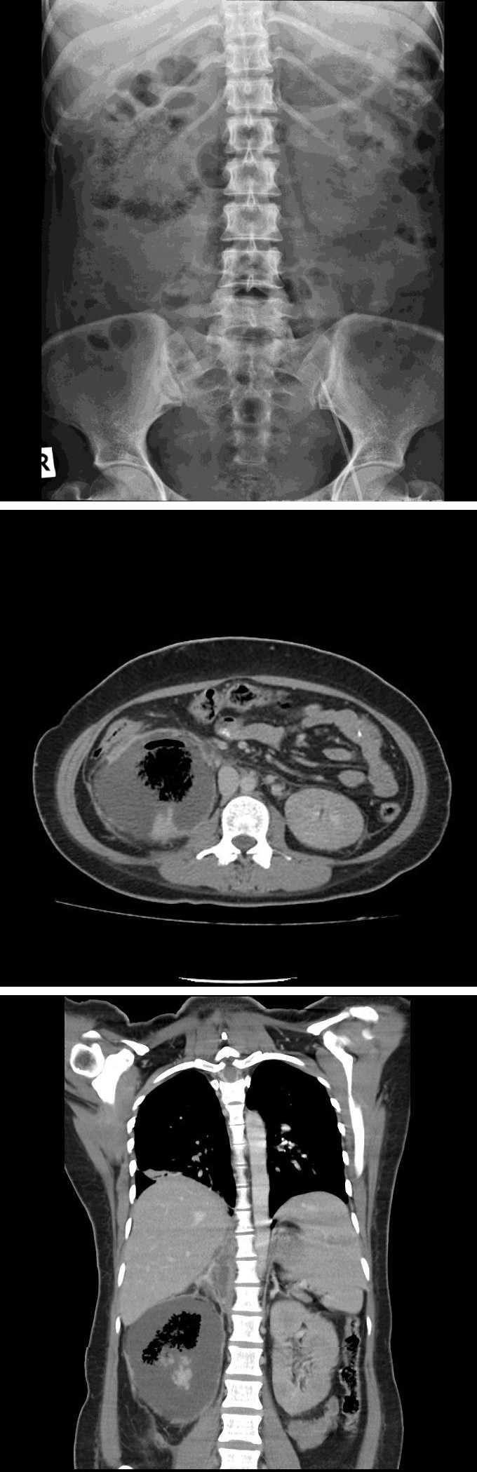
Figure 2(A): CT scan showed gas in the parenchyma of right kidney with right paraspinal abscess collection(Coronal cut).
Figure 2B: CT scan showed gas collection in right kidney (Axial cut). She was diagnosed as acute emphysematous pyelonephritis clinically Class 3B with sepsis and diabetic ketoacidosis. She was started emopirically on rocephine placed on intravenous fluids and insulin infusion pump. She was successfully extubated 48hr later. Neurological assessment of upper limb and lower limb was normal. Blood and urine cultures reported growth E coli. Antibiotics were changed to tazocin(piperacillin/tazobactam) injection and she right nephrostomy inserted. She completed antibiotics for 4weeks and discharged with follow-up at the clinic. She underwent elective right nephrectomy 2 months later (25/12/10) as still having chronic infection (chronic pyelonephritis) of her right kidney and currently recovered well. Her latest renal function is urea 11.9/creat 132. Histopathological findings of kidney tissue with extensive tubular atrophy and variable degree of interstitial fibrosis. Some of the glomeruli show mesangial matrix expansion and necrosis. The urothelium overlying the renal pelvis and calyces is ulcerated in areas. Diffuse mixed acute and chronic inflammatory cells infiltrate consist of neutrophils, lymphoplasmacytic cells, foamy histiocytes, hemosiderin laden macrophages with foci of foreign body type multinucleated giant cells aggregation are also seen within the renal interstitium extending up to the perirenal fat. Multiple cystic spacesconsistent with acute on chronic emphysematous pyelonephritis.
Conclusion
Diabetes mellitus is the major risk factor for emphysematous pyelonephritis, up to more than 80
percent of cases [1, 2, 3, 4, 5].The diagnosis is made by plain films of the abdomen and /or computed tomography (CT) showing air in the renal parenchyma. CT scanning is more sensitive than plain film and it is used to classify emphysematous pyelonephritis and to prognosticate the disease and also in guiding therapy [1]. Presence of thrombocytopenia, acute renal failure, impaired consciousness and shock associated with adverse outcome [1, 6, 7].Overall mortality rate was 13-25 percent. In the past, treatment of emphysematous pyelonephritis or pyelitis usually involved nephrectomy or open drainage along with systemic antibiotics [3, 4, 5]. In more recent reports, successful management with systemic antibiotics together with percutaneous catheter drainage (PCD) of gas and purulent material, as well as relief of urinary tract obstruction (if present), has been described [1, 3, 8, 9, 10]. A systematic review of 10 retrospective studies including 210 patients with emphysematous pyelonephritis noted that mortality associated with medical management plus percutaneous catheter drainage was significantly lower than medical management plus emergent nephrectomy (13.5 versus 25 percent, respectively) [10]. As in our case this patient is estimated Class 3B and complicated with right paraspinal abscess but she did not have neurological deficit. She also has thrombocytopenia, acute renal failure and impaired consciousness upon presention which was associated with mortality and adverse outcome. Patient was treated with broad spectrum antibiotics and right nephrostomy inserted to relief the urinary tract obstruction. Her right paraspinal abscess was treated conservatively with antibiotics. She later on had an elective right nephrectomy as having chronic infection (chronic pyelonephritis) of right the kidney. She made a good clinical recovery. In conclusion, emphysematous pyelonephritis is a rare, life threatening disease and must be suspected in diabetic patients who present with urosepsis. Early imaging is mandatory and treatment is according to class. Treatment is involved medical and surgical management Nephrectomy is preferable if no improvement after antibiotics.
Acknowledgement
Authors are grateful and acknowledge the help of radiology department of Hospital Sultanah Nurzahirah, Kuala Terengganu for their assistance in imaging studies.
References
-
Huang JJ, Tseng CC (2000) Emphysematous pyelonephritis: clinicoradiological classification, management, prognosis, and pathogenesis. Arch Intern Med 160(6): 797-805.
-
Somani BK, Nabi G, Thorpe P, Hussey J, Cook J, et al. (2008) Is percutaneous drainage the new gold standard in the management of emphysematous pyelonephritis? Evidence from a systematic review. J Urol 179(5): 1844-1849.
-
Chen MT, Huang CN, Chou YH, Huang CH, Chiang CP, et al. (1997) Percutaneous drainage in the treatment of emphysematous pyelonephritis: 10-year experience. J Urol 157(5): 1569-1593.
-
Pontin AR, Barnes RD, Joffe J, Kahn D(1995) Emphysematous pyelonephritis in diabetic patients. Br J Urol 75(1): 71-74.
-
Shokeir AA, El-Azab M, Mohsen T, El-Diasty T (1997) Emphysematous pyelonephritis: a 15-year experience with 20 cases. Urology 49(3): 343-346.
-
Falagas ME, Alexiou VG, Giannopoulou KP, Siempos II (2007) Risk factors for mortality in patients with emphysematous pyelonephritis: a meta-analysis. J Urol 178: 880-885.
-
Kapoor R, Muruganandham K, Gulia AK, Singla M, Agrawal S, et al. (2010) Predictive factors for mortality and need for nephrectomy in patients with emphysematous pyelonephritis. BJU Int 105(7): 986- 989.
-
Roy C, Pfleger DD, Tuchmann CM, Lang HH, Saussine CC, et al. (2001) Emphysematous pyelitis: findings in five patients. Radiology 218(3): 647-650.
-
Mydlo JH, Maybee GJ, Ali-Khan MM (2003) Percutaneous drainage and/or nephrectomy in the treatment of emphysematous pyelonephritis. UrolInt 70(3): 147-150.
-
Somani BK, Nabi G, Thorpe P, Hussey J, Cook J, et al. (2008) Is percutaneous drainage the new gold standard in the management of emphysematous pyelonephritis? Evidence from a systematic review. J Urol 179(5): 1844-1849.
- Results of 6-Month Follow-Up of Patients After B-Turp and Thulep
- The Effect of Drinking Water with a High Content of Antimony and Arsenic on the Dynamics of their Distribution in the Kidneys and the Renal Excretory Function in Rats
- Effectiveness and Safety of Tansurethral Thulium Laser Enucleation of the Prostate in the Treatment of BPH: Review
- A Systematic Review on Molecular Pathophysiology Involved in Chronic Kidney Disease and the Role of Animal Models in Drug Discovery to Manage in Chronic Kidney Disease - An Update
- Functional Development of Kidneys in Human Ontogenesis
- Testicular Metastasis: Uncommon Prostate Cancer Case Report