Bilateral Duplex Collecting System with Right Obstructing Stones and Ureterocele - A Case Report with Literature Review
A duplex collecting system (DCS) is a congenital genitourinary tract malformation. It is found bilaterally in 0.3% of excretory urograms. Complete duplication is more uncommon than the incomplete form. Ureterocele is a congenital cystic dilation of the intravesical ureter. An incidentally detection of a rare case of bilateral duplex collecting system in an adult patient with a non-specific abdominal pain is described. The DCS was complete on the right side with the upper ureter moiety ending with an intravesical ureterocele. The right upper ureter moiety was presented along with obstructive stones.
Barbara Brogna1*, Giovanni Castelluzzo2, Paolo Ferravante2 and Carmine Manganiello1
Principe di Napoli 14/A Benevento, 82100, Italy, Email: brognabarbara1@gmail.com obstructive stones.
Keywords: Duplex Collecting System; Ureterocele; Stones; Imaging; Ureteroscopy
Introduction
A duplex collecting system (DCS) is a one of most common congenital genitourinary tract abnormalities, even though it has a rarely occurrence that is reported in only 0.3-0.8% of the population [1, 2]. It develops during the 4th to 5th weeks of gestation when two separate ureteric buds arise from a single Wolffian duct. It is Bilateral Duplex Collecting System with Right Obstructing Stones and Ureterocele - A Case Report with Literature Review characterized by the fusion of the lower and upper pole moieties resulting in an incomplete or complete duplication of the collecting system. This abnormality can cause prenatal hydronephrosis and is usually associated with renal-vesical reflux in pediatric patients. It affects females more than males and may be associated to other anomalies. Nowadays, diagnosis is usually made early, though congenital urinary tract anomalies can be often asymptomatic and incidentally detected in adult patients [3]. Bilateral DCS is a very rare abnormality. It is found in 17-33% of cases and complete ureteral duplication with two separate openings in the urinary bladder is less commonly found than the incomplete forms [1, 2, 3, 4, 5]. Ureterocele is defined as a congenital cystic dilation of the J Urol Nephrol
intravesical ureter. It has an incidence of 1 in 500 to 1 in 12,000 by autopsy and is commonly associated to DCS [6]. Imaging plays a pivotal role in the detection of these anomalies. There are few reports about urinary stones and DCS with coexisting ureterocele.
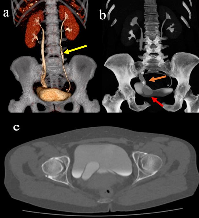
examination found a complete right DCS with the right upper moiety ureter ending with a ureterocele and an incomplete left DCS (Figure 1). Obstructive stones were also visible in the mid and distal portions of the right upper moiety ureter, which was also dilated and showed a delay in the extraction of the contrast medium (Figure 1). Multi-planar reconstruction (MPR), volume rendering (VR) and maximum intensity projection (MIP) were performed. The patient was taken to the operating room for endoscopic ureteral decompression. A retrograde pyelography was also performed. The ureteral meatus of the inferior right ureteral moiety had been easily identified with a normal course of the ureter. The right upper ureter moiety appeared dilated and had difficulty discarging the contrast product from the previous CTU examination. The ureteral meatus of the right upper ureteral moiety was not visible and the incision of the ureterocele was performed using a Collins knife. After this procedure a turbid urinary efflux was also visible. A Right double J stent was placed on the right upper ureter moiety for 4 weeks to reduced complications such as urosepsis. The patient was scheduled for a lithotripsy.
Case Report
A 56-years-old presented at emergency department of our Hospital with low abdominal pain and had no previous history of urinary tract infections or stone diseases. Laboratory results showed the hemoglobin at 13.5 g/dL, white cell count at 12,820/mm3, and platelets at 265,000/mm3. Her Creatinine level was normal at 0.65 mg/dl. No micro/macrohematuria was seen on the urinalysis. An abdominal ultrasound revealed a cyst-like intravesical mass, near the margin of the trigone suspected of ureterocele. A mild dilatation of the right upper renal calyceal system was also described. A Computed tomography urography (CTU) including four- phase (unenhanced, arterial, parenchymal and excretory), was done to rule out any associated anomalies. The CTU
Discussion
A DCS can be complete, when there is a total pyelo- ureteral duplicity with separate ureteric orifice or incomplete when the ureters connect to each other before the distal implantation in the bladder. Unilateral duplication occurs about six times more often than bilateral duplication. Complete ureteral duplication is even more rarely reported than the partial form and follows the Wiegert-Meyer rule. A DCS is found bilaterally in 0.3% of excretory urograms. The upper moiety ureter usually inserts ectopically inferior and medial to the lower pole moiety and is more susceptible to obstruction if associated with ureteroceles. Stasis, ureteral atony and infections predispose to stone formations. The American Academy of Pediatrics classifies a ureterocele as orthotopic (entirely intravesical) or ectopic (if it ends at the bladder neck or at the posterior urethra) [7]. According to Stephens' classification, intravesical ureteroceles are divided in stenotic or non-obstructed, while extravesical ureteroceles are classified as sphincteric, sphincterstenotic, cecoureteroceles, or blind ectopic. Vesicoureteric Reflux (VUR) may occur in the upper moiety or in a combination of moieties. Congenital urinary tract malformations are usually diagnosed during pregnancy or in childhood. However, they can also be incidentally detected by imaging in adult patients and can be mistaken for other causes of abdominal pain such as appendicitis or intestinal obstruction. More common symptoms are obstructive voiding symptoms, urinary retention, urinary tract infections or poor kidney function. Ureterocele is usually discovered by ultrasounds and it usually appears as a sac-like pouch in the bladder. It tends to look like a cobra-head on imaging. CTU is the most valuable imaging tool to diagnose a DCS and it has replaced the ureteropyelography due to its safety, fast imaging and advantages in high quality details in the evaluation of the kidneys and urinary tracts through MPR and 3D reconstructions analysis. The European Society of Urogenital Radiology (ESUR) recommends CTU in the evaluation of the urinary tract malformations and related complications [8]. A DCS usually shows a prolonged delay excretion on CTU because of the malfunction to secrete contrast to the duplicated ureters. The study of Gong, et al. shows that the satisfactory opacification can be achieved 24h after CTU. Intraoperative retrograde pyelography may be used to define the ureterocele anatomy and to assess the presence of VUR. The treatment of a patient with a DCS and an ureterocele may vary based on the clinical and pathophysiological characteristics of each patient. The main aim is to preserve the renal function and reduce septic complications [5, 6, 9, 10]. Primary endoscopic deroofing with double-J stenting is the best initial approach for adequate decompression in a patient with obstructive disease or septic risks. Unroofing, puncture, laser incision, and Collins knife incision are methods used to decompress the ureterocele. An endoscopic incision or puncture is a simple, minimally invasive, safe and efficacy procedure that is easy to perform in an emergency. For an intravesical ureterocele, a small opening is made at the lowest level above the bladder neck. It is easier to decompress an intravesical ureterocele than an ectopic ureterocele. Surgical options are usually preferred in patients who have a non- functioning upper segment or VUR. Recently, there are minimally invasive approaches available including laparoscopic and robotic techniques.
Conclusion
Diagnosis of congenital urinary tract malformations can be a diagnostic challenge in adult patients and should be kept in mind for patients with abdominal pain.
References
-
Yonli DS, Chakroun M, Zaghbib S, Ye D, Bouzouita A, et al. (2019) Bilateral duplex collecting system with bilateral vesicoureteral reflux: a case report. J Med Case Rep 13: 128.
-
Scantling D, Ross C, Altman H (2013) A 52-year-old male with bilaterally duplicated collecting systems with obstructing ureteral stones: a case report. Curr Urol 7(2): 104-106.
-
Davda S, Vohra A (2013) Adult duplex kidneys: an important differential diagnosis in patients with abdominal cysts. JRSM Short Rep 4(2): 13.
-
Sen V, Aydogdu O, Yonguc T, Bozkurt IH, Polat S, et al. (2015) Endourological treatment of bilateral ureteral stones in bilateral ureteral duplication with right ureterocele. Can Urol Assoc J 9(7-8): 511-513.
-
Shokeir AA, Nijman R (2002) Ureterocele: an ongoing challenge in infancy and childhood. BJU Int 90(8): 777-783.
-
Merlini E, Chiesa PL (2004) Obstructive ureterocele- an ongoing challenge. World J Urol 22(2): 107-114.
-
Glassberg KI, Braren V, Duckett JW, Jacobs EC, King LR, et al. (1984) Suggested terminology for duplex systems, ectopic ureters and ureteroceles. J Urol 132(6): 1153-1154.
-
Gong H, Gao L, Dai XJ, Zhou F, Zhang N, et al. (2016) Prolonged CT urography in duplex kidney. BMC Urol 16(1): 21.
-
Xie D, Klopukh B, Nehrenz GM, Gheiler E (2017) Ureterocele: Review of Presentations, Types, and Coexisting Diseases. Int Arch Urol Complic 3(1).
-
Le HK, Chiang G (2018) Long-term management of ureterocele in duplex collecting systems: reconstruction implications. Curr Urol Rep 19(2): 14.
- Results of 6-Month Follow-Up of Patients After B-Turp and Thulep
- The Effect of Drinking Water with a High Content of Antimony and Arsenic on the Dynamics of their Distribution in the Kidneys and the Renal Excretory Function in Rats
- Effectiveness and Safety of Tansurethral Thulium Laser Enucleation of the Prostate in the Treatment of BPH: Review
- A Systematic Review on Molecular Pathophysiology Involved in Chronic Kidney Disease and the Role of Animal Models in Drug Discovery to Manage in Chronic Kidney Disease - An Update
- Functional Development of Kidneys in Human Ontogenesis
- Testicular Metastasis: Uncommon Prostate Cancer Case Report