Rare Presentation of Urethral Condyloma Acuminata: Limited on Meatus with Stricture, Fibrotic Morphology
Penile lesions are frequently encountered in daily urology practice and often difficult to differential diagnosis. These lesions can be detected by various presentations and can be observed in different genital locations. These lesions may be a precursor lesion of a systemic disease or a sexually transmitted disease or a precancerous lesion of the genital organs. In our case, in the examination we detected a sclerotic lesion that covers the penile meatus 360 degrees and causes stenosis with urinary weak stream. Biopsy of the lesion was reported as condyloma acuminata. In this case, we aimed to present the rare presentation and management of Condyloma Acuminata.
Introduction
Penile lesions can be detected as cancer and pre- cancerous lesions or lesions of sexually transmitted disease. Malignant lesions of the penis are rare but in a study conducted in France between 1989 and 2011, this rate was reported as 0.59 / 100.000 [1]. While this ratio is rare before the age of 55, it increases considerably after the age of 75. Human papilloma virus (HPV) especially in certain types (HPV-6, HPV-16, HPV-18), phimosis, and lack of hygiene, chronic irritation, multi-partner ownership, and low socio-economic level are predisposing factors for malignant lesions of the penis [2]. Penile intra-epithelial neoplasia (PeIN) is the most common precancerous lesion. Altough Bowen’s disease, queyrat erythroplasia and in-situ carcinoma are macroscopically different from Pein, they are similar histologically to PeIN [3]. There is a systemic relationship between HPV and these lesions rates 70% and
100%. Especially, it is associated HPV with the basaloid differentiation of PeIN [4]. Although it is very difficult to diagnose, especially in the presence of a lesion involving the urethra, biopsy will guide us in the diagnosis and treatment of these rare cases.
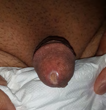
A 66-year-old male patient consulted to our outpatient clinic with complaints of difficulty in urination. The auscultation revealed that the lesion was white, sclerotic and surrounded the penile meatus 360 degrees (Figure 1). It was learned that the lesion had been present for approximately 1 year. We tried to catheterize the meatus with a soft catheter, but failed. Qmax was 5 ml / sec in uroflow test. Trace was interpreted in favor of stenosis. No pathology was detected in urine analysis. No pathology was detected on urinary ultrasound. Although the patient had no previous surgery, he is using anti-hypertensive drug due to hypertension. Punch biopsy was taken from penile meatus under local anesthesia. Pathology was reported as condyloma acuminata. After that, under general anesthesia, the lesion was excised 360
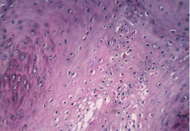
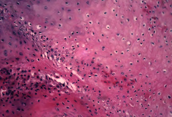
degrees from the penis meatus. The length of the lesion was less than 1cm. Meatoplasty was performed by placing eversion sutures with 5.0 rapid vicryl. Thermocauterization was avoided to create iatrogenic stenosis. Then cystoscopy was performed gently without forcing the tissues. No other lesion was detected in the lower urinary tract. Pathology was reported that condyloma acuminatum is characterized by koilocytic change. Koilocytes are characterized by clear perinuclear halos (Figure 2) wrinkled and hyperchromatic nuclei (Figure 3). Cytoplasm is eosinophilic cellular borders are distinctive, and binucleation is common. The surgical margin was negative. Our patient is followed up without any problem.
Discussion
Approximately, 80% of sexually active men are infected with HPV for a part of their lifes. Most genital HPV infections are asymptomatic, not recognisable, or subclinical, and usually are self-limited. HPV involvement of the urethra is rare, it is usually located in the distal 3 cm area and is difficult to diagnose [5]. In our case, HPV was located in the distal urethral part with imited on the meatus. In the literature, urethral lesions have been reported with different menifestations such as weak stream, urethrorrhagia, and dysuria [6]. In our case, urethral lesion manifested itself with difficulty in urination and weak stream. Vives A, et al. [7] reported that these lesions are usually diagnosed after meatus eversion and verrucous lesions that can be excised. In contrast to the literature, our case was not verrucous but appeared to be sclerotic and fibrotic in the meatus. It is not in a certain part of the meatus and covers the meatus completely 360 degrees. It narrowed the lumen and caused micturition complaints. Surgical excision, electrocautery, cryotherapy, CO2 laser vaporization, 5% fluorouracil gel instillation are the recommended treatments in the literature [8]. We performed surgical excision in our case. We didn’t use electrocoagulation to avoid meatus stenosis. In this case, we presented a different morphologically urethral conduloma involvement according to the literature. In sclerotic lesions of the meatus, we will need to remember the condyloma acuminata.
References
-
Daubisse Marliac L, Colonna M, Trétarre B, Defossez G, Molinié F, et al. (2017) Long-term trends in incidence and survival of penile cancer in France. Cancer Epidemiol 50: 125-131.
-
Hakenberg OW, Comperat EM, Minhas S, Necchi A, Protzel C, et al. (2015) EAU guidelines on penile cancer: 2014 update. Eur Urol 67: 142-150.
-
Lont AP, Kroon BK, Horenblas S, Gallee MP, Berkhof J, et al. (2006) Presence of high-risk human papillomavirus DNA in penile carcinoma predicts favorable outcome in survival. Int J Cancer 119(5): 1078-1081.
-
Lebelo RL, Boulet G, Nkosi CM, Bida MN, Bogers JP, et al. (2014) Diversity of HPV types in cancerous and pre-cancerous penile lesions of South African men: implications for future HPV vaccination strategies. J Med Virol 86(2): 257-265.
-
Von Krogh G, Lacey CJ, Gross G, Barrasso R, Schneider A (2000) European course on HPV associated pathology: guidelines for primary care physicians for the diagnosis and management of anogenital warts. Sex Transm Infect 76(3): 162-168.
-
Pollack HM, DeBenedictis TJ, Marmal JL, Praiss DE (1978) Urethrographic manifestations of venereal warts (condyloma acuminate). Radiology 126(3): 643-646.
-
Vives A, Vazquez A, Rajmil O, Cosentino M (2016) Urethral condlomas in men: experience in 123 patients without previous treathment. Int J STD AID 27(1): 39-43.
-
Debenedicts TJ, Marmar JL, Pradiss D (1977) Intraurethral condyloma acuminata: Management and review of the literature. J Urol 118(5): 767-769.
- Results of 6-Month Follow-Up of Patients After B-Turp and Thulep
- The Effect of Drinking Water with a High Content of Antimony and Arsenic on the Dynamics of their Distribution in the Kidneys and the Renal Excretory Function in Rats
- Effectiveness and Safety of Tansurethral Thulium Laser Enucleation of the Prostate in the Treatment of BPH: Review
- A Systematic Review on Molecular Pathophysiology Involved in Chronic Kidney Disease and the Role of Animal Models in Drug Discovery to Manage in Chronic Kidney Disease - An Update
- Functional Development of Kidneys in Human Ontogenesis
- Testicular Metastasis: Uncommon Prostate Cancer Case Report