Giant Gastrointestinal Stromal Tumor (Gist) of the Stomach and Huge Liver Metastase; Surgical Management
Gastrointestinal stromal tumors (GISTs) represent 85% of all mesenchymal neoplasms that affect the gastrointestinal (GI) tract. These GISTs range in size from small lesions to large masses. Often they are clinically silent until they reach a significant size, so their discovery is usually incidental. Gastrointestinal stromal tumors (GISTs) are relatively common subepithelial tumors that occur most frequently in the stomach, small bowel, esophagus, and omentum. The liver is the most common metastatic site of a GIST [1,2].<br />These tumors are located primarily in the stomach (60% to 70%) and their discovery is often incidental. GISTs range in size from small lesions to large masses [3]. They are clinically silent until they reach a significant size; this is why their discovery is usually incidental. In many cases, GISTs present with abdominal pain, GI bleeding or palpable mass. We report an unusual case of a giant GIST that caused a huge liver metastases.
Case Presentation
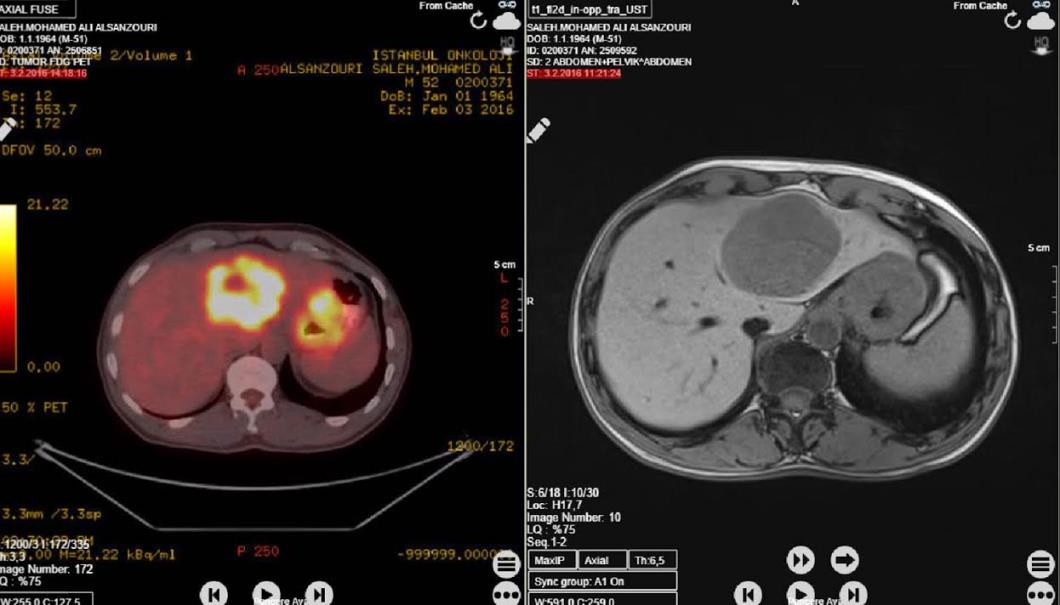
This is a recent case of an exophytic GIST of gastric origin. A 53-year-old male presented with vague upper abdominal pain and feeling of an abdominal lump on and off for two years. There were no other associated general or GI tract symptoms. Clinical examination of the abdomen revealed a well-defined transversely mobile intra-abdominal lump in the right hypochondrium of about 15 × 8cm. All routine blood test results and levels of tumor markers were within the normal ranges. Ultrasonography and computed tomography of the abdomen showed a large mass of 70*35 mm. Huge liver metastasis were seen in abdomen computed tomography (Figure 1). The abdomen was explored electively and a 7 × 4 cm tumor was seen arising from the greater curve of the stomach, exophytically, with a sessile base. There were huge liver metastases were seen in abdominal exploration (Figure 2).
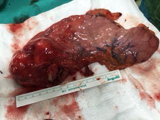
Histopathology of the tumor revealed a 7.5 CM sized neoplasm arising from the gastric submucosa composed of fusiform and epitheloid cells. Mitosis was occasional at 5/50 Immuohistochemistry reported that the tumor cells were positive for C-KİT, CD 34, and S-100, aktın and demsin were negative. The findings favored the diagnosis of epitheloid gastrointestinal stromal tumor of high malignant potential. The resected margin was reported clear of the tumor.
Discussion
Gastrointestinal stromal tumor can occur anywhere in the GI tract. They are submucosal lesions frequently growing endophytically. They also manifest exophytically. Sizes of these tumors have been reported from small 1 cm to large 40 cm diameter excrescences. About 50% to 75% of these originate in the stomach and about 20% in the small bowel, while less frequent sites include the colon and Rectum. Brunner's gland hamartomas of the duodenum mimic the radiological and endoscopic features of GIST [4, 5].
Conclusion
Large sized liver metastases are rare in the case of gastroinetstinal stromal tumor which generally present with varying duration of symptoms before surgery. These tumors may be difficult to distinguish from a number of other benign or malignant spindle cell lesions. Complete surgical resection of the tumor along with adjuvant therapy involving selective receptor tyrosine kinase inhibitors is effective, and should be considered as primary modalities of treatment in high-risk group GIST patients.
References
-
Osada T, Nagahara A, Kodani T, Namihisa A, Kawabe M, et al. (2007) Gastrointestinal stromal tumor of the stomach with a giant abscess penetrating the gastric lumen. World J. Gastroenterol 13(16): 2385-2387.
-
Vij M, Agrawal V, Kumar A, Pandey R (2013) Cytomorphology of gastrointestinal stromal tumors and extra-gastrointestinal stromal tumors: a comprehensive morphologic study. J Cytol 30(1): 8- 12.
-
Zhou L, Liu C, Bai JG, Wei JC, Qu K, et al. (2012) A rare giant gastrointestinal stromal tumor of the stomach traversing the upper abdomen: a case report and literature review. World J Surg Oncol 10: 66.
-
Fülöp E, Marcu S, Milutin D, Borda A (2009) Gastrointestinal stromal tumors: review on morphology: diagnosis and management. Rom J Morphol Embryol 50(3): 319-326.
-
Funahashi H, Okada Y, Sawai H, Wakasugi T, Akamo Y, et al. (2008) Complete extragastric growth in a giant gastrointestinal stromal tumor: report of a case. Int Surg 93(1): 45-49.
- Measuring What Matters: Data Gaps, Certificate of Need Reform, and Pediatric Psychiatric Inpatient Capacity in North Carolina
- Intersecting Epidemics and Climate Vulnerabilities in Conflict- Driven Displacement: Epidemiology, Systemic Challenges, and One Health Gaps in South Sudan
- Advancing Domestic Health Financing for Community Health System Sustainability in South Sudan: The Boma Health Initiative Model (2025–2035)
- Prevalence and Correlates of Post-Exposure Prophylaxis Uptake among Men Who Have Sex with Men in Kisumu County, Kenya
- Medical, Ethical, and Legal Conflicts Surrounding Euthanasia in Argentina. Its Global Implications
- Knowledge and Attitude on Menstrual Hygiene among Adolescent Girls Studying in Secondary Level in Public Schools of Chitwan District, Nepal