Virus in Human Health
Virus discovery as submicroscopic entities, data from the end of the 19th century, which marked the beginning of a new research discipline, the viruses study. Since then, the presence of viruses has been found in cancer and in multiple diseases whose origin was unknown. Viruses have shown their great potential in triggering infections, causing pandemics at a high cost to society as the Spanish Flu in 1918. The challenge of acting at the health emergency due to the outbreak of diseases caused by new viruses, has led to research with the application of new technologies, in order to find strategies, diagnostic methods, treatment and development of vaccines.
Editorial
Presence of virus in plants and animals is dated since the end of nineteenth century with Dimitri Ivanovsky to whom credit is given to recognize a filterable and submicroscopic entity as the cause of tobacco mosaic disease [1, 2]. In 1903 Borrel propose the virus theory of cancer [3] and few years later (1908) an avian leukemia virus was discovered [4]. In 1911 Peyton Rouse reported the isolation of “filterable agent” that was later called the Rouse Sarcoma Virus [5, 6]. The following studies around the Rouse sarcoma virus marked an important starting point in virus research [7]. Finally in a mammary cancer in mice the interaction of three etiological factors (genetic background, hormones and virus) was proposed as clinical disease [8].
Since then, researchers have looked for cancer-related viruses in animals, such as rabbit papilloma-carcinoma, rabbit fibroma and myxoma, murine mammary carcinoma, bovine ocular carcinoma, canine mast cell sarcoma and sheep adenomatosis [8]. In a study of cancer incidence related to infectious agents in the year of 2002, it was founded an association with human papilloma viruses (5.2%), the hepatitis B and C viruses (4.9%), Epstein-Barr virus (1%), human immunodeficiency virus (HIV) together with the human herpes virus 8 (0.9%) and human T-cell lymphotropic virus type I (0.03%) [9]. It has been estimated that between 15% - 20% of all cancers are related with oncogenic viruses [10].
In human, viruses like particles were identified in pre-cancerous, cancerous tissues and serum by electron microscopy, which allowed its morphological description [11, 12, 13, 14, 15, 16, 17]. Currently viral particles are analyzed by different imaging techniques as fluorescence, transmission and scanning electron microscopy, electron tomography, focused ion beam/scanning (FIB/SEM), single molecule tracking, structured illumination microscopy, stimulated emission depletion, stochastic optical reconstruction microscopy, photoactivation localization microscopy, ground state depletion and correlative light electron microscopy (Figure 1) [18]. In clinical samples virus can be identified by viral cultures, ELISA test, immunoprecipitation, in situ hybridization, end point PCR, multiplex PCR, real time PCR, flow cytometry, ViriChip method, and Next-generation sequencing (NGS) [19, 20, 21].
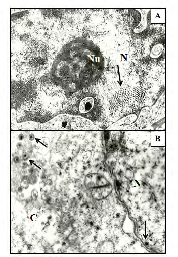
Figure 1: Electron micrograph of viruses. A) Nucleus inclu- sion of papilloma virus in crystalline arrangement in epi- dermal cell (arrows; 19,680X); B) Cytoplasmic and nucleus inclusions of Pseudorrabia virus in kidney cells (arrows; 24,000 X). C: cytoplasm; N: Nucleus; Nu: nucleolus. Uranyl acetate and Lead citrate. Archive photomicrographs from the Experimental Neuropathology Laboratory.
Virus classification has been established by the International Committee on Taxonomy of viruses (ICTV) since 1966; the classification is organized into taxonomic levels that have changed over time according to the knowledge that is being acquired due to technological and molecular advances that have better tools to investigate viruses in more detail [22]. At the beginning ICTV recognized only genera and families, later virus classification developed into a five-rank hierarchy (species, genus, subfamily, family and order) which staying until 2017. In 2019 the virus classification evolved to fifteen-rank structure, which also includes genomic properties and comparative sequences analysis of conserved genes and proteins. In the last decades of the 20th century, other classification was proposed according to their genome type (double- stranded DNA, single-stranded DNA, double- stranded RNA, positive-sense RNA, negative-sense RNA, reverse-transcribing RNA and reverse-transcribing DNA) [2].
The human viroma, which is part of the microbiome, is distributed in different compartments in the body, harboring viral communities that changing during lifespan and in response to environment [23]. It has been estimated that more than ten permanent viral infections are chronically present in the human body [24], which can lead to oncogenesis [10]. In a metagenomics sequencing analysis from human samples only 14% - 87% sequences have been classified [25]. In human, DNA viruses have been found throughout the body in healthy asymptomatic subjects and in illness symptomatic conditions, such as those of the Herpesviridae family (Cytomegalovirus, Lymphocryptovirus, Rhadinovirus, Roseolavirus, Simplexvirus), Papillomaviridae family (Alphapapillomavirus, Betapapillomavirus, Gammapapillomavirus) and Polyomaviridae family (Alphapolyomavirus, Betapolyomavirus, Deltapolyomavirus), among others. Other human viruses with especific distribution in human body have been reported, as is the case of the Coronaviridae family, Paramyxoviridae family, Pneumoviridae family, Orthomyxoviridae family, Parvoviridae family, Astroviridae family, Reoviridae family, Caliciviridae family, Picornaviridae family, Picobirnaviridae family, and Flaviviridae family, among others. Unlike DNA virus, RNA virus tends to cause acute infections. [23].
It has been proposed that virus spread could be by means of horizontal (within a species by postnatal contact), vertical (neonatal o prenatal transmission within a species from mother to young) and by exogenous spread (transmission of virus form one species to another with or without the aid of insect vectors) [8] and it has been observed that individuals that recover from some acute infections can still carry the pathogenic viruses for varying periods of time [26, 27].
Threats to human health due to infections by microorganisms have always been present, which have been controlled by advances in scientific knowledge, however, threats of zoonotic infections have emerged, which have become epidemics and have spread widely, becoming pandemics such as the Spanish Flu in 1918, Asian Flu H2N2 in 1957, Hong Kong Flu H3N2 in 1968, Hantavirus pulmonary syndrome in 1993, SARS CoV-2 in 2002-2003, the Influenza A H1N1 pmd09 in 2009, MERS-CoV in 2012, the polio resurgence in 2014, the Ebola epidemic in West Africa in 2014, the emergence of Zika virus in the Americas in 2016 and currently the SARS-CoV-2 (2019-2020) [28, 29].
Zoonotic virus, severe acute respiratory syndrome- related coronavirus 2 (SARS-CoV-2) according to the ICTV classification, belongs to the order of the Nidovirales, Cornidovirineae suborder, Coronaviridae family, Orthocoronavirinae subfamily, Betacoronavirus genus, and Sarbecovirus subgenus, with a positive-sense single- stranded RNA genome. This novel type of coronavirus made its appearance in late december 2019 in Wuhan China, and from there the infection spread rapidly by human to human transmission [2, 30]. The clinical manifestations of COVID-19 disease are diverse related with respiratory (fever, headache, fatigue, sore throat, nasal congestion, cough, dyspnea, and acute respiratory stress) and neurological disorders, in addition to nausea, vomiting and diarrhea that have also been reported. This pandemic is a new challenge for humanity, but due to the technological advances, the genomic characterization was obtained by next-generation sequencing in patient samples and the scientific community has focused on investigating the origin, structure, genome and biological behavior of this new virus, as well as finding efficient, fast and reliable diagnostic methods, treatments and in vaccines development [31, 32, 33].
The appearance of new infectious diseases is the result of development and globalization, in which the economic exchange between countries, travel, and the invasion and contact with different ecological niches, have confronted humans with new microorganisms, which in turn, due to environmental changes caused by man, have undergone mutations that make them more capable of invading new environments.
References
-
Lustig A, Levine AJ (1992) One hundred years of virology. J Virol 66(8): 4629-4631.
-
International Committee on Taxonomy of Viruses Executive Committee (2020) The new scope of virus taxonomy: partitioning the virosphere into 15 hierarchical ranks. Nat Microbiol 5(5): 668-674.
-
Borrel A (1903) Epithelioses infectieuses et epitheliomas. Ann Inst Pasteur (Par) 17: 81.
-
Ellermann V, Bang O (1908) Experimentelle Leukämie bei Hühnern. Centralbl J Bakt Abt 46: 595-609.
-
Rous P (1911) A sarcoma of the fowl transmissible by an agent separable from the tumor cells. J Exp Med 13(4): 397-411.
-
Temin HM (1969) How do viruses cause cancer? New Horizons for Radiologists Lecture. Radiology 92(5): 931- 938.
-
Rubin H (2011) The early history of tumor virology: Rous, RIF, and RAV. PNAS 108(35): 14389-14396.
-
McAllister MR (1965) Virus and cancer. Calif Med 102(5): 344-352.
-
Parkin DM (2006) The global health burden of infection- associated cancers in the year 2002. Int J Canc 118: 3030-3044.
-
Purushothaman P, Uppal T, Verma SC (2020) Human DNA tumor viruses and oncogenesis. Animal Biotechnology pp: 131-151.
-
Almeida JDA (1963) Classification of virus particles based on morphology. Can Med Assoc J 89(16): 787-798.
-
Almeida JD, Hasselback RC, Ham AW (1963) Virus-like particles in blood of two acute leukemia patients. Science 142(3598): 1487-1489.
-
Burger CL, Harris WW, Anderson NG, Bartlett TW, Kniseley RM (1964) Virus- like particles in human leukemic plasma. Proc Soc Exper Biol Med 115: 151-156.
-
Dmochowski L (1960) Viruses and tumors in the light of electron microscopic studies. A review. Cancer Res 20(7): 977-1015.
-
Dmochowski L, Grey CE, Sykes JA, Shullenberger CC, Howe CD (1959) Studies on human leukemia. Proc Soc Exper Biol Med 101: 686-690.
-
Smith KO, Benyesh Melnick M, Fernbach DJ (1964) Studies on human leukemia. II Structure and quantitation of myxovirus-like particles associated with human leukemia. J Nat Acad Sci 33(3): 557-570.
-
Hummeler K (1963) Symposium on relationship of structure of microorganisms to their immunological properties. V. Relationship of animal virus structures to their immunological properties as determined by electron microscopy. Bacteriol Rev 27(4): 381-390.
-
Florian PE, Rouillé Y, Ruta S, Nichita N, Roseanu A (2016) Recent advances in human viruses imaging studies. J Basic Microbiol 56(6): 591-607.
-
Pineda B, Saniger MM, Chánez-Cárdenas ME, Saniger JM, Bañuelos JG, et al. (2009) Solid-phase assay for the detection of varicella zoster virus. Future Virology 4(6): 543-551.
-
Wang Y, Zhu N, Li Y, Lu R, Wang H, et al. (2016) Metagenomic analysis of viral genetic diversity in respiratory samples from children with severe acute respiratory infection in China. Clin Microbiol Infect 22(5): 458-459.
-
Rodrigo C, Luciani F (2019) Dynamic interactions between RNA viruses and human hosts unravelled by a decade of next generation sequencing. Biochim Biophys Acta Gen Subj 1863(2): 511-519.
-
Simmonds P, Aiewsakun P (2018) Virus classification - where do you draw the line? Arch Virol 163(8): 2037- 2046.
-
Zarate S, Taboada B, Yocupicio-Monroy M, Arias CF (2017) Human Virome. Arch Med Res 48(8): 701-716.
-
Handley SA (2016) The virome: a missing component of biological interaction networks in health and disease. Genome Med 8(1): 32.
-
Norman JM, Handley SA, Baldridge MT, Droit L, Liu CY, et al. (2015) Disease-specific alterations in the enteric virome in inflammatory bowel disease. Cell 160(3): 447- 460.
-
Atmar RL, Opekun AR, Gilger MA, Estes MK, Crawford SE, et al. (2008) Norwalk virus shedding after experimental human infection. Emerg Infect Dis 14(10): 1553-1557.
-
Kapusinszky B, Minor P, Delwart E (2012) Nearly constant shedding of diverse enteric viruses by two healthy infants. J Clin Microbiol 50(11): 3427-3434.
-
Graham BS, Sullivan NJ (2018) Emerging viral diseases from a vaccinology perspective: preparing for the next pandemic. Nat Immunol 19(1): 20-28.
-
Celik I, Saatci E, Eyuboglu AF (2020) Emerging and reemerging respiratory viral infections up to Covid-19. Turk J Med Sci 50(SI-1): 557-562.
-
Salata C, Calistri A, Parolin C, Palu G (2019) Coronaviruses: a paradigm of new emerging zoonotic diseases. Pathog Dis 77(9): 1-5.
-
Carod Artal FJ (2020) Neurological complications due to coronavirus and COVID-19. Rev Neurol 70(9): 311-322.
-
Lu R, Zhao X, Li J, Niu P, Yang B, et al. (2020) Genomic characterization and epidemiology of 2019 novel coronavirus: implications for virus origins and receptor binding. Lancet 22-28 February 395(10224): 565-574.
-
Benvenuto D, Giovanetti M, Salemi M, Prosperi M, De Flora C, et al. (2020) The global spread of 2019-nCoV: a molecular evolutionary analysis. Pathog Glob Health 114(2): 64-67.
- hMPV: Is It Another Covid-19 Like Situation?
- Streptomyces: Sources of Novel Discoveries in Antibiotic Research to Combat Antimicrobial Resistance
- A Review of Mosquitoes (Diptera: Culicidae) and Their Biodiversity, Medical and Veterinary Importance
- Past and Current Immunotherapy in Cancer
- Hematological Cancer and Viral Infection
- The Growing Threat of Antimicrobial Resistance in India: Challenges and Solutions