Genomic Analysis of the First Virological Evidence of a Human Case of Crimean-Congo Haemorrhagic Fever Virus (CCHFV) in Nigeria
Several studies in both humans and animals in Nigeria have presented evidence for the endemicity of CCHFV in Nigeria. In spite of the aforementioned, there has been no confirmed human case of CCHF infection in Nigeria. We present a study giving the first serologic and virological evidence of CCHF in a 15-year-old female, She was admitted in March 2012 into the female medical ward of the University of Maiduguri Teaching Hospital, a tertiary health facility in northeastern Nigeria with a 6 day history of fever, body pain, bloody diarrhoea, and epistaxis. She resides in a peri-urban settlement in old Maiduguri located not far from a major municipal abattoir in Maiduguri, the capital city of Borno state, Nigeria. The abattoir serves as the major animal slaughter house within Maiduguri city. Animals (Camel, Cattle, Goat and Sheep) are brought from within the Stat Borno), neighbouring states and mostly from neighbouring countries such as Cameroun, Chad, Niger, and as far as Sudan, Central Africa. The RNA extracted from this sample (N428) was further characterized by next generation sequencing (NGS) which resulted in complete S, M, and L viral RNA segment sequences. Phylogenetic analysis clustered the S-segment in the Africa 3 clade or phylogenetic group. The S-segment open-reading frame showed close homology with a previous isolate of CCHFV from Nigeria (IbAr10200), as well as isolates from Mauritania (ArD39554) and South Africa (SPU415/85 and SPU128/61/7). The M and L clustered closely with the Sudan ABI-2009 isolate and the Nigeria IbAr10200. The L, M, and S sequences were submitted to GenBank with accession numbers KX238956, KX238957 and KX238958 respectively. This genomic analysis provides the first published evidence of a human case of CCHFV in Nigeria and its phylogenetic context.
Introduction
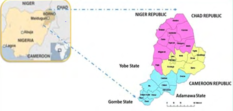
Despite several studies on the seroprevalence of antibodies against CCHFV from humans and animals in Nigeria [1, 2, 3, 4], there has not been any confirmed case of human CCHFV infection reported from any part of the Country. The use of molecular diagnostic tools has resulted in highly sensitive and specific tests for infectious organisms and genetic diseases. These tools, which include polymerase chain reaction (PCR), metagenomics and next generation molecular sequencing, have come in handy for the efficient and rapid identification and characterization of CCHFV strains and other viral Haemorrhagic fever (VHF) agents from suspected cases, vectors and reservoir hosts of these viruses [5, 6, 7]. This study was carried out in Borno state, Nigeria as part of a wider serological and virological surveillance for Haemorrhagic fever viruses in the area. Borno state (Figure 1) is bounded by three Nigerian states (Adamawa, Gombe and Yobe) and three International neighbours (Cameroun, Chad and Niger Republics), where outbreaks of some insect- borne viruses have been reported. Tran’s boundary activities (human and animal movements) have been ongoing. Humanitarian crisis, starting mostly from 2009 as disturbed the ecological balance in the Lake Chad & Sambisa forest area due to Boko Haram insurgency by displacing both animals and humans (creating Internally and Externally Displaced Persons, over 20 million people have so far been affected) [8]. Hence, making Borno state and the Chad basin a favourable ecological niche for some viruses.
Broad Methodology
Summarised Method
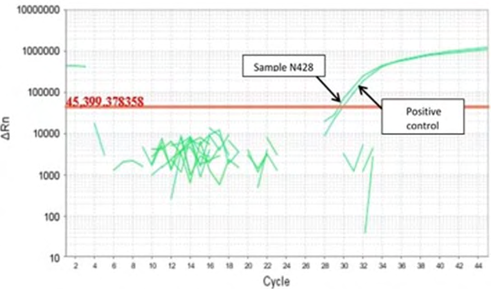
Sera (n=380) collected as part of a serosurveillance Programme for VHFs from undiagnosed febrile (fever of unknown origin) patients in Borno state were analyzed by real time RT-PCR assay for the presence of CCHFV RNA. One positive sample (N428) from a 15-year old female showed a cycle threshold (CT) value of 29.42 and a slope formation typically seen for a positive sample (Figure 2). The RNA from this sample was further characterized by next generation sequencing (NGS) which resulted in complete S, M, and L viral RNA segment sequences.
Detailed Molecular Testing
Extraction of RNA was performed using the MagnaPure 96 small volume RNA kit (Roche). Plates were loaded onto the MagnaPure 96 automated extraction robot and RNA was eluted in 60µl nuclease free water. Target amplification was performed using primers to CCHFv S segment [9] using the Superscript III qRT-PCR Kit (Life Technologies). Analysis was performed using the ABi 7500 (Applied Biosystems) at the following cycling conditions; 50°C for 10 minutes, 95°C for 2minutes followed by 40 cycles of 95°C for 10 seconds,60°C for 40 seconds and a cooling cycle of 40°C for 30 seconds. Temperature cycling was set to maximum ramp speed and data was acquired and analysed using the ABi 7500 on-board software with an automatically selected threshold.
Genomic Characterization
RNA samples were digested with DNAse 1 (Life Technologies) using the off column protocol. The DNAse was inactivated and the samples were cleaned up using a Zymo Clean and Concentrator column and eluted into 6µl mH2O. 5µl of the samples were prepared using the ovation system for amplifying cDNA from total RNA for use in RNASeq applications; samples were prepared following manufacturer’s instructions, with the exception that RNA was denatured for 5 minutes at 85°C prior to first strand synthesis and purified using a Qiagen MinElute Reaction Cleanup Kit and eluted in 15µl mH2O. Illumina library were prepared in house using the Illumina Nextera XT V2 300 cycle kit, 1.5ng of SPIA cDNA from each sample was run following the manufacturer’s instructions with the exception that sample preparation was halted at the stage of Clean Amplified Nucleic acid for final loading, indices were selected using the Illumina experiment manager software. Tagmentation reactions were quantified using a bioanalyser for fragment size analysis and kapa library quantification kit for NGS.
For sequence analysis the fastq files were quality trimmed to an average phred score of 30 across the read. Using Galaxy, the trimmed fastq’s were subjected to a kraken taxonomic screen using a full kraken database [kraken_v2.2_vir_bac_arc_euk] of 8041 complete reference genomes (Bacteria: 3471, Viruses: 4262, Archaea: 307, Eukaryota [Saccharomyces ceravesiae]: and an additional 5055 incomplete nucleotide sequences. Reads were then assembled into contigs, representing an unbiased approach by mapping from assembled fragments. All assembled contigs greater than 500bp or more than 10,000 reads were subject to a nucleotide blast search for CCHF. Reads were then mapped to a CCHF full genome sequence using refseqs for NC_005301.3, NC_005300.2 and NC_005302.1 for L, M and S segments, respectively.
Phylogenetic Analysis
Phylogenetic analyses were performed using MEGA 6. Trees were precomputed using the Neighbour-Joining method then evolutionary history and distances inferred by the Maximum Likelihood method. Maximum Likelihood phylogenetic trees were generated for the open reading frames of the partial L, M and S sequences recovered from next-generation sequencing. All positions containing gaps and missing data were eliminated from the analysis.
Ethical Consideration
Ethical clearance for this study was obtained from the Borno State Ministry of health, Maiduguri.
Results
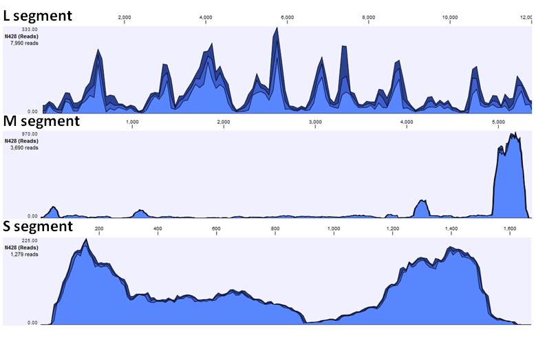
Initial assembly resulted in 1,958 contigs. These contigs were then BLAST searched against the Nairovirus taxon and resulted in 16 contigs typically >95% identity with CCHF Sudan ABI_2009. Near full genome coverage was seen for the sample when mapped to CCHFV (Table 1). These mapping data were also visualized (Figure 3).
| Mapped reads | Minimum coverage | Maximum coverage | Average coverage | Average length (zero coverage regions) | Fraction of reference covered | |
|---|---|---|---|---|---|---|
| S | 1279 | 0 | 225 | 82.539 | 31 | 0.963 |
| M | 3690 | 0 | 970 | 71.247 | 56.2 | 0.948 |
| L | 7990 | 0 | 333 | 66.205 | 8 | 0.999 |
Table 1: Coverage of reads from next-generation sequencing to the segments of CCHF virus.
The genome assembly included three segments; the L Segment [GenBank accession number KX238956], M Segment [GenBank accession number KX238957] and S Segment [GenBank accession number KX238958]. Evolutionary history was inferred using the maximum likelihood method based on the Tamura-Nei model and confidence assessed with the Bootstrap Test with 1000 resamplings.
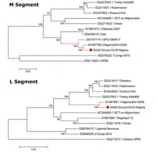
Phylogenetic analysis clustered the S segment in the Africa 3 phylogenetic group. The S segment open-reading frame showed close homology with a previous isolate of CCHF from Nigeria (IbAr 10200), as well as isolates from Mauritania (ArD39554) and South Africa (SPU415/85 and SPU128/61/7). The M and the L clustered closely with the Sudan AB1-2009 isolate and the Nigeria IbAr 10200 isolate.
Phylogenetic analysis clustered the S-segment in the Africa 3 clade or phylogenetic group (Figure 4). The S-segment open-reading frame showed close homology with a previous isolate of CCHFV from Nigeria (IbAr10200), as well as isolates from Mauritania (ArD39554) and South Africa (SPU415/85 and SPU128/61/7). The M and L clustered closely with the Sudan ABI-2009 isolate and the Nigeria IbAr10200 (Figure 5). The L, M, and S sequences were submitted to GenBank with accession numbers KX238956, KX238957 and KX238958 respectively [10].
Discussion and Conclusion
Previous serological results by Bukbuk and co-workers in 2016 [10] have demonstrated the circulation of CCHFV in 4 different local government areas (LGAs) of Borno state, Nigeria with average positivity rates of 10.6% and 3.5% for IgG and IgM responses respectively. While, between September 2011 and February 2012, the same Author in 2014 [7], earlier on reported a prevalence rate of 2.4% for IgG antibodies from 10 LGAs in the same state, using similar ELISA test. This indicates that the virus might be actively circulating in either the arthropod vectors (Ixotid - hard ticks) or animal and human population. This current study has therefore unequivocally demonstrated the virological evidence for the presence and continued exposure to CCHF virus of the human population in Nigeria. This genomic analysis provides the first published evidence of a human case of CCHFV in Nigeria and its phylogenetic context. This study also indicates that CCHF virus and other Haemorrhagic fever viruses could be circulating or are endemic in Borno state, possibly due to the presence of insect vectors and amplifying or reservoirs hosts (humans, domestic & wild animals, migratory birds) in the study area. A detailed virological and vectoral study needs to be conducted in order to elucidate the epidemiology of these viruses in this ecological niche. This could therefore be achieved through the establishment of collaborative study with other institutions within and outside the country.
References
-
Causey OR, Kemp GE, Madbouly MH, David West TS (1970) Congo virus from domestic livestock, African hedgehogs, and arthropods in Nigeria. American Journal of Tropical Medicine and Hygiene 19(5): 846-850.
-
David West TS, Cooke AR, David West AS (1974) Sero- epidemiology of Congo virus (related to the virus of Crimean haemorrhagic fever) in Nigeria. Bulletin of the World Health Organisation 51(5): 543-546.
-
Umoh JU, Ezeokoli CD, Ogwu D (1983) Prevalence of antibodies to Crimean-Congo haemorrhagic virus in cattle in northern Nigeria. International Journal of Zoonoses 10(2): 151-154.
-
Oluwayelu D, Afrough B, Adebiyi A, Varghese A, Eun Sil P, et al. (2020) Prevalence of Antibodies to Crimean- Congo Hemorrhagic Fever Virus in Ruminants, Nigeria. Emerging Infectious Diseases 26(4): 744-747.
-
Drosten C, Kummerer BM, Schmitz H, Gunther S (2003) Molecular diagnostics of viral haemorrhagic fevers. Antiviral Research 57(1-20): 61-87.
-
Fukushi S, Mizutani T, Saijo M, Kurane I, Taguchi F, et al. (2006) Evaluation of a novel vesicular stomatitis virus pseudotype-based assay for detection of neutralizing antibody responses to SARS-CoV. J Med Virol 78(12): 1509-1512.
-
Bukbuk DN, Fukushi S, Tani H, Yoshikawa T, Taniguchi S, et al. (2014) Development and validation of serological assays for viral hemorrhagic fevers and determination of the prevalence of Rift Valley fever in Borno State, Nigeria. Trans R Soc Trop Med Hyg 108(12): 768-773.
-
UNOCHA (2016) Lake Chad Basin: crisis overview. New York: United Nations Office for the Coordination of Humanitarian Affairs.
-
Atkinson B, Chamberlain J, Logue CH, Cook N, Bruce C, Dowall SD, et al (2012). Development of a real-time RT-PCR assay for the detection of Crimean-Congo hemorrhagic fever virus. Vector Borne Zoonotic Dis. 12(9): 786-793. doi: 10.1089/vbz.2011.0770. Epub 2012 Jan 4. PMID: 22217175.
-
Bukbuk DN, Dowall SD, Lewandowski K, Bosworth A, Baba SS, et al. (2016) Serological and Virological Evidence of Crimean-Congo Haemorrhagic fever virus circulation in the human population of Borno state, northeastern, Nigeria. PLOS Neglected Tropical Diseases 10(12): e0005126.
- hMPV: Is It Another Covid-19 Like Situation?
- Streptomyces: Sources of Novel Discoveries in Antibiotic Research to Combat Antimicrobial Resistance
- A Review of Mosquitoes (Diptera: Culicidae) and Their Biodiversity, Medical and Veterinary Importance
- Past and Current Immunotherapy in Cancer
- Hematological Cancer and Viral Infection
- The Growing Threat of Antimicrobial Resistance in India: Challenges and Solutions