Total Abdominal Hysterectomy for a 36 Weeks Size Fibroid Uterus through a Supra Pubic Transverse Incision
Introduction: Uterine fibroids are the commonest Gynecological benign tumor which affect reproductive age women. Surgical management of large fibroid uterus create multiple challenges to both surgeon and the patient. This is a cases of 36 weeks size fibroid uterus, grown up to the level of xiphisternum which treated with total abdominal hysterectomy through a suprapubic transverse incision. Case: 45-year-old, previously healthy multi-parous women presented with progressive abdominal distention for one to two years. Clinical and Ultrasound scan confirmed the fibroid uterus. She successfully underwent total abdominal hysterectomy through a supra-pubic transverse incision. Conclusion: Management of a fibroid uterus depends on patient’s age, fertility wishes, uterine preserving desires, symptomatology, site and size of the fibroids. Successful hysterectomy can be done through a supra-pubic transverse incision even for a 36 weeks size fibroid uterus in carefully selected cases.
Introduction
Uterine leiomyomas or fibroids are the commonest benign gynecological tumor which may affect up to 80% of women before the menopause [1]. Prevalence of fibroids among women between 30 to 50 years is 20 to 30% [2]. Even though most of the women with fibroid uteruses are asymptomatic, fibroid uterus will create a huge negative impact on women’s life due to symptoms like menorrhagia, dysmenorrhea, pelvic pain disorders, infertility, or early pregnancy complications [3]. In rare instances some of these fibroids metastasize even though they are benign which called as benign metastasizing leiomyoma [4]. Prevalence of fibroid uterus is very low in women below the age of 20 years [5].
Even though exact mechanism for the fibroids are not yet known, presence of high number of estrogen and progesterone receptors in these smooth muscle cells explained the increase prevalence of these tumors during reproductive age and drastically reduction in prevalence after menopause [6]. Higher concentration of aromatase, estrogen receptors and progesterone receptors have been detected in leiomyoma cells compare to normal myometrial cells [7]. Other factors that may affect the growth of fibroids are obesity, early menarche, pregnancy and exogenous estrogens. Even though some authors suggest a connection between red meat and broiler chicken consumption with fibroids, current evidence are not enough to prove it [8]. Mutations of chromosome 6, 7, 12, and 14 have been found in leiomyoma cells even though mechanism of subsequent hyperproliferation was not described yet [9]. Prevalence of fibroid uterus (with relatively larger size, detection at younger ages, symptomatic) is higher among African women [10].
Best diagnostic tool for fibroid uterus is Ultra Sound Scan (USS) due to its high diagnostic accuracy, easy availability and cost effectiveness. Magnetic resonance imaging (MRI) is the best tool to assess the pelvic soft tissues which can be used to distinguish leiomyosarcoma from fibroids [11]. Treatment for the fibroid uterus depends on patient’s age, fertility wishes, site and size of the fibroids.
When considering the abdominal incision techniques in open Gynecological surgeries, low transverse incision techniques (Pfannenstiel incision) have gained popularity compare to mid line incisions due to less post-operative pain, less risk of incisional hernia and cosmetic acceptability. But mid line incision may need when good access is required or in a case of ovarian malignancies [12].
Case
45-year-old mother of 4 children (delivered vaginally), who was previously healthy presented with progressive abdominal distention over the last 1 to 2 years. She initially attributed this to her laxed pendulous abdomen following her four pregnancies. She developed difficulty in breathing and she was found to have and abdomino-pelvic mass. She was referred to a Gynecologist. She had regular menstrual cycles without heavy menstrual bleeding, secondary dysmenorrhea, inter menstrual bleeding or post coital bleeding. She denied any loss of appetite or loss of weight. She didn’t have any family history of breast, uterine, ovarian or colorectal carcinomas. She had never undergone any kind of abdominal surgeries.
On examination she was averagely built lady without any features of anemia. Her abdominal examination revealed abdominopelvic mass which compatible with a 36 weeks size gravid uterus, extending up to the level of xiphisternum. Characteristics of this mass suggested a presence of fibroid uterus. She had a very laxed abdomen. Trans abdominal ultrasound scan of the abdomen and pelvis confirmed the presence of multiple fibroid uterus. A cluster of intra-mural fibroids noted arising from the fundus of the uterus without distorting the endometrial cavity. Largest fibroid was 25cm in diameter. Rest of the uterus was normal. Bilateral ovaries were normal. There was no free fluid in the peritoneal cavity. Even though the requirement of an MRI aroused to exclude sarcomatous changes in a this much of larger fibroid uterus, it was not done as she didn’t have risk factors for leiomyosarcoma (tamoxifen therapy and past pelvic irradiation) and MRI was not freely available. Patient was counseled regarding the findings and available management options. She agreed for total abdominal hysterectomy through a laparotomy without any pre-surgical GnRH (Gonadotrophin releasing Hormone) therapy to reduce the size of the uterus. She was informed about the requirement of mid line laparotomy. She agreed for that and all the other possible complications.
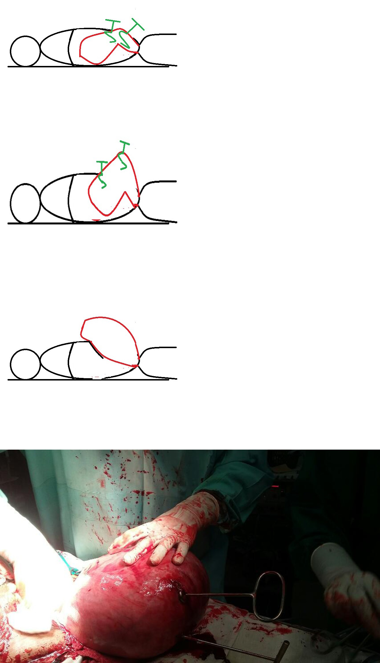
Her abdomen was re-examined after giving general anesthesia which confirmed the presence of good mobility of the mass. About 4 inches size supra-pubic transverse incision was made and opened into the peritoneal cavity. Surgeon’s hand was inserted into the peritoneal cavity and thoroughly explored around the fibroids to identify the anatomy and to exclude adhesions. Then the incision was extended laterally and good access to the fibroids was obtained (Figure A). A myoma screw was carefully inserted into the fibroid at its superior most approachable point through the transverse incision (Figure B). Then it was pulled up and towards caudal end of the patient. Then another myoma screw was inserted to the fibroid above the previous fibroid and pulled in the same direction until more and more area of the fibroid exposed through the transverse incision (Figure C). Then the previous myoma screw was removed and re inserted above the last one (Figure D). These steps were repeated until fundus of the uterus delivered out through the incision (Figure E). After delivering the whole uterus out of the peritoneal cavity, a routine total hysterectomy and bilateral salpingectomy was done while preserving the both ovaries (Figure F). Ovaries were anchored to ipsilateral round ligament to prevent future torsions and dyspareunia. She had an uneventful recovery and histology confirmed the radiological diagnosis of fibroid uterus.
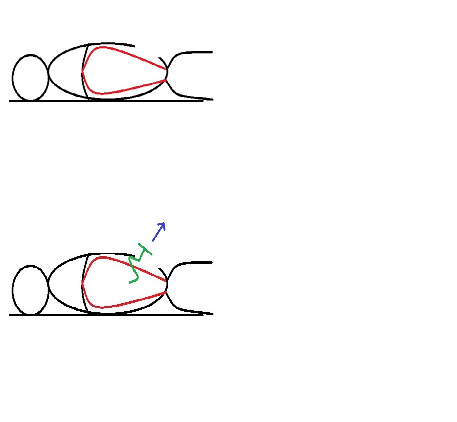
A: getting a good exposure to the uterus through a supra- pubic transverse incision B: insertion of a myoma screw at the superior most approachable point of the fibroid
C: insertion of the second myoma screw above the first one while pulling out from the first myoma screw D: removal of first myoma screw and re-insertion above the second one
E: uterus delivered out though the incision F: intra operative view of the uterus Figure 1: steps of delivering the fibroid uterus out from the peritoneal cavity.
Discussion
This patient had delay in seeking treatments until her uterine fibroids grown up to the level of xiphisternum. As she had a laxed pendulous abdomen due to her multiparity compare to a nulliparous woman, she may had attributed the symptoms of fibroid uterus to that. Other than that, she didn’t have any menstrual problems as this was an intra-mural fibroid. Performing an MRI scan in this patient is justifiable to exclude sarcomatous changes as it is the best imaging tool to visualize soft tissues [11]. But other than its large size she didn’t have any risk factors for a leiomyosarcoma like past pelvic radiation and tamoxifen therapy. Even though more widely available compare to MRI, computerized sonography is not recommended for the further evaluation of fibroid uterus [13].
As she was a multiparous lady who had completed the family without any uterine preserving desire, she can undergo hysterectomy, even though many non-surgical methods are available. The best treatment modality for her is hysterectomy due to its large size, not having future fertility wishes, not having any desire to retain the uterus, not affecting hormonal cycle and zero recurrence rate [14]. Uterine artery embolization and MRI guided focused ultrasonography are another two non-surgical options. But the suitability for this kind of large fibroid uterus is not well established [15]. Other thing is she needs subsequent visits to manage the complications of the procedures like post- embolization syndrome and recurrences of fibroids. Medical management options like GnRH (Gonadotrophin releasing hormone) analogues can be used for a short period or as a pre-surgical treatment to reduce the size of the fibroid and reduce the intra-operative bleeding. By doing that a larger fibroid uterus which need a mid-line incision, can be delivered during the surgery through a transverse incision as it reduces the fibroid size. But in case of myomectomy, the enucleation of the fibroid will be difficult as pre-operative GnRH treatment obscure the tissue planes. Same time she will have to wait another few months to get done the surgery. But if pre-operative GnRH therapy become successful, hysterectomy can be done by laparoscopically as well with expert hands. But care should be taken to prevent spillage of myoma particles with in the peritoneal cavity during morcellation, even though sarcomatous changes could have been excluded by pre-operative MRI. Because spillage of particles even from a benign fibroid can cause disseminated peritoneal leiomyomatosis [16].
Selecting a most appropriate incision for an open hysterectomy in a large fibroid uterus is challenging. Disadvantages of a supra-pubic transverse incision in this patient is limited access to the upper abdomen as fibroid had extended up to the xiphisternum level. The problem could have become worse if there were adhesions with the fibroid. The second thing is difficulty in delivery of the fibroid out from the peritoneal cavity. But pre-operative imaging confirmed the exact nature and location of fibroid (intra-mural fibroid cluster arising from the fundus of the uterus). Same time, examination under general anesthesia demonstrated good mobility of the mass which clinically exclude dense adhesions. All these facts are in favor of supra-pubic transverse incision instead of midline incision which increases the post-operative pain, risk of incisional hernias and poor patient satisfaction of patient about the cosmetic outcome. Delivery of the fibroid uterus out from the peritoneal cavity before starting the hysterectomy is the routine practice in case of large fibroid uterus. In this case this step was done by using two myoma screws alternatively.
Conclusion
Uterine leiomyomas are the commonest Gynecological tumors among reproductive aged women some times present with larger sizes which warrants exclusion of sarcomatous changes by pre-operative imaging with MRI. Mode of treatment depends on various factors. Performing a total abdominal hysterectomy for a large fibroid uterus through a supra-pubic transverse incision can be done successfully after selecting a suitable patient with a virgin abdomen with proper pre-operative fibroid mapping and examination after giving anesthesia.
Declarations
Ethical approval: As this is a case report which do not contain any patient identification details, ethical approval is not required. Informed consent: Informed written consent was obtained from the patient.
Author contribution: The author confirms sole responsibility for study conception and design, data collection, analysis and interpretation of results, and manuscript preparation.
References
-
Baird DD, Dunson DB, Hill MC, Cousins D, Schectman JM (2003) High cumulative incidence of uterine leiomyoma in black and white women: ultrasound evidence. Am J Obstet Gynecol 188(1): 100-107.
-
Laughlin SK, Baird DD, Savitz DA, Herring AH, Hartmann KE (2009) Prevalence of uterine leiomyomas in the first trimester of pregnancy: an ultrasound-screening study. Obstet Gynecol 113(3): 630-635.
-
Fortin C, Flyckt R, Falcone T (2018) Alternatives to hysterectomy: the burden of fibroids and the quality of life. Best Pract Res Clin Obstet Gynaecol 46: 31-42.
-
Walawe Nayaka S, Karunarathna SMG 2023 Benign Metastasizing Leiomyoma. J Gynecol 8(3): 000262
-
Grapsa D, Smymiotis V, Hasiakos D, Kontogianni-Katsarou K, Kondi-Pafiti A (2006) A giant uterine leiomyoma simulating an ovarian mass in a 16-year-old girl: a case report and review of the literature. Eur J Gynaecol Oncol 27(3): 294-296.
-
Bekker G, Gavrilescu T, Rickets-Holcomb L, Puka- Khandam P, Akhtar A, et al. (2004) Symptomatic fibroid uterus in a 15-year-old girl. Int Surg 89(2): 80-82.
-
Rein MS, Barbieri RL, Friedman AJ (1995) Progesterone: a critical role in the pathogenesis of uterine myomas. Am J Obstet Gynecol 172(1 Pt 1):14-18.
-
Ernest A, Mwakalebela A, Mpondo BC (2016) Uterine leiomyoma in a 19-year-old girl: Case report and literature review. Malawi Medical Journal 28(1): 31-33.
-
Ligon AH, Morton CC (2001) Leiomyomata: heritability and cytogenetic studies. Hum Reprod Update. 7(1): 8-14.
-
Baird DD, Dunson DB, Hill MC, Cousins D, Schectman JM (2003) High cumulative incidence of uterine leiomyoma in black and white women: ultrasound evidence. Am J Obstet Gynecol 188(1): 100-107.
-
Karasick S, Lev-Toaff AS, Toaff ME. (1992) Imaging of uterine leiomyomas. AJR Am J Roentgenol 158(4): 799- 805.
-
Burger JWA, van‘t Riet M, Jeekel J (2002) Abdominal incisions: Techniques and post-operative complications. Scandinavian Journal of Surgery 91(4): 315-321.
-
Casillas J, Joseph RC, Guerra JJ Jr. (1990) CT appearance of uterine leiomyomas. Radiographics 10(6): 999-1007.
-
Moroni RM, Vieira CS, Ferriani RA, Reis RM, Nogueira AA, et al. (2015) Presentation and treatment of uterine leiomyoma in adolescence: a systematic review. BMC Womens Health 15: 4.
-
Goldberg J, Pereira L, Berghella V (2002) Pregnancy after uterine artery embolization. Obstet Gynecol 100(5 Pt 1): 869-872.
-
Al-Talib A, Tulandi T (2010) Pathophysiology and Possible Iatrogenic Cause of Leiomyomatosis Peritonealis Disseminata. Gynecol Obstet Invest 69(4): 239-244.
- The Need for Partner Education and Mental Health Support During Pregnancy and the Postpartum Period
- Application of Combined PGT-A and PGT-M for Reproductive Management in a Couple Carrying GCDH Mutations with Prior Affected Offspring: A Rare Case Report
- The Effect of Using a New Technique Karman Injector (Elif Technique) on the Healing Process of Wound Infection-Case Series
- GSM: Counseling Points to Discuss with Women Fearful of Vaginal Estrogen
- Antenatal Diagnosis of Meckel Syndrome: A Case Report
- Discrimination and Workplace Harassment (Mobbing) against Women in the Post-Pandemic Era