A Case of Postnatally Diagnosed Sirenomelia in a Patient with Poorly Controlled Type 2 Diabetes Mellitus
Sirenomelia, also known as mermaid syndrome, is a rare constellation of anomalies of caudal fetal development associated with fusion or complete absence of the lower limbs. The prognosis for neonatal survival is poor. Diagnosing this condition can pose a challenge due to associated oligo/anhydramnios. First-trimester transvaginal and transabdominal anatomy ultrasound performed between 11-13.6 weeks of gestational age (WGA) can evaluate fetal anatomy for anomalies. Transvaginal (TVUS) ultrasound offers the best opportunity to detect this condition. We present a case of a 1240g baby born from a 34yo with poorly controlled type 2 diabetes (T2DM) delivered via repeat cesarean section. Antenatal management for presumed previable pre-labor rupture of membranes (PROM) was provided. Postnatally, the infant had unexpected defects consistent with Sirenomelia. The neonatal exam showed webbed legs due to fused knees and toes, microcephaly, two-vessel umbilical cord, ambiguous genitalia, and bilateral renal agenesis. After confirming bilateral renal agenesis, we provided palliative care. The neonate died 12 hours after birth from cardiac arrest.
Introduction
Sirenomelia, or Mermaid Syndrome, is a rare congenital anomaly of caudal fetal development, causing a fusion or complete absence of lower limbs. It affects approximately
0.98 - 4 per 100,000 live births [1, 2, 3, 4]. The prognosis is poor due to associated severe anomalies [2, 4, 5, 6, 7]. Most newborns with Sirenomelia die within the first few hours of life due to the sequelae of renal agenesis, pulmonary hypoplasia, cardiac defects, absent urinary bladder, esophageal atresia, rectal atresia, diaphragmatic hernia, omphalocele, imperforate anus, absent external genitalia, ambiguous genitalia, lumbosacral/pelvic bone abnormities, and spina bifida [1, 4, 5, 6, 8, 9, 10]. Rarely cases with less severe anomalies have been shown to survive after a multidisciplinary surgical approach [11].
The exact mechanism by which this pathology arises is not known, but several risk factors have been associated with Sirenomelia including maternal hyperglycemia, extremes of maternal age, monozygotic twinning, exposure to teratogens, and/or heavy metals and a previously affected child [8, 10, 12, 13].
Objectives/Purpose
We present a case of a 34-year-old with a history of poorly controlled type 2 diabetes mellitus. Anhydramnios was found at 17 weeks gestation. She reported leakage of fluid and was managed as presumed pre-viable premature rupture of membranes. Anhydramnios obscured anatomic views. Sirenomelia with renal agenesis was diagnosed postnatally, illustrating the importance of earlier transvaginal detailed ultrasound in acoustically challenging high-risk patients.
Case Report
A 34-year-old G5P3013 with history of cesarean section x three, body mass index >45, andT2DM presented for maternal fetal medicine consultation due to poorly controlled T2DM (HgbA1c 9.2) at 13.6wga by a dating transabdominal ultrasound obtained at that visit. A crown rump length measured 13.6wga and normal amniotic fluid seen around the singleton fetus. Poor maternal acoustics inhibited anatomic evaluation via transabdominal approach. The patient was scheduled for a detailed anatomic survey at 17wga. Cell free DNA for sex chromosome aneuploidy, trisomy 21, trisomy 18, and/or trisomy 13 and showed low risk testing with an XX fetus. The 17wga detailed anatomy ultrasound (76811) showed anhydramnios with severe fetal growth restriction (FGR) and multiple suspected congenital anomalies including ventriculomegaly with dangling choroid, possible AV canal defect, abnormal curvature of cervical and lumbar spine. The cavum septum pellucida and fetal kidneys could not be demonstrated.
Upon questioning, she reported leaking of fluid for several days. A physical exam revealed no pooling or ferning, with a negative nitrazine test. She was counseled on the poor prognosis due to presumed previable PPROM with multiple fetal anomalies and was offered induction of labor pursuant to Georgia law, which allows termination of a medically futile pregnancy, but she desired expectant management and declined induction of labor. She received latency antibiotics at 20wga. She had no findings of chorioamnionitis. Follow- up ultrasound at 21.6wga showed continued anhydramnios, severe FGR, suspected holoprosencephaly, atrioventricular canal defect, at least unilateral renal agenesis, and abnormal cervical and lumbar spine curvature with breech fetus.
She reiterated her desire for maximal fetal interventions despite the anticipated poor neonatal prognosis. Consultation with pediatric palliative care and the Neonatal Intensive care team was provided due to the anticipation of poor neonatal prognosis. Growth ultrasounds every 4 weeks and weekly biophysical profiles (BPP) with umbilical artery (UA) Doppler starting at 28wga were planned. She agreed with inpatient management starting at 23.0wga and was given betamethasone for induction of lung maturity, and magnesium sulfate for neuroprotection.
Inpatient maternal/fetal monitoring for intraamniotic infection, maternal sepsis, placental abruption, and preterm labor was provided. Stricter glycemic control was attempted with limited success. Her glucose control continued to be challenging secondary to steroid administration, diet noncompliance, and the patient’s frequent insulin refusal, but inpatient hospitalization was otherwise uncomplicated. She remained inpatient until a repeat cesarean section at 34.0wga. During her hospitalization, she again met with the neonatal intensive care unit (NICU) team who reiterated the prognosis was poor.
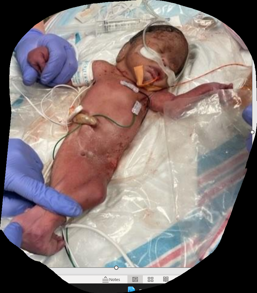
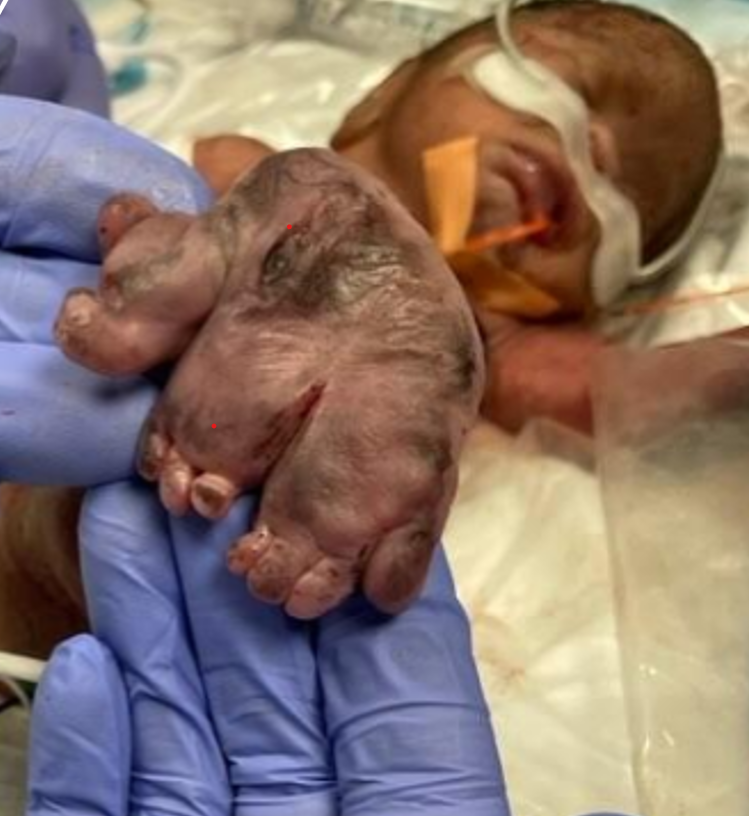
At delivery, the fetus had APGARS of 2 and 8 at one and five minutes, respectively, with a fetal weight of 1240gm. The neonatal care team provided positive pressure ventilation. At 30 seconds of life, an attempted at intubation was unsuccessful in the delivery room. Continued positive airway pressure was provided in the interim until another intubation was attempted in the NICU. Physical exam demonstrated microcephaly with a palpable defect in the cranial bones, asymmetric chest expansion, two-vessel cord, ambiguous genitalia, imperforate anus, webbed legs with knee joints fused bilaterally, fused feet with 11 total toes, no primitive reflexes, and no palpable femoral pulses (Figures 1 & 2).
In the NICU, the infant continued to demonstrate poor oxygen saturation despite successful intubation. A chest x-ray showed bilateral pneumothoraxes requiring thoracenteses. Abdominal ultrasound showed renal agenesis without fluid in the bladder. No further futile interventions were provided after diagnosing renal agenesis. Around the 12th hour of life, the neonate went into cardiac arrest.
The family declined autopsy but desired further genetic testing to understand the recurrence risk for this condition. Microarray, whole exome sequencing, and mitochondrial sequencing were negative for pathogenic findings. Her postpartum course was uncomplicated. She was counseled that she has a recurrence risk of 3-5% for Sirenomelia and that poor glucose control could contribute to the pathology. We recommend weight loss and improved glucose control prior to a future pregnancy.
Discussion
While there are reports of a few surviving neonates [11, 14] babies with Sirenomelia generally have poor outcomes and typically die within 1 to 2 days following birth [8]. Several classification systems for Sirenomelia have been developed over the years. The Stocker-Heifetz system is currently the most widely used classification system. The system classifies Sirenomelia into seven types based on skeletal structures of the lower limb (type 1 to 7) [15]. Our case likely demonstrated a type I or II classification. Accurate classification requires an X-ray and/or CT scan. Postmortem imaging can assist in this classification system. The patient declined this examination [16].
Sirenomelia was previously considered a severe form of caudal regression syndrome. While the topic is still under debate, multiple researches now consider these two conditions as separate disease processes [17, 18, 19]. Sirenomelia is likely secondary to the interaction of genetic and environmental factors which occur before embryogenesis is complete leading to compromised vasculature formation in the lower body of the embryo due to vascular steal [12, 18, 20]. Murine models have demonstrated several genes to cause or be associated with Sirenomelia. Poorly controlled diabetes mellitus is another important risk factor. It is postulated that the increased free oxygen radicals production in poorly controlled maternal diabetes, exert a teratogenic effect in embryonic development leading to this anomaly [1, 3, 5, 8, 21].
Most cases are sporadic, but the chance of recurrence in 3-5% of cases highlights the genetic contribution to development of this syndrome [6]. Women with pre-existing diabetes should attempt to improve glucose control prior to conception to reduce the risk of poor pregnancy outcomes. Obese diabetics are at increased risk of fetal malformation and are acoustically difficult to image in the standard window at 18-22 wga.
International Society of Ultrasound in Obstetrics and Gynecology (ISUOG) has guidelines for the assessment of fetal anatomy in the first trimester using transvaginal and transabdominal imaging to complete an anatomic evaluation. ISUOG further recommends that all patients should have access to anatomy assessment between 11-13.6 wga to allow for anomaly detection. Earlier detection can improve care coordination. It allows more time for genetic testing, parental counseling, and decision-making. A transvaginal ultrasound in conjunction with transabdominal ultrasound around 13.6 wga may represent the best opportunity to evaluate diabetic fetopathy in obese women. It would likely have revealed the defects seen in this case prior to onset of anhydramnios and could have led to a prenatal diagnosis. This condition is difficult to diagnose because oligo/anhydramnios has developed by the traditional anatomy ultrasound window at 18-22 wga. The lack of an acoustic windows and adequate visualization of anatomic structures obscures the diagnosis. An earlier diagnosis in our case would have resulted in improved counseling about the medical futility of aggressive support and would have prevented an unnecessary hospital stay. Improved understanding of anatomic defects in the first trimester will result in a less ambiguous discussion about the likely lethal nature of this condition and for some women will allow a timely decision about pregnancy termination when more reproductive options are available.
Conclusion
Sirenomelia remains a fatal condition with multisystem involvement. It poses a diagnostic challenge. First trimester transvaginal detailed ultrasound can be performed to detect serious congenital anomalies. Earlier diagnosis offers a wider range of reproductive options and provides more time for families to consult with specialists and make decisions regarding management.
References
-
Al Hadhoud F, Kamal AH, Al Anjari A, Diejomaoh MF (2017) Fusion of lower limbs with severe urogenital malformation in a newborn, a rare congenital clinical syndrome: case report. Int Med Case Rep J 10: 313-317.
-
Kavunga EK, Bunduki GK, Mumbere M, Masumbuko CK (2019) Sirenomelia associated with an anterior abdominal wall defect: a case report. J Med Case Reports 13(1): 213.
-
Ramphul K, Mejias SG, Ramphul-Sicharam Y (2018) Mermaid Syndrome: A Case Report in Mauritius. Cureus 10(2): e2210.
-
Morales-Roselló J, Loscalzo G, Buongiorno S, Jakaitė V, Perales-Marín A (2022) Sirenomelia, case report and review of the literature. J Matern-Fetal Neonatal Med 35(6): 1203-1206.
-
Turgut H, Ozdemir R, Gokce IK, Karakurt C, Karadag A (2017) Sirenomelia associated with Hypoplastic Left Heart in a Newborn. Balkan J Med Genet 20(1): 91-94.
-
Sikandar R, Munim S (2009) Sirenomelia, the Mermaid syndrome: case report and a brief review of literature. J Pak Med Assoc 59(10): 721-723.
-
Kucuk Ş, Kucuk İG (2020) Sirenomelia (Mermaid Syndrome): A Case Report. Turk Patoloji Derg 36(3): 256-260.
-
Tamene A, Molla M (2022) Sirenomelia: A case report. SAGE Open Med Case Rep. 10: 2050313X221092560.
-
Fadhlaoui A, Khrouf M, Gaigi S, Zhioua F, Chaker A (2010) The Sirenomelia Sequence: A Case History. Clin Med Insights Case Rep 3: 41-49.
-
Vijayaraghavan SB, Amudha AP (2006) High-Resolution Sonographic Diagnosis of Sirenomelia. J Ultrasound Med 25(4): 555-557.
-
Messineo A, Innocenti M, Gelli R, Pancani S, Lo Piccolo R, et al. (2006) Multidisciplinary Surgical Approach to a Surviving Infant with Sirenomelia. Pediatrics 118(1): e220-e223.
-
Garrido-Allepuz C, Haro E, Gonzalez-Lamuno D, Martinez-Frias, Bertocchini F, et al. (2023) A clinical and experimental overview of sirenomelia: insight into the mechanisms of congenital limb malformations. Disease Models & Mechanisms 4(3): 489-499.
-
Orioli IM, Amar E, Arteaga-Vazquez J, Bakker MK, Bianca S, et al. (2011) Sirenomelia: an epidemiologic study in a large dataset from the International Clearinghouse of Birth Defects Surveillance and Research, and literature review. Am J Med Genet C Semin Med Genet 157C(4): 358-373.
-
Stanton MP, Penington EC, Hutson JM (2003) surviving infant with sirenomelia (mermaid syndrome) associated with absent bladder. J Pediatr Surg 38(8): 1266-1268.
-
Stocker JT, Heifetz SA ( 1987) Sirenomelia. A morphological study of 33 cases and review of the literature. Perspect Pediatr Pathol 10: 7-50.
-
Russo A, Reginelli A, Pignatiello M, Montella M, Toni G, et al. (2021) Sirenomelia: The role of post - Mortem diagnostic imaging. J Pediatr Surg Case Rep 71: 101921.
-
Perez‐Aytes A, Montero L, Gomez J, Paya A (1997) Single aberrant umbilical artery in a fetus with severe caudal defects: Sirenomelia or caudal dysgenesis. Am J Med Genet 69(4): 409-412.
-
Boer LL, Morava E, Klein WM, Schepens-Franke AN, Oostra RJ (2017) Sirenomelia: A Multi-systemic Polytopic Field Defect with Ongoing Controversies. Birth Defects Res 109(10): 791-804.
-
Duncan PA, Shapiro LR, Klein RM (1991) Sacrococcygeal dysgenesis association. Am J Med Genet 41(2): 153-161.
-
Twickler D, Budorick N, Pretorius D, Grafe M, Currarino G (1993) Caudal regression versus sirenomelia: sonographic clues. J Ultrasound Med 12(6): 323-330.
-
Pandey D, Divedi P, Mishra P, Mishra P (2014) Sirenomelia: Case report and discussion of its prenatal diagnosis. J Basic Clin Reprod Sci 3(2).
- The Need for Partner Education and Mental Health Support During Pregnancy and the Postpartum Period
- Application of Combined PGT-A and PGT-M for Reproductive Management in a Couple Carrying GCDH Mutations with Prior Affected Offspring: A Rare Case Report
- The Effect of Using a New Technique Karman Injector (Elif Technique) on the Healing Process of Wound Infection-Case Series
- GSM: Counseling Points to Discuss with Women Fearful of Vaginal Estrogen
- Antenatal Diagnosis of Meckel Syndrome: A Case Report
- Discrimination and Workplace Harassment (Mobbing) against Women in the Post-Pandemic Era