Benign Vulvar Conditions: Comprehensive Overview
Benign ailments of organs surround a large spectrum of non-diseased environments that influence the genitalia of outdoor girls. These environments can cause pain, aches, and misery for the damaged objects. This review supports a concise survey of mild vulvar ailments and their dispassionate presentations, sickness, and management. Lichen sclerosis (LS) is a non-meansfinishing angering condition characterized by tingling, pain, and atrophic adjustments. Early acknowledgments and the state of affairs are sent via email to save you from never-ending confusion. Lichen planus provides accompanying mauve, polygonal papules, and erosions, and its management involves syndrome aid and immunosuppressive remedies, which are unavoidable. Vulvar dermatoses, together with touch rash and rash, frequently present with pruritus and call for identification and removal of determinants. Vulvar intraepithelial neoplasia (VIN) is characterized by pre-malignant changes in the vulvar skin. Welltimed disease and invasion are essential for the development of vulvar tumors. Bartholin gland cysts and abscesses can result in pain, lumps, seepage, and marsupialization. Vestibulodynia and vulvodynia are caused by incessant vulvar pain, making it essential to use multidisciplinary techniques for efficient management. Understanding favorable vulvar illnesses and their distinct dispassionate visages is vital for proper disease management and tailored treatment plans. This records healthcare thought providers to determine appropriate care, relieve signs, and upgrade the reputation of the boom for stirred people.
Introduction
A significant proportion of women experiencing vulval symptoms do not have gynecological or venereal issues but rather suffer from vulval skin conditions [1, 2, 3]. Consequently, e many primary care physicians continue to refer such patients to gynecological departments. All gynecologists need to gain a functional understanding of vulval skin disorders. Essential for discussion, gynecologists need to give thoughts specifically designed for gynecologists, with the emphasis that correctly identifying common skin conditions and employing simple, safe, and appropriate treatments is not only beneficial for their patients but also personally fulfilling At many hospitals, mixed clinics were fashioned wherein patients with vulval symptomatology could be diagnosed and treated correctly in a one-prevent fashion, saving time and expense. Combined clinics act as critical referral centers for different experts. Regardless of the modifications delivered about by using more open journalism, many patients, not always the older ones are embarrassed through vulval signs and are every so often tragically late in the behavior thoughts records offering. Whatever can be accomplished to mitigate this case is proper, and an open, mild, and tactful technique isn’t the best. However, the important first step toward assisting them with records taking records and offering family.
History Taking
The embarrassment and delicacy of the scenario may be lessened by means of enquiring first about the historical past dermatological history—noting pores and skin c; sensitivity to soaps and irritants; beyond records of atopy (eczema in early life, bronchial asthma, and hay fever); allergies, e.g. nickel, the most common marker of bent-to-touch dermatitis and more relevantly perfumes, creams, and latex; a Past and circle of relatives history of psoriasis (scaly plaques on scalp, knees, and elbows); and signs and symptoms suggestive of Seborrhoeic dermatitis (dandruff, paranasal rash) via this time, the patient have to be extra at ease and a more fluent history of the present grievance can be received. Most significantly: What are the main symptoms of itching or pain? What is its frequency? Vestibules
- Urethral opening
- Vaginal aperture
- Epithelial surface: color, texture and palpation with cotton-tipped swab Perianal Areas
- An examination of the vulva is not complete without an inspection of the whole perineum, including the perianal skin (embryo logically derived from the cloaca) Biopsy of the Vulva: This is a simple procedure requiring a local anesthetic should be performed as an outpatient procedure, taking some 10 min. A punch biopsy is all that is usually needed using either a 3 or 4-mm dermatopunch It is important to state the exact site that is biopsied in the vulva, as the histology will differ according to the Area [4] was sampled.
Indications for Biopsy Include
- Difficulty in establishing a clinical diagnosis.
- All blistering disorders: separate punch biopsy for Immuno fluorescence should be taken and placed in special transport media.
- All pigmented lesions.
- Inflammatory lesions that do not respond as expected to anti-inflammatory drugs in order to exclude neoplasia.
- Persistently erosive lesions.
Procedure
- 10 minutes before the biopsy, an application of local anesthetic cream (e.g., lignocaine plus prilocaine or lignocaine gel) from the interlabial sulcus inwards will blunt the pain of an injection of a local anesthetic.
- The area is cleaned with diluted chlorhexidine antiseptic.
- Plain lidocaine (1%), with or without adrenaline, instilled with a fine needle.
Surgical Procedures 4: Punch biopsy. A 3 or 4-mm punch will usually be adequate. The punch is driven full thickness through the epithelial surface. The surrounding skin is pressed and the plug will pop outwards. This can be ‘harpooned’ with a fine needle. Taking care not to damage the overlying epithelium, the plug is lifted and snipped off at its base with scissors. Inflammatory Skin Diseases of the Vulva- Lichen sclerosus.
Terminology: The old terms kraurosis vulvae, leukoplakia, and leukoplastic vulvitis are no longer valid but were undoubtedly applied to cases of lichen sclerosus, lichen planus, and cicatricial pemphigoid in the past. Likewise, vulval dystrophy and vulval squamous cell hyperplasia are unhelpful terms that should no longer be used.
Definition: Lichen sclerosus is an unfavorable inflammatory circumstance with a predilection for genital pores and skin [5] (Figure 1).
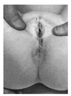
Figure 1: Pre-pubertal lichen sclerosus – at the age of 5. Note the well demarcated whitening, rubbery oedema of labia minora and purpura. Such changes can be mistakenly taken as evidence of sexual abuse. Note fissuring around the anal canal. This causes painful defaecation and hence retention so that constipation is a frequent presenting symptom in girls.
Aetiology: There’s mounting proof that lichen sclerosus is an autoimmune disease going on in genetically predisposed people. In a single study, 44% had sizeable auto-antibodies, 22% were family records and 21% were further autoimmune sickness [6]. Vitiligo and alopecia areata is no longer uncommon in the vulval health center and myxoedema isn’t on occasion identified in compliance with-up patients.
Pathophysiology: The autoimmune assault is due to lymphocytes within the upper epidermis, and each dermo- epidermal junction above and the epidermis below suffers; there’s liquefactive degeneration of the basal cellular layer with the destruction of melanocytes and stimulation of dermal fibroblasts to provide a giant sheet of homogenized collagen in the upper dermis [7, 8]. This produces the bodily symptoms of vibrant whitening and scarring—the ‘primary’ lesion being a white spot regularly with follicular indentation The overlying epidermis sometimes responds with compensatory epidermal proliferation, causing thickening and hyperkeratosis . This continuous ‘wounding’ process of the dermo-epidermal junction by the autoimmune lymphocyte attack and the prolonged stimulation of repair processes is hypothesized by some to be the trigger for the increased incidence of squamous cell carcinoma arising in the setting of lichen sclerosus (and lichen planus) [9, 10].
Ncidence: Lichen sclerosus is seen in both sexes at any body site and in all races, but most commonly affects the genital skin of white women. The peak ages for presentation are childhood and around or after menopause. The true incidence is unknown; prevalence has been estimated at between 1 in 300 and 1 in 1000 of the population.
Presentation in Childhood: Phimosis due to lichen sclerosus is the chief reason for young boys to require medical circumcision. Lichen sclerosus affects the perianal skin in females (but not males) and fissuring around the anal canal causes painful defecation and hence retention. Constipation is a frequent presenting symptom in girls (Figure 1).
Presentation in Adults: Affected women present with pruritus and only rarely are Pain or dyspareunia is a prominent complaint [5]. The condition is commonly misdiagnosed as ‘recurrent candidiasis and many years may pass until the correct diagnosis is made. It is salutary that many of those newly referred have long histories as well as physical signs that must have been established over many years.
Clinical Findings
The first changes occur on the labia minora and clitoral hood. Close examination shows the normal supple pink lips to become swollen with rubbery edema and take on a dull, creamy color. Similar changes may extend to the fourchette where small tears may be seen. If unchecked inflammation progresses destroying melanocytes and stimulating scarring. The whole perineum and genitocrural folds may become bright white with progressive shrinkage and resorption of normal vulval architecture. The labia minora become ‘plastered down’ and the clitoris buried as the hood tightens over it. The surface becomes wrinkled, there is ‘cigarette- paper scarring’ and the poorly supported blood vessels rupture easily, causing purpura
- Have pores and skin adjustments been seen or felt? Are there any lesions elsewhere?
- Has there been any vaginal discharge, and what is its nature?
- What medicines or different remedies have been hired, and what has been the reaction to these?
Examination: Accurate strong lighting fixtures, a skilled chaperone and an appropriately placed examination couch are vital; some authorities use a colposcope; however, the 4× lens magnifying loop is equally powerful. A scientific method is recommended and should begin with a preferred view of the vulva, looking at the pores, skin, and hair of the mons pubis and labia majora.
Widespread View (a) Hairs
- Distribution and extent (e.g. alopecia areata, evidence of virilization)
- Greatness and situation (e.g. color, damaged hairs) from friction)
- Infestation (b) Skin Color
- Pigmentary disturbance (e.g. vitiligo)
- inflammation, present or absent (c) Skin Texture
- Abnormal Thickness (e.g. lichenification or atrophy) (d) Pores and Skin Surface
- Integrity
- Excoriation
- Erosions (e) Palpation
- Tenderness or underlying loads (e.g. cysts) 1. Labia minora (a) Presence or absence (b) Developmental abnormality 2. Clitoral Area (a) Hood (b) Clitoris: normal size and surface 3. Vestibule (a) Urethral opening (b) Vaginal aperture (c) Epithelial surface: color, texture, and palpation with a-tipped swab five Perianal vicinity An examination of the vulva isn’t always complete without an inspection of the complete perineum, which includes the perianal skin (embryo logically derived from the cloaca) Biopsy of the vulva This is an easy system requiring a neighborhood anesthetic and must be completed as an outpatient method, which takes a few minutes. A punch biopsy is typically performed. Using either a three- or 4-mm d, it is very important to determine the exact website that is biopsied in the vulva, as the histology will vary in keeping with the region sampled.
The Symptoms of a Biopsy include the Following:
- Problems in establishing a clinical diagnosis.
- All blistering problems: A separate punch biopsy for immuno fluorescence must be taken and positioned in specific shipping media.
- Pigmented lesions.
- Inflammatory lesions that don’t respond as anticipated to anti-inflammatory capsules so that neoplasia can be excluded.
- Constantly Erosive Lesions.
Procedure
Ten minutes earlier than the biopsy, the utility of neighborhood anesthetic cream (e.g., lignocaine plus prilocaine or lignocaine gel) from the interlabial sulcus inwards blunts the ache of an injection of a nearby anesthetic. The vicinity was cleaned with a diluted chlorhexidine antiseptic. Undeniable lidocaine (1%), with or without adrenaline, was instilled using an excellent needle.
Surgery- punch biopsy. A 3- or 4-mm punch is typically adequate (Plate 51.1, facing p. 562). The punch is driven completely by thickness via the epithelial floor. The encompassing skin is pressed, and the plug will pop outwards. This may be‘harpooned’ with an exceptional needle. Taking care not to damage the overlying epithelium, the plug was lifted and snipped off at its base with scissors. Inflammatory skin illnesses of the vulva, Lichen Sclerosus.
Terminology: The antique terms kraurosis vulvae, leukoplakia, and leukoplastic vulvitis are no longer legitimate; however, they have been implemented in lichen sclerosus, lichen planus, and cicatricial pemphigoid in the past. Similarly, vulval dystrophy and vulval squamous mobile hyperplasia are unhelpful terms that should no longer be used.
Definition: Lichen sclerosus is a detrimental inflammatory condition with a predilection for genital pores and skin [5] (Figure 1).
Aetiology: There may be mounting evidence that lichen sclerosus is an autoimmune disorder going on in genetically predisposed people. In one look, using dermatological % had wide spread car-antibodies, 22% had an incidence history, and 21% had an additional autoimmune ailment. Vitiligo and alopecia areata are not uncommon in vulval health centers, and myxoedema is not occasionally diagnosed in follow-up patients.
Pathophysiology: An autoimmune attack occurs via lymphocytes in the upper dermis, and each dermo- epidermal junction above and the dermis underneath suffer; liquefactive degeneration of the basal cell layer occurs with the destruction of melanocytes and stimulation of dermal fibroblasts to supply a large sheet of homogenized collagen inside the upper epidermis [6, 8]. This produces the physical signs of shiny whitening and scarring—the ‘number one’ lesion being a white spot frequently with follicular indentation. The overlying epidermis occasionally responds with compensatory epidermal proliferation, inflicting thickening and hyperkeratosis.
This non-stop ‘wounding’ procedure of the dermo- epidermal junction by using the autoimmune lymphocyte attack and the prolonged stimulation of techniques is hypothesized by a few to be the cause for the elevated occurrence of squamous cellular carcinoma springing up in the putting of lichen sclerosus (and lichen planus) [9, 10]. Lichen sclerosus is visible in all sexes at any website and in all races; however, the maximum generally influences the genital pores and skin of white girls. The heights for presentation were early life and around or after menopause. The genuine incidence is unknown; it has been estimated at 1 in 300 and 1000 of the population Presentation in Early Life: Imosis due to lichen sclerosus is the leading motive for younger boys to require medical circumcision. Lichen sclerosus influences the perianal pores and skin in girls (but no longer men) and fissuring around the anal canal causes painful Defecation and subsequent retention Constipation is a frequent symptom in girls (Figure 1).
Presentation in Adults: Affected women present with pruritus, and the simplest rare complaint is pain or dyspareunia, a prominent complaint. The condition is typically misdiagnosed as ‘recurrent candidiasis, and many years may also be skipped until a perfect prognosis is achieved. It is salutary that many of those newly referred to have lengthy histories in addition to bodily signs that need to be set up over a few years.
Scientific Findings: Primary adjustments occur in the labia minora and the clitoral hood. Near examination suggests the regular supple crimson lips to emerge as swollen with rubbery edema and take on a stupid creamy shade. Comparable adjustments may extend to the fourchette, wherein small tears can be observed. If unchecked Inflammation progresses, destroying melanocytes and stimulating scarring.
The entire perineum and genitocrural folds may come to be vivid white with innovative Shrinkage and resorption of the normal vulval structure. The labia minora become ‘plastered down’ and the clitoris buriedeeks as the hood tightens over it. The floor becomes wrinkled, there is ‘cigarette-paper scarring’ and the poorly supported blood vessels rupture without difficulty, inflicting purpura and ecchymoses Such modifications can be mistakenly taken as evidence of sexual abuse in young girls [11].
Continuous resorption of the labia minora tightens the introitus position. Once in a while alarmingly; but, the vaginal mucosa is always spared and subsequently can be utilized in reconstruction operations when essential. Each time a patient with lichen sclerosus reviewed the dermis punch overlying the scarred white areas should be closely scrutinized; the search is for the squamous cell carcinoma that not often supervenes (or approx. 3–4%): This may start as a continual erosion, hyperkeratotic papule or a fleshy nodule, which may additionally ulcerate and infiltrate deeply—any suspicious vicinity or continual erosion must be biopsied [12] (Plate 51.7, dealing with p. 562). About 0.33 of patients with lichen sclerosus have a thickened epidermis (hypertrophic lichen sclerosus) and even though this more often than not resolves with topical corticosteroid treatment, it is far in any setting that squamous cell carcinoma generally arises . The histological pattern of lichen sclerosus-associated squamous cell carcinoma consists of epidermal hyperplasia and differentiated intraepithelial neoplasia (dysplastic modifications limited to basal layers) [13].
Treatment
The modern-day treatment of choice was a remarkably strong topical corticosteroid (e.g., clobetasol propionate ointment). The specific routine is important: small (30g) tubes of ointment are favored. The first tube has to suffice for 3 months. A small, ‘pea-sized’ amount was applied at night to the vulva. The frequency must be nightly for 4 weeks. Nights for 4 weeks, after which two times weekly for 4 weeks change. On review at three months, the affected person is advised to use the drugs for 2–3 nights in succession if signs and symptoms recur. Non-responders must be cautiously reviewed, as some might also show other extra-resistant situations, including lichen planus or cicatricial pemphigoid. Topical testosterone has no effect [14]. The region of more moderate non- steroidal, anti-inflammatory, immunomodulating topicals (e.g. tacrolimus and pimecrolimus) arguable the author feels that care has to be exercised with marketers that could theoretically accelerate cancer trade, using those dealers has to likely be confined to the rare sufferers now not responding to extraordinary-amazing Topical corticosteroids [15].
Extra-Genital Lesions: Those are rare in guys but can be discovered in 5-10% of girls common websites encompass upper lower back, shoulders, hips, he and strain websites (lichen sclerosus is one of the koebnerizing dermatoses, acting on websites for previous pores and skin damage, such as scars, vaccination, and radiation. pores and skin lesions might also show follicular hyperkeratotic plugs, giving a ‘nutmeg grater’ sense, and after the Plugs have dislodged prominent follicular ‘delling is characteristic. Such changes help differentiate lichen sclerosus from other white lesions consisting of morphea, scleroderma, scars, or vitiligo.
Histology
The most obvious exchange was the faded sector of acellular hyaline scarring within the higher epidermis. Below is a heavy bluish band of infiltrating lymphocytes, giving a ‘sandwich effect.’ The dermis above is flattened with a lack of rete ridges and melanocytes. 30% of sufferers have overlying epidermal hyperplasia Lichen Sclerosus and Malignancy: There’s a bimodal occurrence of vulval squamous cell carcinoma, and younger ladies with vulval intraepithelial neoplasia (VIN) generally wear oncogenic traces of human p (HPV), and older without papilloma virus, proof of HPV contamination. Even though there is undeniably an extended occurrence of squamous cell carcinoma in patients with lichen sclerosus, this has been exaggerated within the beyond and is absolutely no justification for prophylactic mutilating surgical procedures. In practice, the incidence is properly below five percent. When pathological specimens of squamous cell carcinoma and concomitant lichen Sclerosus was discovered in >50% of cases.
Lichen planus ‘The blue rash’ (lichen planus) is characterized by small, purplish polygonal papules with vibrant surfaces. They are most frequently found in the inner wrists, axillary folds, and genitalia [16]. Annular lesions and lesions appearing in recent scratch marks (Koebner phenomenon) are further diagnostic recommendations. The lesions are intensely itchy and nearly as awful as scabies. The inner cheeks should be searched for white lace-like regions and the tongue for asymmetrical ‘bald’ patches of papillary loss with a bluish hue.
Aetiology: The cause is unknown, but similar to lichen sclerosus, it entails a lymphocyte-mediated attack. In this example, it is far more carefully centered on basal keratinocytes at the d junction and does not motivate the hyaline band of dermal scarring. A heavy band of lymphocytes lies under the basal keratinocytes, several of which die through apoptosis, leaving their corpses as colloidal bodies. Sea of liquefactionermo-epiderma degeneration there is pigmentation. Incontinence with tattooing of the epidermis via pigment weighted macrophages. The overlying ridges are effaced in a ‘noticed-teeth’ pattern. As in lichen Sclerosus, there can be overlying hyperkeratosis without dysplasia.
Pathophysiology: Even though the cause is unknown, lichen planus appears in all likelihood to indicate any other autoimmune diseases. T-lymphocytes mount an immunological attack on basal keratinocytes, and the circumstances are associated with a few patients with different autoimmune conditions consisting of the number one biliary sclerotype. Moreover, similar ‘lichenoid’ changes can be seen in bone marrow transplant patients with graft- versus-host ailments. Lichen planus may be brought about by tablets (e.g., β-blockers, gold). Familial cases have been defined and there may be some proof of its affiliation with HLA-DR1.
- Diagnostic Test Listing • Intensely itchy blue papules (take a look at wrists) might also seem in scratch marks, frequently annular lesions • check internal cheeks for lace-like pattern • Number one lesion: flat-topped brilliant polygonal papule, which may also show Wickham’s striae • Nail adjustments can rarely arise Age: 25–40 year olds, rare in adolescence and antique age
- Scientific Placement and Evolution • Desirable prognosis: natural history commonly 9–18 months, although a few may have many years of illness. • Lesions leave pigmented tattoos that last 9–1 years.
- Control • Punch biopsy for affirmation • Raised lively papules should respond well to (grade I) awesome-strong topical corticosteroid (e.g. clobetasol propionate) • Second-line remedy: topical Immunomodulators (e.g. tacrolimus (zero.1%)) Erosive lichen planus Vulvo- vaginal-gingival syndrome (syndrome of Hewitt) and Pelisse) [17, 18].
This rare circumstance causes vulval pain due to erosion of the labia minora and vestibule. The labia majora and closing skin are unaffected, but the anal, oral, and vaginal mucosa is often present. This situation hastily leads to scarring, often with synechiae, and the alarming lack of ordinary architecture with atrophy, fusion, scarring, and burying of the clitoris.
There can be characteristic milky striae on the margins, vaginal examination is frequently very painful, and a vaginal speculum must be in contact with bleeding, and erosions are seen with synechiae and stenosis. Vaginal shortening can also arise. The etiology is unknown; however, histology suggests an aggressive shape of lichen planus (although biopsies can show the handiest non-particular ulceration if samples are not taken from the advancing fringe of lesions).
Aetiology: There may be an association with HLA-DQB 0201. 1. Diagnostic Test List (a) Significantly painful vulva (b) Bleeding dyspareunia (c) Vaginal discharge (d) Eroded inner lips of labia minora and introitus (e) Marginal milky striae (f) Vaginal erosions, contact bleeding (g) Gingivae denuded and ulcerated ± synechiae (h) (Conjunctiva spared, cf cicatricial pemphigoid) 2. Evolution (a) Negative diagnosis, continual relapsing situation (b) poor reaction to remedy (c) Burnt-out instances go away, leaving sizeable scarring
Management
That is quite hard. Swabs should be collected intermittently to search for treatable secondary contamination. Example: β hemolytic streptococcus or candida. Antiseptic emollient soaks can soothe (for example, Emulsiderm and Dermal). Specific treatment is aimed at the lymphocyte assault, for example, topical steroids of incredible power (e.g. Clobetasol propionate or diflucortosone 0.3%, Nerisone forte) Oily cream). They can be generously applied to dissolvable seaweed dressings (for example, Sorbsan), applied without delay to the vulva or wrapped around a tampon inserted into the introitus for 15-30min. Day-by-day attendance at an expert dermatological nursing day care status quo for every week or so may be very beneficial.
The more recent non-steroidal remedies, for example, topical tacrolimus, can produce advantages in some but aggravate others; theoretically, these may initiate neoplastic alternate. The transport of steroids into the vagina is surprising easy. A steroid cream can be introduced with an applicator. Steroid suppositories are available and steroid foams are used for proctitis and colitis (e.g. hydrocortisone acetate foam (10%)). Vaginal synechiae and stenosis may be managed with the aid of Gynecologists and dermatologists collectively, as put up-operative topical or once in a while, oral steroids ought to be used together with dilators to try to arrest re-sealing.
Systemic Treatment: Oral steroids may be attempted; however, these are regularly disappointing. Standard immunosuppressive regimens with azathioprine, Cyclosporine, etc., have not proved useful.
Eczem: Eczema and dermatitis are phrases used anonymously in situations that cause epidermal irritation of a non-scarring and reversible nature. In urticaria, sudden leakage of tissue fluid from dilated capillaries fills the dermis to form wheals underneath an unchanged epidermis (hives, nettle rash), which then fills the epidermis with fluid. Histologically, this is termed spongiosis and in extreme instances, it may additionally ooze onto the surface (‘moist eczema’) or pools in the vesicles (vesicular eczema).
The dermis and underlying epidermis are infiltrated by inflammatory cells (lymphocytes, eosinophils, basophils, and polymorphic leukocytes), and epidermal macrophages (Langerhans cells) probably play a pivotal role in marshaling such inflammation. The affected epidermis becomes hyper proliferative and parakeratotic so that scaling is visible (which in wet vulval regions becomes a macerated greyish white ‘dulling’ of the ordinary crimson coloration) . Vulval eczema is frequent and happens in diverse scientific conditions, for example, atopic pores and skin ailment, Seborrhoeic eczema, irritant dermatitis, touch allergic dermatitis, and frictional eczema, for example, lichen simplex chronicus.
Seborrheic Dermatitis: This is a not-unusual itchy, red, scaly eruption with a preference for face and scalp pores and skin. An extended history of intermittent Dandruff is typically drawing close. Sufferers display poorly demarcated crimson scaly patches within the seborrheic areas of the scalp, behind ears, sideburns, and nasolabial gutters. The eyebrows and outside auditory meati often show scaling on an erythematous base. A share of sufferers shows comparable adjustments in different wet areas (critical chest, axillae, perineum, and groins. Lady patients can also complain of vulval itching and could be labeled as having continual recurrent candidiasis, even though they won’t have any history of discharge. In diffuse cases, the prognosis may be different. Cult and search beneath the hairs in the labium for some antibodies exist for pink, dry, thickened scaly dermatitis should be made. More extreme cases display extension onto the pubic mound and genitocrural folds.
Aetiology: A genetic tendency to seborrheic dermatitis is probable t in over 15% of the population and may arise at any age, with peaks in infancy (cradle cap) and early maturity. Growing proof links the circumstance to an individual’s reactivity to commensal lipophilic yeasts (Malassezia furfur), whose populace is the finest in greasy and occluded websites.
Treatment
Successful treatment involves first quelling though,, by using (1) the avoidance of irritants and (2) using anti- inflammatories (e.g. cleaning soap substitutes and topical corticosteroids, grade III or on occasion, grade II) and then curbing the yeast population with anti-fungals (e.g. imidazole. topicals). Seborrheic dermatitis versus psoriasis. Text ebook examples of both situations provide contrasting bodily signs, but often, especially in flexural areas such as the vulva, a mixed image is found. Continuous plaque psoriasis gives the acquainted well-demarcated dry, scaly, meaty plaques over the extensor surfaces (e.g., elbows, knees, sacrum, and scalp).
In flexural areas, the scales emerge as macerated and less obvious, but lesions are still well demarcated. Such crisp demarcation is a diagnostic pointer to psoriasis, whereas, in seborrheic dermatitis, lesions soften imperceptibly into the surrounding skin. Pure seborrheic dermatitis itches, whereas psoriasis is either asymptomatic or occurs every so often, mainly while fissured. (Such fissures might also enlarge up the natal cleft). While faced with a red perineum, the subsequent might also help differentiate.
Irritant Dermatitis: Vulval skin differs from pores and skin somewhere else by being more porous, like its male counterpart, the scrotum, each for transpiration and less complicated penetration [19]. This leads to a susceptibility to number one irritant dermatitis from, for instance, bubble tubs, disinfectants, deodorants, and infective vaginal discharges, feces, or and drugs together with anti-wart treatment and topical cytotoxics. The diagnosis may or may not be apparent, as patients do not always effortlessly apprehend or volunteer the relevant history.
Management
Treatment includes identity and withdrawal of the irritant, using emollient cleaning soap substitute’s instance, aqueous cream BP, or Emulsiderm, probably, and the use of corticosteroids grade IV or antifungal eczema corticosteroid for 7-10 days.
Contact Allergic Eczema: The opportunity of touching allergic eczema is often overlooked, probably, because it is a lot more unusual than in perianal pores and skin [20]. However, the pores and skin over the pubic mound and labia majora can truly appear to be in contact dermatitis behind schedule (type IV) allergy is examined via Patch exams are examined at 48 h. Vulnerable offender allergens include medicaments (e.g. neomycin, nearby anesthetics, ethylene diamine, or even some corticosteroid molecules, which includes hydrocortisone acetate or clobetasol propionate) rubber (e.g. in condoms) or different chemical substances (e.g. spermicides and perfumes). Management entails suitable patch testing by specialist dermatological units and the avoidance of the culprit allergen. Grade II or, in intense instances, grade I topical corticosteroids are wished, although in very severe instances, systemic Steroids are used (e.g., prednisolone 30mg in the afternoon for five days). In
acute weeping eczema, potassium permanganate soaks for some days are very beneficial.
Allergic Contact Urticaria of the Vulva: Contact urticaria to latex is a growing problem, especially for regular wearers of rubber gloves, including fitness professionals [21]. Sufferers generally make the prognosis themselves and observe lip swelling right now after blowing up balloons. In such individuals, latex condoms can cause instantaneous, uncomfortable urticarial swelling of the labia. Contact urticaria to seminal fluid has been defined as both to the semen itself or to treat other allergens (e.g., penicillin) carried inside the seminal fluids [22]. Unique immunotherapy against semen has been defined [23].
Lichen Simplex Chronicus: A vicious circle may be set up in which scratching (for some reason) ends in thickened, itchy patches of pores and skin. In addition, scratching worsens the situation, and finally, a subconscious scratching addiction can grow. It may be brought about via heat (e.g., in bed, after warm bathing), irritants (e.g., soaps, bubble baths, etc.), or psychological elements. The habit is difficult to wreck, particularly because sufferers are largely blind to their scratching behavior. Commonplace websites for L. simplex Chronicus consist of areas without difficulty on hand to the dominant hand, for example, the right side of the lower back of the neck, right calf, and the right labia major (and right aspect of the scrotum). The most common scientific state of affairs is that of a patient complaining bitterly of pruritus vulvae who, on preliminary examination, seems to have an everyday vulva.
However, a search underneath the hairs on the labium major may also display the normal lichenified, thickened, darkish pink scaly patch. If the affected person is asked to point with one finger at the worst area, she can usually pinpoint the lesion. Management involves reassurance and an explanation of the ‘itch-scratch’ cycle. This frequently heightens at times of stress, and without a doubt, a confrontational approach is seldom beneficial. An emollient and short-time period grade I or II topical corticosteroids can help break the dependency. Behavioral therapy may be mainly aimed at helping to extinguish the habit of scratching [24].
Psoriasis: In both scientific and lay minds, psoriasis is rightly associated with scaly plaques on the extensor surfaces of the knees. Elbows and scalp instead of the perineum. but genital psoriasis may be very commonplace. The genetic tendency to psoriasis in all likelihood exists in some five percent of all populations, with a point incidence of a few 2%. Psoriasis isn’t commonly itchy, but sufferers with vulval lesions may additionally cause discomfort or pain. There is mostly secondary vulvodynia (q.v.).
Lesions can cause problems in prognosis because, at this occluded website, the same old silvery scales emerge as macerated and the plaque appears as an infected ‘beefy- red’ lesion. The threshold, however, stays crisply described and the plaques have a symmetrical look. Any place inside the perineum can be affected but the vaginal mucosa is usually spared. Perianal pores and skin may additionally be affected, extending up the natal cleft where painful fissures every so often arise. Corroborative lesions have to be sought somewhere else. As with many sufferers with vulval symptomatology, a close inspection of the scalp can be very beneficial. In this example, revealing nicely demarcated scaly plaques are revealed. The classical psoriatic areas of the elbows, knees, sacrum, and scalp should be checked. Nails show symptoms of psoriasis in about 10% of patients with thimble pitting, sub-ungual hyperkeratosis,and onycholysis. However, genital lesions may be the most severe manifestation of psoriasis in some patients.
The etiology is unknown; however, there is a genetic predisposition, and lesions can be precipitated (1) using streptococcal contamination (commonly guttate psoriasis) and (2) trauma (the Koebner phenomenon). Histopathology shows marked thickening of the epidermis (acanthosis), with epidermal ridges projecting deep down into the epidermis with rounded, burgeoning pointers. No nuclear atypia is seen. Thickened dermis may additionally show patches of spongiosis with neutrophil infiltration and overlying parakeratosis. The underlying blood vessels were regularly grossly dilated with continuous surrounding inflammatory cells.
Management
Complete rationalization and reassurance of the noninfectious nature of psoriasis are mandatory. A mild explanation is that scratching exacerbates and that the use of emollients and cleaning soap substitutes can soothe lesions at this occluded, warm website. Secondary contamination, for example, with Candida albicans, should be determined. Anti-psoriatic topical treatments, such as dithranol and calcipotriol, can also cause excessive irritation at occluded websites. However, others can be beneficial, including tar, tacalcitol, calcitriol, and topical corticosteroids, which are no stronger than grade II (for example, 1:4 B). Ointment RD with or without 5% coal tar solution in patients with intense and severe psoriasis, systemic remedies are occasionally used (e.g., weekly oral methotrexate, retinoids, or cyclosporin).
Flexural sites can be expected to respond equally well. For example, sufferers need to be under the supervision of a specialist sanatorium for such prescriptions to be considered, and positive medicines can exacerbate psoriasis (for example, lithium, chloroquine, and beta blockers). Reiters’s ailment (circinate ulcerative vulvitis) the associated signs of uveitis and arthritis point to the diagnosis. Circinate balanitis is well recognized but the corresponding vulvitis could be much rarer [25]. Lesions can be eroded, ulcerative, or scaly. Histologically, these modifications are similar to those observed in pustular psoriasis. Pigmentary changes of vulval skin.
White patches: Vitiligo is a common autoimmune ailment with strikingly symmetrical regions of vibrant white depigmentation, normally on peri-orificial websites, specifically around the genitalia. It is regularly additionally determined within the axillae and cosmetically feared at the face and arms. Lichen Sclerosus lesions can be bright white but are not difficult to differentiate from vitiligo through scarring and purpura. Sub inflammatory pigmentation is the most common cause of dark patches in the vulva. This is most often followed by destructive irritation, which includes lichen planus and, much less often, lichen sclerosus.
Pigments are accrued inside the dermis using macrophages, which can be gradually dispersed. Larger asymmetrical patches that followed a constant drug eruption for several months. Idiopathic pigmentation (of Laugier) [26] is well recognized; there are no records of antecedent inflammation, and histology does not display melanocytic proliferation; however, multiplied pigmentation in the basal location and Pigmentary incontinence have been reported. Such the lesions were fixed, and a biopsy confirmed their benign nature.
Ulcerating and blistering (bullous) disorders such lesions generally cause pain in preference to itchiness, and their maximum is uncommon. It is vital to study other mucous membranes to provide evidence of the disease. A biopsy with a unique Immuno-fluorescent pathology is regularly required to establish the analysis, and dermatological advice should be sought.
Aphthae: Oral aphthae are common and widely known; their vulval equivalents are frequently unrecognized or recognized as recurrent herpes.’ Patients with vulval ulcers are usually unmarried. Spherical with a yellowish floor and pink rim, the treatment of recurrent aphthae is difficult. 5% Lignocaine Ointment can deliver symptomatic comfort and some patients respond to topical tetracycline. Erythema multiforme, an acute pore and skin response sample, typically lasts about a fortnight, and a few instances are confined to the mucosae (oral, ocular, and genital), but these are our best friends observed via small, round areas of erythema on the arms and feet that have an ordinary target-like morphology. In severe cases, big bullous lesions may appear from time to time, which is the main reason chronic scarring can arise (the Stevens-Johnson syndrome). Extreme cases are common in HIV disease Erythema multiforme can arise eight–10 days after antigenic stimulation, such as viral contamination with the herpes simplex virus or drug exposure; however, in half of the patients, no cause was observed.
Management: The trigger antigen needs to be sought, followed by putative drug avoidance or suppression of recurrent herpes simplex virus with long-term low-dose acyclovir, if necessary. Oral and ocular involvement should be evaluated and handled. Cutaneous lesions may be handled with a grade I topical corticosteroid for 1 to 2 weeks. quick guides of systemic steroids can reduce fever, toxicity, and nearby pain; as an example, prednisolone 40 mg in line with day lowering over 6 days. Toxic epidermal necrolysis (TEN) (Lyell’s syndrome) is a more excessive form of superficial reactive infarction that may result in good-sized and even deadly skin loss [27]. Such lesions are common in HIV patients.
Allergy to drugs is the standard purpose (non-steroidal anti-inflammatory marketers, Carbamazepine, Phenytoin, Co-Trimoxazole, Dapsone, Complement, and sulfur capsules). Management requires early diagnosis and speedy cessation of the triggering drug, as well as early transfer of patients to a high-dependency treatment unit. Previously, High-dose systemic steroids have been used but may be associated with elevated mortality due to serious infections.
Intravenous immunoglobulins have been proposed and are currently undergoing clinical trials. Experienced nurses require a nearby remedy for both ocular and vulval pores and skin lesions to prevent synechiae and permanent scarring. The Sickness is associated with a high mortality rate. A less excessive and more superficial variant is visible in childhood as a result of staphylococcal exotoxin—the Staphylococcal Scalded Pores and Skin Syndrome [28].
Fixed drug eruption (FDE) in this condition, asymmetrical patches of intense inflammation arise on the skin and mucosal surfaces every time the offending drug is administered. Patients do not often have an eruption with the medication, which can be a proprietary one, for example, codeine in an analgesic or a phenolphthalein laxative [29]. Other capsules included paracetamol, non-steroid anti- inflammatory drugs, tetracycline, griseofulvin and cytotoxics. Undertaking consequences in response at exactly the same websites whenever beginning within 24hours Restoration often leaves darkly pigmented patches as proof for many months.
Management
Records and bodily symptoms should advocate the diagnosis; however, identifying the trigger pills can take time, and an allergic reaction is lifelong.
Bullous Pemphigoid: This is an uncommon autoimmune bullous sickness affecting both pores and skin. Mucous membranes, including the vulva it is more common in older girls, but sometimes occurs in young people [30]. The circumstance is due to a circulating antibody, which reacts in opposition to the dermo-epidermal junction. This may be identified with the aid of oblique or direct Immuno- fluorescent strategies. There may be associated with different autoimmune illnesses, including lichen sclerosus. For management, dermatological recommendations should be continually acquired. Nearby, high-quality topical steroids are a completely extensive and useful resource to remedy; however, systemic steroids are usually required with or without cytotoxic pills.
Cicatricial Pemphigoid: This is a rare variant of pemphigoid in which mucous membrane involvement is the most prominent feature. This ends in scarring, specifically toughness inside the vulva and conjunctivae. Furthermore, older girls were the most regularly affected. Circulating antibodies are not constantly located, but direct immuno fluorescence of a biopsy specimen is effective [31].
Pemphigus Vulgaristumors: This is an unprecedented, severe, Immunologically-mediated bullous eruption that affects the mucous membranes and skin. Blisters are fragile and short-lived, resulting in characteristic erosion. Most of the patients were teenagers. Semitic individuals are specifically affected. Dermatological recommendations have to be sought and excessive-dose systemic steroids, cytotoxics, or mycophenolate mofetil are generally required. Benign familial pemphigus (Hailey-Hailey disorder) is an unprecedented genetically determined disorder in which friction causes erosion, most commonly of the flexural pores and skin, such as the neck, axillae, and vulva. Histology is a useful diagnostic tool. Benign tumors of vulval pores and skin congenital tumours.
Vascular Birthmarks: These are not uncommon. Capillary nevi were also observed. Beginning and does not fade. They have no useful sequelae, and remedies, for instance, with lasers, ought to depend on cosmetic concerns.
Cavernous Haemangioma (Strawberry Naevus): Such lesions commonly appear in the first six weeks can grow swiftly and can arise on genital websites. Sometimes they wreck down and may be a site of ingress of serious infection. In fashionable, the step-by-step treatment occurs spontaneously over a 10–12months period; however, early verification in specialized Dermatological Laser devices has to be taken into consideration when lesions threaten functional cosmetically.
Angiokeratomata: These appear as tiny, ecstatic, dark red/blue vascular-seasoned liberation with overlying hyperkeratosis. They occur in only 2% of women. Comparable lesions are visible on scrotal skin [32]. Acrochordia (pores and skin tags). These are extremely common in all frictional and flexural websites, along with axillae, eyelids, and groins. They can reach pretty large sizes, and if they twist on the stalk was painfully thrombosed. Remedies using scissor amputations.
Vestibular/Labial Papillomatosis: It is a variant of everyday life. Surprisingly, prominent thickening and folding of the labial epithelium may be stimulated during puberty or pregnancy. Such changes are often mistaken for HPV contamination, specifically as both styles of lesions display ‘aceto-whitening’ after the use of 4% acetic acid. (Warts are focal, asymmetrical, and scattered.) Biopsy outcomes can also be puzzling, as the glycogen-rich pale cells of the hypertrophic vestibular epithelium stain pale and can be improper for viral koilocytes by using a green pathologist.
- Keratinous Cysts: Closed and open comedones are not unusual in Labia majora. Acniform irritation or calcification can also occur.
- Venous Varicosities: These are asymptomatic but may also extend at some stage after pregnancy. Vestibular Mucinous Cysts: These are not uncommon in adults but are occasionally seen in youth and are harmless.
Papillary Hidradenomata: Those are sweat gland adenomas with apocrine fluctuation. They arise most normally inside the anogenital location of center-aged white women. A company-asymptomatic papule or nodule is determined on the labia majora or any part of the perineum. Every so often, multiple lesions arise. Excision is required for histological affirmation.
Syringomata: Those are unusual eccrine duct tumors, which can be generally asymptomatic, with a couple of bilateral and symmetrical tumors going on on the labia majora. They can be destroyed using electro-desiccation under local anesthesia [33].
Giant Venous Ectasia
This is a florid shape of venous varicosity in the labia. Those dilate in addition at some point in pregnancy and might cause problems during shipping. Vulval cutaneous manifestations of underlying systemic disorder
- Crohn’s disorder
- Pyoderma gangrenosum
- Behcet’s Syndrome
- Necrolytic migratory erythemas (NME)
- Acrodermatitis enteropathica
- Acanthosis Nigricans
- Crohn’s disease As soon as visible, the ulceration, fissuring, sinuses, and fistulae of the perineum accompanying Crohn’s disease aren’t without difficulty forgotten (Plate 51.19, going through p. 562). Perineal involvement can certainly precede, now and again over many years, the recognition of an inflammatory bowel ailment. Cutaneous lesions in the anogenital region are the most common lesions and are visible in up to 30% of Crohn’s disease patients. If the lesions are clear of the anal margin, they’re regularly called ‘metastatic lesions’. Vulval lesions are frequently of this nature [34]. The maximum usual presentation is chronic unilateral labial edema; later extra-familiar ulceration, fissuring, and fistulae can also arise [35].
Histology frequently suggests granulomatous infection but is not specific, and prognosis may be difficult, particularly in the absence of demonstrable bowel disease. Hidradenitis suppurativa can be clinically differentiated through the presence of bridged commed ones and ordinary axillary and unfashionable auricular lesions. Treatment is problematic. Nearby measures with antiseptic soaps and topical anti- inflammatory steroids can help. However, regularly, systemic treatment is required. There may be some evidence that suppression or elimination of inflammatory bowel can assist. Oral steroids without or with azathioprine or cyclosporin can help. Recently, there have been encouraging anecdotal reports of the advantages of Infliximab.
Pyoderma gangrenosum: In this circumstance, violaceous purulent cutaneous ulceration normally takes place at the shins, but lesions can be visible at the vulva [36]. The etiology is unknown; however, lesions regularly coagulate, and thus, any attempt at surgical remedy is regularly accompanied with the aid of a speedy extension of the disorder. Underlying inflammation is observed in some 50% of cases, along with rheumatoid arthritis, ulcerative colitis, Crohn’s disease, myeloproliferative issues, and paraproteinaemia. The histology is inflammatory but non-precise, and the diagnosis is typically clinical. The condition typically responds promptly to systemic steroids, but on occasion, different retailers are required to halt the development (e.g., dapsone, azathioprine, minocycline, and cyclosporin).
Behçets Syndrome: In the nineteen-thirties, Behçet described a triad of oral and genital ulcerations with ocular uveitis. The condition can affect many different systems, and the diagnostic standards have been refined. Recurrent oral ulceration is compulsory and ought to be observed using the following: recurrent genital ulceration, eye lesions, cutaneous lesions, and a wonderful pathogen test. The causative agent can be viral. and the pathogenesis of the lesions is probably vascular. There is an affiliation with HLA-B5 in a few components of the sector.
Onset is commonly earlier than the age of 50. Oral ulcers are indistinguishable from recurrent aphthae. Vulval ulcers normally occur at the labia minora, are long-lasting, recurrent, painful, and heal with scarring. Cutaneous lesions consist of sterile pustules following trauma consisting of a needle prick (pathergy) or pyodermatous plaques and erythema nodosum. The histology may display non-particular ulceration; however, hence occasionally, thrombosed arterioles are seen underneath lesions. The aim is to halt the development of blindness and neurological headaches. Vulval ulcers may respond to topical tetracycline and corticosteroids. Resistant, excessive ulceration commonly heals with rather low doses of thalidomide; however, supplies are hard to obtain, and the drug is teratogenic. Treatment may be complicated via neuropathy but is pleasant when dealt with in skilled centers. Recently intralesional Recombinant human granulocyte/macrophage colony-stimulating aspect (rhGM-CSF) has been reported to heal huge ulcers. Necrolytic migratory erythema (glucagonoma syndrome) This vanishingly rare syndrome comprises a distinct peri-orificial migrating erosive eruption related to pancreatic islet-cellular glucagonoma. The perineum is most severely affected, but peri-oral lesions are visible. There may be associated glossitis and regular diabetes. Prognosis is made by locating a raised glucagon level. Topics are usually middle-aged women. However, the hyperlink between the pancreas and skin remains unexplained. Theories consist of a deficiency of amino acids, fatty acids, and zinc.
Acrodermatitis Enteropathica: This circumstance is generally seen in neonates or youngsters, and red, eroded vesiculo-pustular lesions particularly have an effect on the genital area but additionally on the peri-oral skin. Zinc deficiency is the purpose, and lesions heal swiftly as soon as that is administered. Deficiency can result from a recessively inherited disorder or, for instance, from total parenteral nutrition, prematurity, gut-pass surgery, or a penicillamine remedy for Wilson’s disease.
Acanthosis Nigricans: Acanthosis Nigricans can cause weight problems without or with insulin resistance and are thought to have an increased hormone-like impact. There are symmetrical velvety brown thickened pores and skin spreading out from the labia majora onto the groins and extending peri-anally, regularly with comparable lesions in the axillae. There can be accompanying pores and skin tags. Rarely are such adjustments visible in thin people and are dependable and ominous markers of underlying malignancy. Vulvodynia and Vestibulodynia [37, 38]: The concept of the various pain syndromes has gone through massive debate and scrutiny over the last decade. Preferred practitioners, professionals, and sufferers alike have been at a loss for words through records of disabling and extensive signs and symptoms of pain in the absence of a visible or histological abnormality. Neurologists and pain physicians have substantially contributed to the information on such pain syndromes. The names used depend upon the anatomical web page affected: glossodynia (mouth), anodynia (perianal), vulvodynia (vulva), and vestibulodynia (vulval vestibule). The notion of contact or ache depends on complicated neurological pathways, from pores and skin receptors via sensory nerves and spinal ganglia to important ache-reception areas. Such pathways are also stimulated via higher centers; worry about pain is an effective exacerbator.
The ache may be very real; knowledge of its cause and treatment make it extra tough. Sufferers with out-of- body pathology generally tend to fall into two categories: corporations. Older sexually inactive girls tend to have excessive intermittent signs and symptoms—often the use of phrases such as ‘burning’ or ‘aching’—and cannot tolerate difficult chairs or prolonged sitting. Factor tenderness isn’t always a particular function on examination with cotton buds. Dysesthetic Vulvodynia is used to describe this institution. Younger girls alternatively frequently revel in vulval aches with contact best. On examination, tenderness can be elicited at 5 to 7 o’clock inside the vulval vestibule. These sufferers suffer from superficial dyspareunia and incapacity to tolerate toxins, consequently the term vestibulodynia [39, 40, 41]. A proportion of each patient has a bitch of urethral ache and dysuria. They will be thought to have recurrent urinary tract infections— cultures are constantly poor. The urethral signs and symptoms are thought to be part of the same pain syndrome.
In addition, it’s now been diagnosed with the aid of skilled vulvodyniologists that vulvodynia may be prompted by using explain using a preceding inflammatory dermatosis, for example, lichen planus or psoriasis (secondary vulvodynia).
Management
Experience is required to help such sufferers. Cautious and affected person, paying attention to signs and symptoms, and a gentle, confident method are required. Excessive investigation can undermine this and needless biopsies or cystoscopies can themselves cause, in addition to post- operative pain. A ‘neurological sensory’ primarily based rationalization can assist significantly. The use of topical local anesthetics, including Lidocaine 5% ointment, helps and might build self-assurance and permit continuing sexual members of the family. The older, customary tricyclic antidepressants and centrally appearing drugs such as gabapentin and carbamazepine form the mainstay of the doctor’s arsenal. Amitriptyline (sedating), Imipramine, and Nortriptyline are the retailers of first choice [42]. It can help to explain why such capsules are used in better dosages for the remedy of melancholy; however, lower dosages might also be okay for the suppression of vulval pain.
Sufferers ought to be started on 10mg nightly, growing by means of 10mg each week until the pain remedy is done. If, at 90, there may be no reaction, this remedy should be abandoned and any other tried. The dosage, at which pain remedy is executed, needs to then persist for a complete 3–6 months before a sluggish withdrawal. Sufferers can be helped by learning that pain physicians use such drugs to alleviate pain in different instances and certainly co-session with such colleagues can help.Fashionably, younger patients respond properly to topical treatments on their own, and with time, the medications can be stopped. Older girls may require more prolonged periods of treatment.
Research Method
On this basis, a complete review of the present literature on benign vulvar illnesses was conducted. The research technique involved the systematic examination of clinical databases, case reviews, and relevant literature posted within the past decade. The inclusion standards for deciding on facts were primarily based on their relevance to the subject. This approach allowed us to gather a wide spectrum of statistics and insights regarding benign vulvar diseases.
Result
The outcomes of our research screen for a variety of benign vulvar illnesses, each with its own wonderful personal traits. We located versions in the superiority and occurrence of those conditions, with some, which include lichen Sclerosus and lichen planus, being exceptionally uncommon, while others, like vulvar fibroma, are rarer. Scientific displays vary, with patients experiencing symptoms along with itching, burning, pain, and skin modifications. Diagnosing those situations can be difficult, regularly necessitating a medical examination, a biopsy, and various exams for accurate confirmation.
Discussion
The findings of this study underscore the significance of knowledge about benign vulvar diseases. Their occurrence and varying medical presentations highlight the need for accurate diagnosis and powerful management. Healthcare companies face the assignment of differentiating between these situations due to their diverse symptomatology, and the sensitive nature of the vulvar area calls for careful diagnostic procedures.
Treatment options are available for benign vulvar illnesses, with choices dependent on the specific circumstance and its severity. Those may additionally consist of topical steroids, immuno suppressants, surgical procedures, and different interventions. Beyond the bodily factors, it’s important to don’t forget the psychosocial impact of those conditions. Patients may additionally enjoy emotional misery, stigma, and bad effects on shallowness and relationships.
Conclusion
In the end, this study paper offers a comprehensive review of benign vulvar diseases, emphasizing the importance of early diagnosis, effective control, and continued research in this field. It’s our wish that this overview will contribute to extended focus, improved affected person care, and ongoing exploration of these critical aspects of women’s health.
Acknowledgment
The completion of this research project would not have been possible without the contributions and support of many individuals and organizations. We are deeply grateful to all those who played a role in the success of this project We also thank my mentor, Naweed Imam Syed, Prof. Department of Cell Biology at the University of Calgary, and Dr. Sadaf Ahmed Psychophysiology Lab, University of Karachi, for their invaluable input and support throughout this study. Their insights and expertise were instrumental in shaping the direction of this project Declaration of Interest I at this moment declare that I have no pecuniary or other personal interest, direct or indirect, in any matter that raises or may raise a conflict with my duties as a manager of my office Management Conflicts of Interest The authors declare that they have no conflicts of interest. Financial support and sponsorship No Funding was received to assist with the preparation of this manuscript.
References
-
Leibowitch M, Staughton RC, Neill SM, Barton S, Marwood R (1997) An Atlas of Vulval Disorder A blend Dermatological, gynecological, and neurological approaches 1st (Edn.), CRC Press.
-
Ridley CM, Neill SM (1999) The Vulva. 2nd (Edn.), Wiley– Blackwell.
-
Edwards MD, Lynch PJ (2017) Genital Dermatology Atlas. 3rd (Edn.), Lippincott Williams and Wilkins.
-
Burns A, Breathnach S, Cox N, Griffiths C (2004) The genial, perianal, and umbilical regions. 7th (Edn.), Rook’s Textbook of Dermatology.
-
Wallace HJ (1971) Lichen Sclerosus etatrophicus. Trans St John’s Hosp Dermatol Soc 57(1): 9-30.
-
Fung MA, Boit PE (1998) Light microscopic criteria for the diagnosis of early vulvar lichen sclerosus: a contrast with lichen planus. Am J Surg Pathol 22(4): 473-478.
-
Meyrick TRH, Ridley CM, McGibbon DH, Black M (1988) Lichen sclerosus et atrophicus and autoimmunity. Br J Dermatol 118(1): 41-46.
-
Marren P, Dean D, Charnock M, Wojnarowska F (1997) Basement membrane area in lichen sclerosus: an immuno histological look at Br J Dermatol 136(4): 508- 514.
-
Leibowitch M, Neill S, Pelisse M, Moyal BM (1990) The epithelial adjustments related to squamous cell carcinoma of the vulva: an evaluation of the clinical, histological, and viral findings in 78 ladies. Br J Obstet Gynecol 97(12): 1135-1139.
-
Vilmer C, Cavalier BB, Nogues C, Trassard M, Doussal V (1998) Analysis of alterations adjacent to invasive vulvar carcinoma and their relationship with the associated carcinoma: a study of 67 cases. Eur J Gynecol Oncol 19(1): 25-31.
-
Carli P, Cattaneo A, Magnus A, Biggeri A, Taddei G, et al. (1995) Squamous cellular carcinoma bobbing up in lichen sclerosus: a longitudinal cohort study. Eur J Cancer Prev 4(6): 491-495.
-
Jenny C, Kirbyhe P, Furquay D (1989) Genital lichen sclerosus is unsuitable for toddler sexual abuse. Pediatrics 83(4): 597-599.
-
Crum CP, McLachlin CM, Tate JE, Mutter GL (1997) Pathobiology of vulval squamous neoplasia. Curr Opin Obstet Gynecol 9(1): 63-69.
-
Sideri M, Origini M, Spinachi L, Ferrari A (1994) Topical testosterone as a remedy for vulvar lichen sclerosis. Int J Gynecol Obstet 46(1): 53-56.
-
Bunker CB, Neill S, Staughton RC (2004) Topical tacrolimus, genital lichen sclerosus, and risk of squamous cell carcinoma. Arch Dermatol 140(9): 1169.
-
Lewis FM, Shah M, Harrington CI (1996) Vulval involvement in lichen planus: a look at 37 women Br J Dermatol 135(1): 89-91.
-
Pelisse M, Leibowitch M, Sedel D, Hewitt J (1982) A new vulvovaginogingival syndrome. Plurimucous erosive lichen planus. Am Dermatol Venereol 109(9): 797-798.
-
Pelisse M (1996) Erosive vulvar lichen planus and desquamative vaginitis Semin Dematol 15(1): 47-50.
-
Elsner P, Wilhelm D, Maibach H (1990) Physiological skin surface water loss dynamics of human vulvar and forearm skin. Acta Derm Venereol (Stockh) 70(2): 141- 144.
-
Goldsmith PC, Rycroft RJ, White IR, Ridleyet MC, Neill SM, et al. (1997) Contact sensitivity in women with anogenital dermatoses. Contact Dermatitis 36(3): 174- 175.
-
Elsner P, Martius J (1993) Vulvovaginitis. 1st (Edn.), Marcel Dekker, pp: 85-110.
-
Green RL, Green MA (1985) Post-coital urticaria in a penicillin-sensitive patient: possible seminal switch of penicillin. J Am Med Assoc 254(4): 531.
-
Boom BW, Toorenenbergen AW, Nierop G, Schroeff JG (1991) A case of seminal fluid allergy was effectively dealt with with immunotherapy in a sooner or later rush procedure. J Dermatol 18(4): 206-210.
-
Bridgett CK, Noren P, Staughton RC (1996) Atopic pores and skin disease: A guide for practitioners Petersfield: Wrightson Biomedical.
-
Edwards L, Hansen RC (1992) Reiter’s syndrome of the vulva Arch Dermatol 128(6): 811-814.
-
Dupre A, Viraben R (1990) Laugier’s disease. Dermatologica 181(3): 183-186.
-
Meneux E, Paniel BJ, PougetF, Revuz J, Roujeau JC, et al. (1997) Vulvovaginal sequelae in poisonous epidermal necrolysis. J Reprod Med 42(3): 153-156.
-
Keefe R, Dagg JH, Mackie RM (1987) The staphylococcal scalded pores and skin syndrome in aged immuno compromised sufferers. Br Med J 295(6591):179-180.
-
Ackroyd JF (1985) Constant drug eruptions. Br Med J 290: 1533-1534.
-
Wakelin SH, Allen J, Wojnarowska F (1995) Childhood bullous pemphigoid – report of a case with dermal fluorescence on salt‐split skin. Br J Dermatol 133(4): 615-618.
-
Setterfield J, Bhogal B, Shirlaw P (1996) A complete look at the scientific immuno pathological and immuno genetic findings in cicatricial pemphigoid. Br J Dermatol 136 (Suppl 47): 13.
-
Blair C (1970) Angiokeratoma of the Vulva. Br J Dermatol 83(3): 409-411.
-
Carter J, Elliott P (1990) Syringoma-an unusual cause of pruritus vulvae. Aust N Z J Obstet Gynecol 30(4): 382- 383.
-
Trimble CL, Hildesheim A, Brinton l, Shah KV, Kurman RJ (1996) Heterogeneous etiology of squamous cell carcinoma of the vulva. Obstet Gynecol 87(1): 59-64.
-
Urbanek M, McKee PH, Neill SM (1996) Vulval Crohn’s: difficulties in diagnosis. Clin Exp Dermatol 21(3): 211- 214.
-
McCalmont CS, Leshin B, White W, Greiss FC, Jorizzo JL (1991) Vulvar pyoderma gangrenosum. Int J Gynecol Obstet 35(2): 175-178.
-
Danielsson I, Sjoberg I, Wikman M (2000) Vulvar vestibulitis: medical, psychosexual and psychosocial aspects, a case-control study. Acta Obstet Gynecol Scand 79(10): 872-878.
-
Bergeron S, Binik Y, Khalife S, et al. (2001) A randomized assessment of institutional cognitive-behavioral therapy, surface electromyographic biofeedback, and vestibulectomy within the treatment of dyspareunia resulting from vulvar vestibulitis. Pain 91(3): 297-306.
-
Bergeron S, Binik YM, Khalife S, Pagidas K (1997) Vulvar vestibulitis syndrome: A crucial review. Clin J Pain 13(1): 27-42.
-
Jong JM, Lunsen RHW, Robertson EA, Stam LNE, Lammes FB (1995) Focal vulvitis: a psychosexual problem for which surgery is not the answer. J Psychosom Obstet Gynecol 16(2): 85-91.
-
Jadresic D, Barton S, Neill S, Staughton R, Marwood R (1993) Psychiatric morbidity in women attending a clinic for vulval problems--is there a higher rate in vulvodynia? Int JSTD AIDS 4(4): 237-239.
-
Leijon G, Boivie J (1989) valuable publication on stroke pain: a controlled trial of amitriptyline and Carbamazepine. Pain 36(1): 27-36.
-
Volmink J, Lancaster T, Gray S, Silagy C (1996) Treatments of postherpetic neuralgia A systemic review of randomized managed trials randomized. Fam Pract 13(1): 84-91.
- The Need for Partner Education and Mental Health Support During Pregnancy and the Postpartum Period
- Application of Combined PGT-A and PGT-M for Reproductive Management in a Couple Carrying GCDH Mutations with Prior Affected Offspring: A Rare Case Report
- The Effect of Using a New Technique Karman Injector (Elif Technique) on the Healing Process of Wound Infection-Case Series
- GSM: Counseling Points to Discuss with Women Fearful of Vaginal Estrogen
- Antenatal Diagnosis of Meckel Syndrome: A Case Report
- Discrimination and Workplace Harassment (Mobbing) against Women in the Post-Pandemic Era