Gallic and Citric Acid Present in the Peels of Tropical Fruits as an Alternative in the Fight against Cancer
Most drugs exert their effects by interacting with proteins that serve specific biological functions. For instance, if a protein is involved in energy production, a drug may bind to it—what chemists refer to or inhibit its energy-producing capabilities. In both scenarios, the drug disrupts the dynamic equilibrium of the organism, leading to a biological effect. With this context in mind, this study utilized the Autodock Vina (integrated into Chimera) program to evaluate the affinity of two compounds found in citrus peels: citric acid and gallic acid. These compounds were tested against a protein known to inhibit the alpha subunit of the tubulin dimer, which is recognized for its ability to kill cancer cells. The findings revealed that gallic acid exhibits a greater affinity to pironetin, a compound identified as the ideal inhibitor drug for this subunit. This suggests that gallic acid could be a potential candidate for treating tumor cell lines.
Introduction
Cancer refers to a group of diseases characterized by uncontrolled cell growth. Currently, cancer is considered a significant public health issue, ranking among the leading causes of mortality in Mexico and worldwide. To date, there is no treatment that is 100% effective and/or safe. Additionally, cancer cells can develop resistance to the drugs used to inhibit their replication [1, 2]. Among the drugs employed to combat this disease are cell cycle inhibitors, which target a dimeric protein that forms a more complex structure known as the mitotic spindle. The proteins that constitute this structure are referred to as the alpha and beta subunits of tubulin. This protein dimer assembles to facilitate cell division during the replicative phase. Compounds that stabilize the formation of the mitotic spindle, such as vinca alkaloids extracted from the flower of the plant species Catharanthus roseus and paclitaxel derived from the bark of the tree Taxus brevifolia, induce cell death during the replicative stage, making them viable alternatives for cancer treatment. Unfortunately, tumor cells can develop resistance to these compounds [2]. It is important to note that these drugs interact with the beta subunit of tubulin. Therefore, there is a pressing need to explore new drugs that exhibit inhibitory or stabilizing activity against the alpha subunit, particularly for cancer cell lines that have developed resistance. Consequently, the objective of this study was to evaluate the theoretical affinity of gallic acid and citric acid for the alpha subunit of the tubulin dimer.
Materials and Methods
The chemical structures of gallic acid and citric acid were optimized using density functional theory to determine their spatial arrangements (Figure 1), specific, we worked out the different molecular systems individually using amorphous cell tool under Material Studio software (MSS) then to get the geometry energy optimization to obtain the molecular systems under their lower energy state; getting the most real approximation of gallic and citric acids as well as a pironetin structure [3].
The geometry optimization for the energy minimization processes of the different nano-molecules were carried out using MSS Dmol3 computational code in sense to obtain the most stable structures for every system. The functional, for the density functional theory (DFT), used, at every case, was the GGA-PW91 (gradient-corrected functionals-Perdew and Wang, 1992) and the input data for the initial calculation as follows. The chosen core treatment was all electrons, atomic orbital basis was set to double numerical plus polarization d-function for non-hydrogen atoms, k-point set don´t apply for these calculations, the precision used in the numerical integration for the Hamiltonian was Fine and the electronic Self-Consistence-Field (SCF) tolerance was set to Fine (10-
6). The basis file used was 3.5, the Global orbital cutoff was Fine quality. All measurements were obtained at vacuum environment (not considering a solvent medium).
The crystallographic structure of tubulin was obtained from the Protein Data Bank (PDB ID: 1TUB) and prepared using UCSF Chimera software (version 1.17.3). All ligands, water molecules, and heteroatoms present in the model were removed, and the DockPrep tool was subsequently applied to optimize the receptor structure, adding hydrogens, assigning Gasteiger-type partial charges, and correcting possible topological errors. The ligands of interest (caffeic acid, citric acid, and pironetin) were constructed and conformationally optimized as described above. Molecular docking was performed with AutoDock Vina (integrated into Chimera) using a blind docking approach, without prior restriction to the active site, with a search box covering the entire protein surface. For each ligand, multiple conformations were generated, and the model with the lowest affinity energy was selected for analysis. The resulting complexes were visualized using BIOVIA Discovery Studio Visualizer (version 21.1.0.20298).
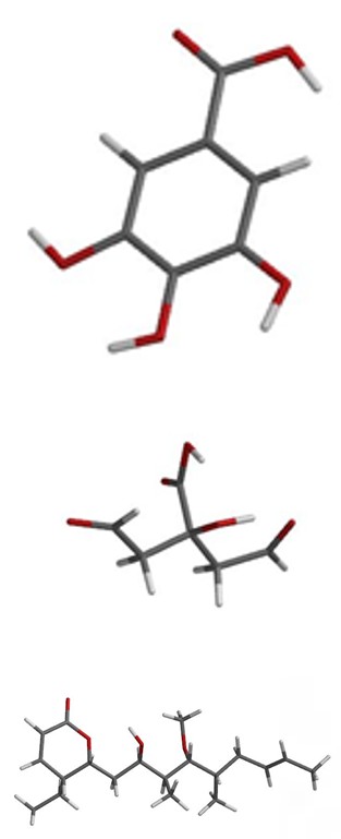
a) Gallic acid
b) Citric acid
Results and Conclusions
Table 1 shows the energy of the structures optimized using density functional theory (DFT), while in Figure 1, we can see the geometric structures from which the coupling analysis was started.
| Energies Kcal mol-1 | Kinetic | Electrostatic | Eigenvalue | Orbital # |
|---|---|---|---|---|
| Citric Acid | -2,564.75 | -993.42 | -132.575 | HOMO (84) |
| -57.536 | LUMO (85) | |||
| Pironetin | -4,393.03 | -5,200.81 | -120.122 | HOMO (178) |
| -44 | LUMO (179) | |||
| Gallic Acid | -3.729 | -2.086 | -122.198 | HOMO (88) |
| -40.31 | LUMO (89) |
Table 1: The energy of the structures optimized.
Molecular docking analysis indicates that gallic acid exhibits a greater affinity to pironetin, obtaining values of -7.0 and 6.2 kcal/mol respectively. This affinity is notably higher than that of citric acid, which has an affinity constant of -5.9 kcal/mol (Table 2). It is important to note that these comparisons are relative, as a higher affinity correlates with a lower interaction energy value, measured in kilocalories. In the pironetin cases, the complex with maximum affinity was found near lysine 352, an amino acid identified as a target of pironetin. Additionally, the mechanism of action of pironetin was expertly described by Usui, et al. using cloning techniques in conjunction with mass analysis [4]. However, the location of gallic and citric acids in the complex with pironetin seems to indicate that it has a binding site different from pironetin, but near to Lys 352 as you can see in the diagram of the interactions, showing key tubulin residues (Figures 2-4).
| Tubulin versus | Score |
|---|---|
| Citric Acid | -5.9 |
| Gallic Acid | -7 |
| Pironetin | -6.2 |
Table 2: Complex energy kcal/mol.
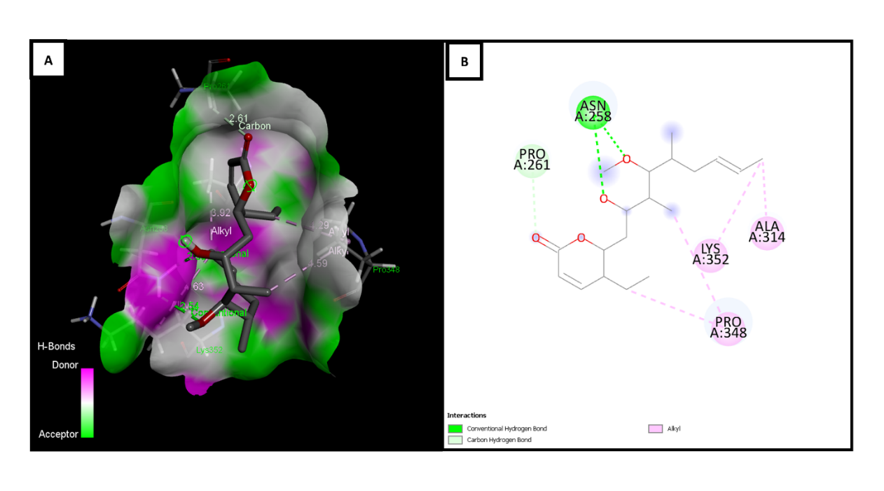
Figure 2: Molecular docking versus tubulin (PDB: 1TUB) and pironetin. (A) 3D representation of the complex, visualizing the interactions of pironetin with tubulin residues. Conventional hydrogen bonding (green), carbon bonds (light green), and Pi- alkyl interactions (light purple) are highlighted. (B) 2D diagram of the interactions, showing key tubulin residues (Asn258, Pro261, Lys352, Ala314, Pro348) and their interactions with pironetin, where conventional hydrogen bonds are represented in green and Pi-alkyl interactions in light purple.
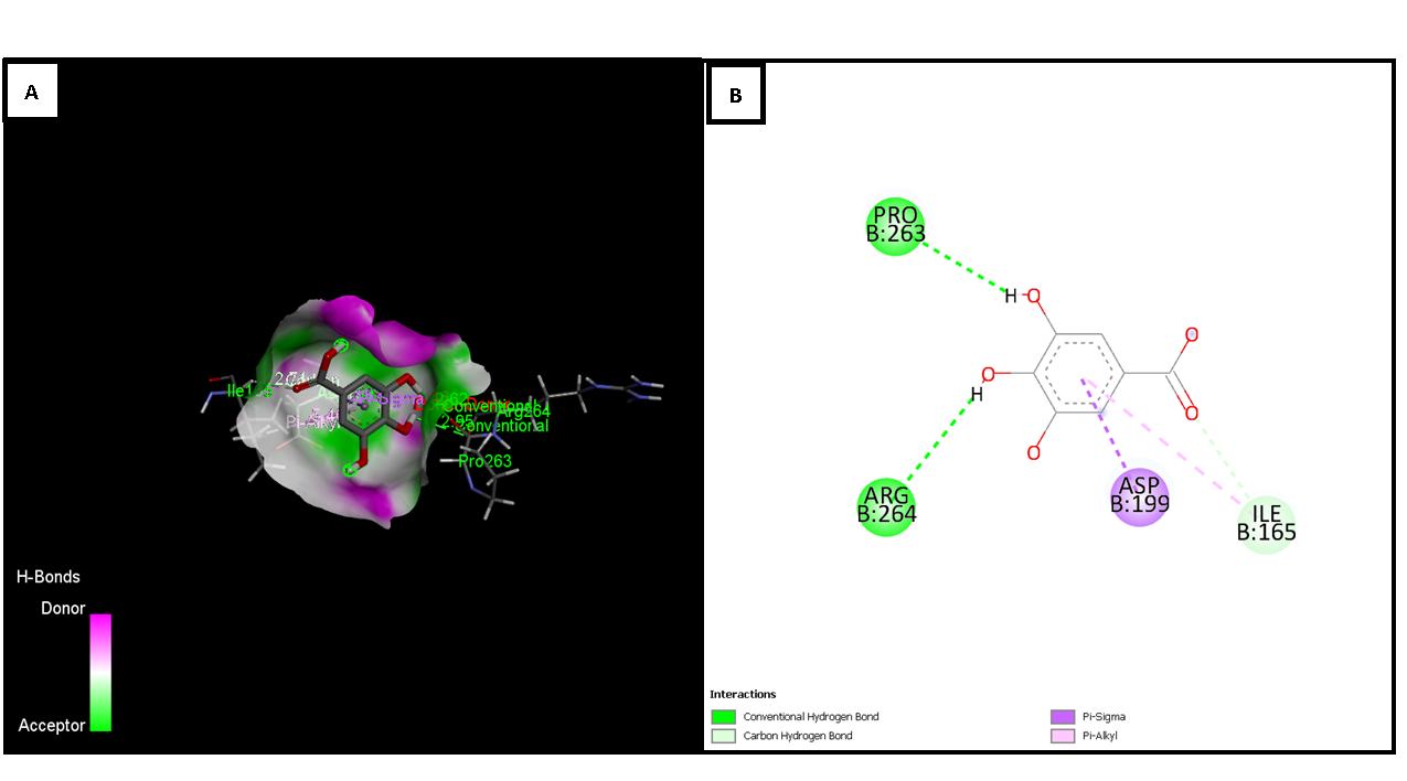
Figure 3: Molecular docking between tubulin (PDB: 1TUB) and gallic acid. (A) 3D representation of the complex, visualizing the interactions of gallic acid with tubulin residues. Conventional hydrogen bonds (green), carbon bonds (light green), and Pi- sigma interactions (purple) are highlighted. (B) 2D diagram of the interactions, showing key tubulin residues (Pro263, Arg264, Asp199, Ile165) and their interactions with gallic acid, where conventional hydrogen bonds are represented in green, carbon bonds in light green, and Pi-sigma interactions in purple.
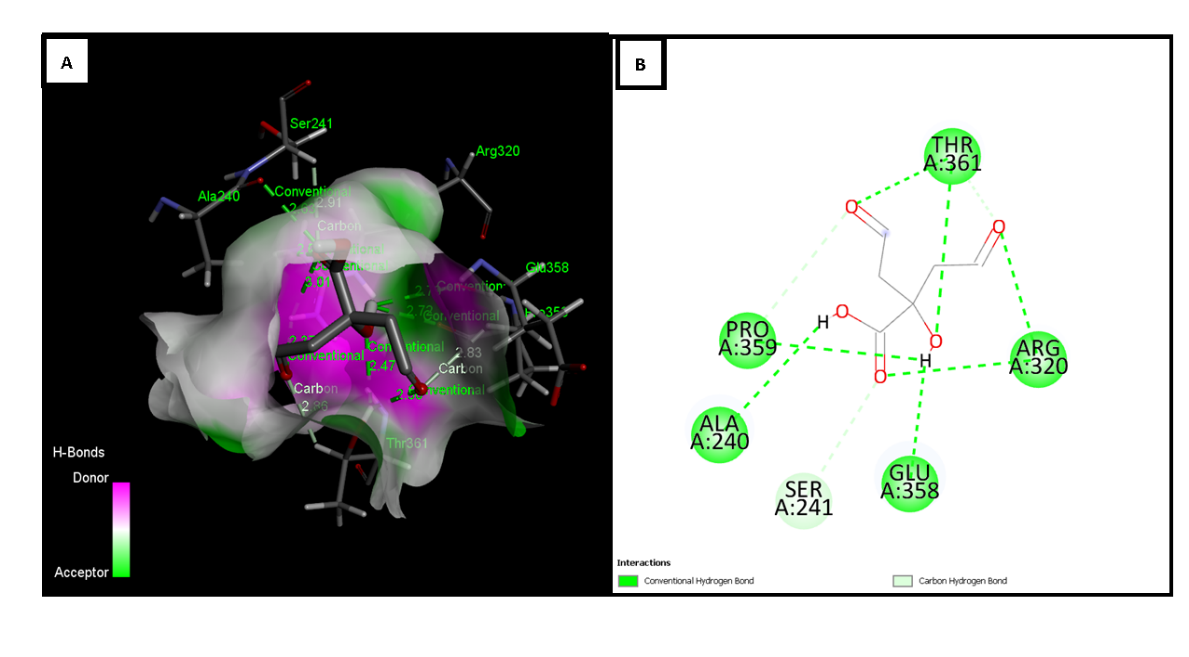
Figure 4: Molecular docking between tubulin (PDB: 1TUB) and citric acid. (A) 3D representation of the complex, visualizing the interactions of citric acid with tubulin residues. Conventional hydrogen bonding (green) and hydrogen bonds with carbon atoms (light green) are highlighted. (B) 2D diagram of the interactions, showing key tubulin residues (Pro359, Ala240, Ser241, Glu358, Arg320, Thr361) and their interactions with citric acid, where conventional hydrogen bonds are represented in green and hydrogen bonds with carbon atoms in light green.
In conclusion, gallic acid found in the peels of tropical fruits [5] has the potential to interact with the alpha tubulin due to its energy similarity to pironetin. This interaction suggests that gallic acid may inhibit tumor cells during their replicative stage, with a possible binding site near lysine 352, making it a strong candidate for in vitro testing.
Acknowledgments
Part of this project is supported by the Secretaría de Educación, Ciencia, Tecnología e Innovación de la Ciudad de México, under the project funded (SECTEI/148/2024) with folio number 3617c24, and by the Universidad Autónoma de la Ciudad de México under the annual budget for 2024.
References
-
Mendoza-Espinoza JA, López-Vallejo F, Fragoso-Serrano M, Pereda-Miranda R, Cerda-García-Rojas CM (2009) Structural reassignment, absolute configuration, and conformation of hypurticin, a highly flexible polyacyloxy- 6-heptenyl-5,6-dihydro-2H-pyran-2-one. J Nat Prod 72(4): 700-708.
-
Chel-Guerrero LD, Sauri-Duch E, Fragoso-Serrano M, Pérez-Flores LJ, Gómez-Olivares JL, et al. (2018) Phytochemical Profile, Toxicity, and Pharmacological Potential of Peels from Four Species of Tropical Fruits. Journal of Medicinal Food 21(7): 734-743.
-
Mendoza-Espinoza JA, Rangel-Sánchez E, López-Chávez E, García-Quiroz A, López-Barrera JA, et al. (2024) Microtubules as an anticancer target: The case of a Dihydropyrone. In: Martínez Cano E, et al. (Eds.), An Introduction to Bioscience Research. Universidad de Guadalajara, Centro Universitario de Los Lagos, Mexico, pp: 31-38.
-
Usui T, Watanabe H, Nakayama H, Tada Y, Kanoh N, et al. (2004) The anticancer natural product pironetin selectively targets Lys352 of alpha-tubulin. Chem Biol 11(6): 799-806.
-
Díaz-Sánchez C, Mendoza-Espinoza JA, Ponce-Sánchez C, Díaz de León-Sánchez F (2023) Chemical Profile of Polyphenolic Compounds Present in the red and white varieties of Tunillo (_Stenocereus stellatus_) fruit. Advances in Pharmacology & Clinical Trials (APCT) 8(4): 1-4.
- Treating the Forehead Lines with Combination of Forehead and Glabellar Botulinum Toxin Among Japanese Patients
- Clinical Evaluation of Patients Suffering from Breast Cancer & Determination of Treatment Therapies and Better Strategies Related to Breast Cancer
- Medieval Recipes by Al-Zahrāwī for Heart Palpitations Treatment
- Etiology and Prescription Errors of Myocardial Infarction in Different Health Care Systems of Azad Kashmir
- Early Diagnosis and Multidisciplinary Management of Turner Syndrome: A Paediatric Case Study
- Pheochromocytoma: Therapeutic Agents against the Disease and Chromatographic Methods for their Determination in Biological Fluids