Facial Nodule in a Child, What you can think of?
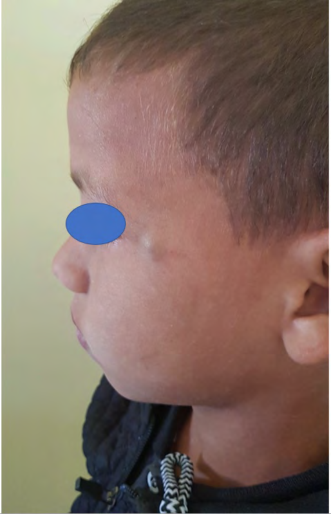
Nodules of the face in children are not an uncommon diagnosis. Apart from benign vascular tumors, other nodules of the face pose a diagnostic and therapeutic challenge. Juvenile xanthogranuloma is a clinical form of non-Langerhansian histiocytosis, the true incidence of which is not well known [1]. It presents as one or more firm erythematous or yellowish nodules rarely bluish, as in our patient, located preferably on the head and neck. Most cases begin in infancy with the possibility of congenital presentation or in adulthood [2]. Extracutaneous involvement is rare, often involving the eye, but also the lungs, heart, brain, liver and bone. The search for extracutaneous involvement is not systemic, but is recommended in the presence of multiple cutaneous lesions in a child under 2 years of age or in the presence of warning signs. The diagnosis of juvenile xanthogranuloma is basically clinical. However, it is sometimes necessary to confirm it by histology [1]. In cases described in the literature, biopsy without complete resection has been associated with an increase in lesion size with bleeding [3]. Regarding the treatment, JXGs normally follow a benign course, with spontaneous remission in about 3 to 6 years, and usually do not require treatment. Surgical excision or laser treatment may be proposed for cosmetic purposes. In ocular JXG, topical corticosteroids are usually used. In symptomatic systemic forms, treatment with surgery, radiotherapy and/or chemotherapy may be considered [1]. We report a case of a child with a nodule of the face diagnosed as juvenile xanthogranuloma. A 9-year-old child from a non-consanguineous marriage, without any pathological history, consulted for a painless bluish lesion on the tail of the left eyebrow, evolving for 6 months, without any other associated sign. On examination, we found a 0.5 cm firm nodule of regular contour,with a bluish surface on the tail of the left eyebrow. In this situation, we suspected a dermoid cyst, a pilomatricoma, a venous malformation and finally a xanthogranuloma. A complete surgical removal of the nodule was recommended in our patient. On pathological examination, we noted the presence of a dense infiltrate at the dermal level made of siderophages and macrophages with foamy cytoplasm containing lipids, multinuclear cells of the "Touton" type with a ring of nuclei surrounding an eosinophilic center and presenting in the periphery some eosinophilic granulocytes, in favor of a juvenile xanthogranuloma. We did not recommend any further examinations or investigations in our patient. After one year of follow-up, we did not note any recurrence.
Consent
The examination of the patient was conducted according to the Declaration of Helsinki principles.

Conflicts of interest
The authors do not declare any conflict of interest.
References
-
Hernandez-San Martin MJ, Vargas-Mora P, Aranibar L (2020) Juvenile Xanthogranuloma: An Entity With a Wide Clinical Spectrum. Actas dermo-sifiliograficas 111(9): 725-733.
-
So N, Liu R, Hogeling M (2020) Juvenile xanthogranulomas: Examining single, multiple, and extracutaneous presentations. Pediatric dermatology 37(4): 637-644.
-
Pajaziti, L, Hapçiu SR, Pajaziti A (2014) Juvenile xanthogranuloma: a case report and review of the literature. BMC research notes 7: 174.
- Epithelioid Granuloma; 3cases with Different Clinical Features
- Advancing Representation in Dermatology Clinical Trials: Ethical, Scientific, and Regulatory Imperatives for Inclusion Across all Fitzpatrick Skin Types
- A Case of Atopic Dermatitis with Concurrent Psoriasis Vulgaris: Successful Treatment with Upadacitinib
- Innovation Lifting Eyeshadow: A Synthesis of Makeup and Optical Illusion
- Distinguishing Superficial Actinic Porokeratosis from Actinic Keratosis with UVF Dermoscopy: A Case Report
- High Mobility Group Box 1 (HMGB1) in Cutaneous Inflammation: An Immune Modulator Bridging Cellular Stress, Ferroptosis and Danger Signaling