A Large Erosion of the Scalp
Erosive pustular dermatosis of the scalp (EPDS) is a rare inflammatory disease affecting elderly people with photodamaged scalps. The typical signs include pustules, erosions, and crusts mainly occurring after a well-identified or unsuspected local trauma. The diagnosis may be difficult because it resembles and is often associated with most common diseases affecting the scalp and has no specific histologic patterns. The current article presents a case about an initially misdiagnosed EPDS. Our patient presented an atypical form, without any pustules, associated with an actinic keratosis (AK) histology compatible with the clinical picture. He was treated with topical corticosteroids with a complete remission in one month. The case underlines the importance of considering EPDS as a differential diagnosis of common diseases, such as AK and skin cancer, to avoid treatments that would worsen it (cryotherapy, photodynamic therapy, surgery…) and lead to scarring.
Case Report
A 76-year-old man presented a 3- month history of unhealing erosion of the scalp despite local treatment, topical and systemic antibiotics. He had a history of actinic keratosis (AK) and a recent trauma. The biopsy of the edges showed an actinic keratosis. When he was referred to our clinic, he presented a 10cm erosion associated with two
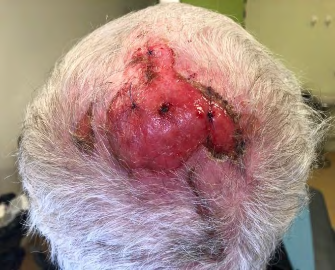
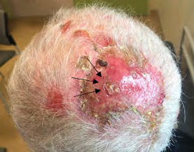
recent satellite erosions (Figure 1). Further biopsies were performed to rule out a transformation into carcinoma. Two weeks later, the patient presented the same erosion with crusts, and rare pustules (Figure 2). The diagnosis of erosive pustular dermatosis of the scalp (EPDS) was suspected. The second set of biopsies showed ulceration without tumour cells.
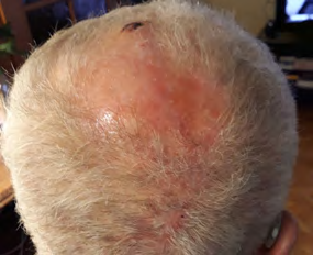
However, dermal elastosis with atrophic hair follicle, suppurative folliculitis and a lymphocytic and neutrophilic infiltrate were found (Figure 3) and were consistent with the diagnosis of EPDS. The patient was then treated with topical corticosteroids leading to full remission in one month.
![Figure 3: Histologic patterns of the lesion (magnification x 100). A. Suppurative folliculitis. B. Atrophic hair follicle. C. Lymphocytic and neutrophilic infiltrate. D. Elastotic derma Erosive pustular dermatosis of the scalp (EPDS) is a subtype of localized amicrobial pustulosis and is considered as a rare inflammatory disease affecting older women and to a lesser extent bald men [1] (sex ratio: 2/1). The triggering factor seems to be a local trauma, mostly iatrogenic, (including surgery, radiotherapy, laser, cryotherapy, and photodynamic therapy) on photo- damaged scalps [1,2,4,5]. It is characterized by sterile painful pustules, erosions and crusts. The diagnosis may be difficult, as it is frequently associated with other frequent sun-related diseases affecting the same area such as AK and cutaneous carcinomas, and because there are no specific histologic features [2,3].](/fulltextimages/8661/fig_3.png)
Figure 3: Histologic patterns of the lesion (magnification x 100). A. Suppurative folliculitis. B. Atrophic hair follicle. C. Lymphocytic and neutrophilic infiltrate. D. Elastotic derma Erosive pustular dermatosis of the scalp (EPDS) is a subtype of localized amicrobial pustulosis and is considered as a rare inflammatory disease affecting older women and to a lesser extent bald men [1] (sex ratio: 2/1). The triggering factor seems to be a local trauma, mostly iatrogenic, (including surgery, radiotherapy, laser, cryotherapy, and photodynamic therapy) on photo- damaged scalps [1, 2, 4, 5]. It is characterized by sterile painful pustules, erosions and crusts. The diagnosis may be difficult, as it is frequently associated with other frequent sun-related diseases affecting the same area such as AK and cutaneous carcinomas, and because there are no specific histologic features [2, 3].
Piccolo V. et al. (3) reported indeed a case series of EPDS with an initial misdiagnosis. They showed that among all misdiagnoses the most common were skin malignancies including KA, basal cell carcinomas and squamous cell carcinomas.
Physicians should be aware of these atypical clinical presentations. Our case illustrates how EPDS can be easily misdiagnosed if the most likely presumption of a precancerous skin growth or skin cancer is not challenged enough.
To conclude, identifying EPDS is crucial to initiate the adapted treatment to prevent cicatricial alopecia and avoid unnecessary therapeutics that can worsen it, such as surgery of precancerous lesions or carcinomas [2].
References
-
Karanfilian KM, Wassef C (2021) Erosive pustular dermatosis of the scalp: causes and treatments. Int J Dermatol 60(1): 25-32.
-
Marsh RL, Spohn GP, Kaffenberger JA (2020) Erosive pustular dermatosis of the scalp. Dermatol Online J 26(8): 13030.
-
Piccolo V, Russo T, Bianco S, Ronchi A, Alfano R, et al. (2019) Erosive Pustular Dermatosis of the Scalp: Why Do We Miss It? Dermatology 235(5): 390-395.
-
Saridakis S, Giesey RL, Ezaldein HH, Scott JF (2020) Erosive pustular dermatosis of the scalp following surgical procedures: a systemic review. Dermatol Online J 26(4): 13030.
-
Roche-Kubler B, Monnin C, Aubin F, Dupond AS (2015) Erosive pustular dermatosis of the scalp and thigh associated with skin graft recipient and donor sites. Eur J Dermatol 25(3): 269-271.
- Epithelioid Granuloma; 3cases with Different Clinical Features
- Advancing Representation in Dermatology Clinical Trials: Ethical, Scientific, and Regulatory Imperatives for Inclusion Across all Fitzpatrick Skin Types
- A Case of Atopic Dermatitis with Concurrent Psoriasis Vulgaris: Successful Treatment with Upadacitinib
- Innovation Lifting Eyeshadow: A Synthesis of Makeup and Optical Illusion
- Distinguishing Superficial Actinic Porokeratosis from Actinic Keratosis with UVF Dermoscopy: A Case Report
- High Mobility Group Box 1 (HMGB1) in Cutaneous Inflammation: An Immune Modulator Bridging Cellular Stress, Ferroptosis and Danger Signaling