Cutaneous Lupus Erythematosus in Men
Background: Systemic lupus erythematosus is an autoimmune systemic disease in which skin involvement is particularly frequent, representing 80% of cases, with a clear female predominance. The clinical features, especially the cutaneous manifestations in men, are therefore rarely evaluated. The objective of this study is to describe clinical presentation and outcome of cutaneous lupus erythematosus in men. Materials and Methods: Retrospective descriptive study conducted in the dermatology department of the Hassan II university hospital of Fez-Morocco, including 12 male patients with cutaneous lupus erythematosus. Results: The mean age of the patients was 39 years, with age extremes ranging from 17 to 61 years. A history of chronic smoking was found in 5 patients. Photosensitivity was noted in 4 patients. The average disease duration was 6.3 years, varying from 1 to 12 years. 10 patients were phototype IV of Fitzpatrick and 2 patients of phototype III. The clinical cutaneous subtypes reported were discoid lupus in 83% of the cases (10 patients), 1 case of lupus tumidus and 1 case of subacute annular lupus. The ANA were positive in 4 patients. Extracutaneous systemic manifestations were found in 2 patients, such as arthralgia. Conclusion: It is important to raise awareness among physicians regarding cutaneous lupus erythematosus in men, its clinical and prognostic characteristics and its therapeutic implications.
Introduction
Systemic lupus erythematosus (SLE) is a chronic systemic autoimmune disease mainly affecting women [1]. The increased rate of SLE in females implicates hormones as essential in disease manifestations, and this influence of sex hormones is also seen in animal models of the disease [2]. Cutaneous Lupus Erythematosus (CLE) is a nomination applied to patients with lesions produced by lupus erythematosus, whether the disorder is exclusively cutaneous or part of a systemic disease [3]. Skin involvement occurs in 80% of cases throughout the disease course and is now well known by physicians [4]. However, this disease remains relatively rare in men. The clinical and prognostic characteristics of this entity have therefore not yet been clearly established [1, 4]. Thus, there are gender disparities in this disease that ought to be evaluated. The objective of this study is to describe clinical presentation and outcome of cutaneous lupus erythematosus in men.
Patients and Methods
This research is a retrospective descriptive study, in which the variables under study were documented from the medical records of male patients with cutaneous lupus erythematosus who attended the outpatient consultation of the dermatology department of the University hospital Hassan II of Fez Morocco. In addition, the medical reports of hospitalized patients were also included. In total 12 patients were recruited in 2014-2020 from the time of the first consultation in the department. The database includes patient-level information about sociodemographic characteristics, clinical manifestations mainly cutaneous involvement with dermoscopic description of lesions and association with systemic disease.
Results
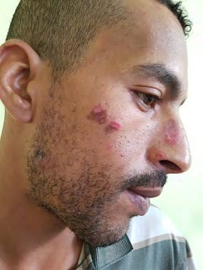
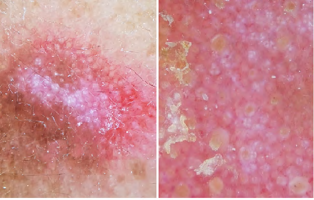
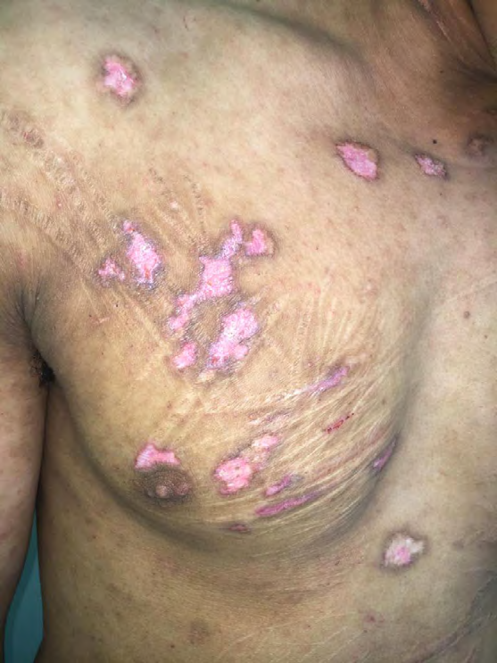
The average age of the patients was 39 years old, with extremes of age ranging from 17 to 61 years old. A history of smoking was found in 5 patients. The average disease duration was 6.3 years, varying from 1 to 12 years. Photosensitivity was reported in 4 patients. 10 patients were phototype IV of Fitzpatrick and 2 patients of phototype III. The clinical cutaneous subtypes reported were 10 cases of discoid lupus, 1 case of lupus tumidus and 1 case of subacute annular lupus. Areas of involvement were predominantly the head and neck in all patients with disoid lupus, clinically presenting as sharply demarcated, erythematokeratotic, atrophic or scarring lesions. Dermoscopy of these lesions typically showed Perifollicular whitish halo, Follicular keratotic plugs, Telangiectatic vessels, White scales, Structureless whitish areas. Among those patients two had a particular localization in the genitalia and also over old tattoos and scars showing a Koebner’s phenomenon. One case of lupus tumidus with infiltrated erythematous plaques located on the face and trunk. We also reported one case of subacute lupus with annular erythematous patches of variable size with a central clear area involving the face, neck, upper back and the arms. Dermoscopy of these lesions showed erythema in the central area and linear vessels and crusts at the periphery. Only two patients had extracutaneous signs consisting of intermittent arthralgias. ANA were positive in 4 patients.
Discussion
Systemic lupus erythematosus (SLE) is a chronic systemic autoimmune disease resulting from abnormal interactions between T and B cells (3) [1]. Within females of childbearing age, the ratio of females to males is nine to one. This ratio is lower before puberty and decreases in the elderly [2]. The increased rate of SLE in females implicates hormones as essential in disease manifestations, and this influence of sex hormones is also seen in animal models of the disease [1, 2]. Several studies have described clinical features of SLE in men, cutaneous features have rarely been evaluated [5, 6]. The prevalence of cutaneous lupus, particularly in men, is higher than that of systemic lupus. The age of onset varies between 30 and 50 years old, with rare cases in children and exceptionally in elderly subjects [2, 5]. Our series is in consisten with the literature, as we noted a peak in the age group between 30-50 years with a only one 17-year-old adolescent who presented with discoid lupus evolving for 4 years. Smoking seems to play a role in the pathogenesis of lupus in both sexes with a higher prevalence in men [7]. In our series, 5 patients had a history of active smoking. Cutaneous lesions constitute 4 of the 17 new criteria established by the Systemic Lupus International Collaborating Clinics (SLICC) [3]. Discoid lupus erythematosus (DLE) is the most common subtype in males with a sex ratio of 2F1M [3, 8]. Photosensitivity seems to be less present compared to acute and subacute forms [8]. Aligning with this data, 83% of our patients had discoid lupus, only 3 of which had photosensitivity in our series. Dermoscopy of DLE has widely been described [9]. Typical dermoscopic features are Perifollicular whitish halo, Follicular keratotic plugs, Telangiectatic vessels, White scales, and structure less whitish areas [9]. We reported all of these structures in all of the patients with DLE. As seen in two of our patients, less common localizations like the genitalia and over tattoos or scars in the form of koebner’s phenomenon might be difficult to distinguish from other differential diagnoses [10, 11]. Other forms of cutaneous lupus have also been described in men but with a lower proportion. Petty AJ, et al. analyzed the clinical and histological characteristics of induced cutaneous lupus in men. The subacute form was predominant, most often induced by drugs including anti-hypertensives and proton-pump inhibitors. There was no significant difference between the two groups systemic features or autoantibody positivity [12]. In our patient no drug intake was reported and antinuclear antibodies (ANA) were positive. Schwartzman-Morris reviewed several cohorts comparing the clinical manifestations of systemic lupus erythematosus in men and women. SLE in men was associated with a shorter duration of disease. Systemic manifestations are relatively rare but when present, were more severe in men. Among these manifestations cardiovascular, neurological and renal involvement were the most reported [2, 4]. The cutaneous manifestations were dominated by discoid lupus and lupus tumidus [2]. In our series, only two patients had extracutaneous involvement consisting of intermittent arthralgias and 4 patients had positive ANA.
Conclusion
Several groups have studied the sex disparities in this disease and have suggested gender, along with ethnicity, age of disease onset, or autoantibody profiles as a means to identify SLE subgroups [2, 8]. It is important to raise awareness among physicians regarding cutaneous lupus erythematosus in men, its clinical and prognostic characteristics and its therapeutic implications. As gender differences may affect drug action and availability, tailored treatments for males and females might improve outcomes and overall prognosis for both genders [2].
Consent
The examination of the patient was performed in accordance with the principles of the Declaration of Helsinki.
Conflicts of Interest
The authors declare no conflicts of interest.
References
-
Rider V, Abdou NI, Kimler BF, Lu N, Brown S, et al. (2018) Gender Bias in Human Systemic Lupus Erythematosus A Problem of Steroid Receptor Action? Front Immunol 9: 611.
-
Schwartzman-Morris J, Putterman C (2012) Gender Differences in the Pathogenesis and Outcome of Lupus and of Lupus Nephritis. Clin Dev Immunol 2012: 9.
-
Moura Filho JP, Peixoto RL, Martins LG, Melo SD de, Carvalho LL de, et al. (2014) Lupus erythematosus considerations about clinical, cutaneous and therapeutic aspects. An Bras Dermatol 89(1): 118-125.
-
do Socorro Teixeira Moreira Almeida M, da Costa Arcoverde J, Jacobino MNB, Coimbra Neto AR. (2011) Male Systemic Lupus Erythematosus, an Overlooked Diagnosis. Clin Pract 1(4): e103.
-
Jarrett P, Thornley S, Scragg R (2016) Ethnic differences in the epidemiology of cutaneous lupus erythematosus in New Zealand. Lupus 25(13): 1497-1502.
-
Stefanidou S, Benos A, Galanopoulou V, Chatziyannis I, Kanakoudi F, et al. (2011) Clinical expression and morbidity of systemic lupus erythematosus during a post-diagnostic 5-year follow-up a malefemale comparison. Lupus 20(10): 1090-1094.
-
Ekblom-Kullberg S, Kautiainen H, Alha P, Leirisalo-Repo M, Julkunen H (2013) Smoking and the risk of systemic lupus erythematosus. Clin Rheumatol 32(8): 1219-1222.
-
Cooper EE, Pisano CE, Shapiro SC (2021) Cutaneous Manifestations of “Lupus” Systemic Lupus Erythematosus and Beyond. Rothschild BM, éditeur. Int J Rheumatol 2021: 1-19.
-
Lallas A, Apalla Z, Lefaki I, Sotiriou E, Lazaridou E, et al. (2013) Dermoscopy of discoid lupus erythematosus Dermoscopy of discoid lupus erythematosus. Br J Dermatol 168(2): 284-8.
-
Romiti R, Anzai A, Nico M (2014) Genital discoid lupus a rare manifestation of cutaneous lupus erythematosus. Lupus 23(7): 707-710.
-
Kluger N, Andraud M, Lartigau-Roussin C, Sultan-Bichat N (2021) The Koebner phenomenon on tattoos and piercings in a patient with cutaneous lupus a case report and review of the literature. Acta Dermatovenerol Alp Pannonica Adriat 30(1): 43-46.
-
Petty AJ, Cardones AR, Marano AL (2020) Analysis of clinical characteristics of drug-induced cutaneous lupus erythematosus in men. J Am Acad Dermatol 83(5): 1455- 1457.
- Epithelioid Granuloma; 3cases with Different Clinical Features
- Advancing Representation in Dermatology Clinical Trials: Ethical, Scientific, and Regulatory Imperatives for Inclusion Across all Fitzpatrick Skin Types
- A Case of Atopic Dermatitis with Concurrent Psoriasis Vulgaris: Successful Treatment with Upadacitinib
- Innovation Lifting Eyeshadow: A Synthesis of Makeup and Optical Illusion
- Distinguishing Superficial Actinic Porokeratosis from Actinic Keratosis with UVF Dermoscopy: A Case Report
- High Mobility Group Box 1 (HMGB1) in Cutaneous Inflammation: An Immune Modulator Bridging Cellular Stress, Ferroptosis and Danger Signaling