A Case with Photodistributed Erythema Multiforme
Erythema multiforme is a type IV hypersensitivity reaction characterized by acute onset erythematous edematous target like papules and plaques. It has been associated with infections, especially herpes simplex virus, drugs and rarely autoimmune diseases. Unlike other causes, photo-distriubuted idiopathic erythema multiforme develops after exposure to UV and settles in sun-exposed areas. The diagnosis is often based on history, clinical examination, histopathology and phototesting. The management of EM is treating the underlying etiology, sympthomatic topical treatment and antihistaminic drugs.
Case Report
A 51-year-old woman presented with pruritic rashes on the face, neck and forearms, which had started a few days ago. There was no family or personal history of skin disorders, systemic disease, herpes simplex virus (HSV) infection or a

history of drug ingestion in association with the beginning of lesions. Physical examination revealed erythematous papules and target like plaques in the sunexposed distribution on the face, neck and the forearms (Figures 1 and 2). She had no mucosal involvement.
The results of laboratory tests, including ANA, anti- DNA, anti SSA, anti-SSB, serological tests for HSV and all other laboratory results were within normal values. She did not accept to have a phototest thus photoprovocation tests could not performed. A punch biopsy specimen taken from the targetoid lesion revealed basal vacuolar degeneration in the epidermis, apoptotic keratinocytes, superficial edema in
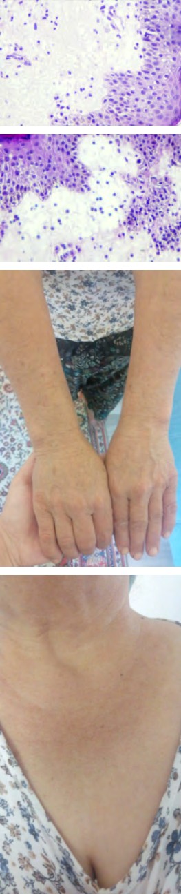
the dermis, formation of bulla in the subepidermal area, and a perivascular lymphoplasmocytes infiltrate in the dermis (Figures 3 and 4). She was treated with a high potency of topical corticosteroid and antihistaminic drug. At the follow- up, the lesions cleared after 2 weeks, postinflammatory hyperpigmentation was not observed (Figures 5 and 6).
Figures 3 and 4: Variable epidermal spongiosis and dermal edema. Diffuse lymphocytic infiltrate in the papillary dermis.
Figures 5 & 6: The V area of the neck and the dorsal aspects of the forearms after 2 weeks of treatment.
Discussion and Conclusion
Erythema multiforme (EM) is an acute, self-limited skin disease characterized by the abrupt onset of symmetric fixed red papules, some of which evolve into typical and/ or occasionally “atypical” papular target lesions [1]. The etiopathogenesis of which has not been fully elucidated, which may develop after drug administration and infections. HSV infection is the most common cause of EM development [1].
Erythema multiforme is classified among the photosensitivity diseases. In some cases, lesions appear on sun-exposed areas. Therefore, the term photosensitive or photodistributed erythema multiforme (PEM) is used. We observed approximately 20 cases in the literature review [2, 3, 4]. Triggers identified were drugs (55%), HSV infection (22.2%), and polymorphous light eruption (5.5%). The remaining cases were classified as idiopathic [2].
Phenylbutazone, triclocarban, afloqualone, bufexamac, paclitaxel, simvastin, pravastatin, paroxetine, naproxen, itraconazole, vandetanib have been reported as drugs responsible for PEM [2, 3, 4]. Drug-induced PEM usually occurs once. However, if the disease is misdiagnosed or the drug is continued, the disease can become chronic, with recurrent rashes triggered by sun exposure over many years [2]. The time from the start of treatment to the appearance of lesions can vary. Periods ranging from 15 days to 4 years have been reported in the literature. There is usually no personal or family history of photodermatosis or recurrent HSV infection [2]. Lesions are located on sun-exposed areas and systemic symptoms and mucosal involvement are seen. Lesions due to topical drugs are usually seen at the application site [5, 6].
The second common cause of PEM is HSV infection. Some HSV cases have been reported in the literature [2, 7]. While the main herpes labialis is the causative agent, cases have also been described in patients with genital herpes and other areas of herpes. HSV-related PEM occurs in young men with no personal or family history of photodermatosis and who report not using medication. It is characterized by recurrent episodes that occur once or twice a year, normally during the sunniest months of the year. Herpes lesions precede the skin rash about 7 to 10 days. The eruption affects sun-exposed areas and is usually not accompanied by systemic or mucosal involvement [2].
Cases secondary to polymorphous light eruption have also been reported in the literature [2]. Several cases of PEM have been reported in patients without herpes reactivation, drug use, or any other known trigger like our case.
In the pathogenesis of PEM; UV radiation can contribute to lesions by inducing the release of inflammatory mediators such as kinins, prostaglandins and histamine that will increase vascular permeability, thus facilitating the passage of skin antigens into the bloodstream and their formation in sun exposed areas [2]. Subclinical reactivation of HSV has been identified in the etiology of a few cases of idiopathic PEM. HSV DNA was detected by polymerase chain reaction in 40% of cases of recurrent, apparently idiopathic EM. Some of the cases responded to prophylactic antiviral therapy, suggesting that they were triggered by subclinical HSV infection [2].
The differential diagnosis of erythema multiforme should include EM-like PMLE, subacute cutaneous lupus erythematosus, EM-like contact dermatitis, paraneoplastic pemphigus, IgA pemphigus, pruritic urticarial papules and plaques of pregnancy, syphilis, fixed drug eruption, ecthyma gangrenosum, erythema chronicum migrans, and granuloma annulare [2].
The goal of treatment in EM is to eliminate the causative agent and treat the symptoms with antihistamines and topical and/or systemic corticosteroids. Using topical sunscreens is also important in the case of PEM. We treated our patient with topical steroid and oral antihistaminic.
In our case, the clinicopathologic findings were similar to PEM. We wanted to present because PEM cases are rarely seen in the literature.
References
-
Wolfram Hötzenecker, Christina Prins and Lars E. French.
-
Rodríguez-Pazos L, Gómez-Bernal S, Rodríguez-Granados MT, Toribio J (2013) Photodistributed Erythema MultiformeEritema multiforme fotodistribuido. Actas Dermosifiliogr 104(8): 645-653.
-
Patri A, Gabriella F, Megna M, Lauro W, D’Onofrio P, et al. (2021) Itraconazole‐induced photodistributed erythema multiforme. Dermatologic Therapy 34(2): e14901.
-
Caro-Gutierrez D, Muruzabal MUF, Fuente EG, Franco AP, Estebaranz JLL (2014) Photo-induced erythema multiforme associated with vandetanib administration. JAAD 71(4): E142-E144.
-
Leroy D, De Raucourt S, Deschamps P (1987) Drug- induced erythema multiforme with photodistribution and genital lesions. Photodermatol 4(1): 52-54
-
Kurumaji Y (1998) Photo Koebner phenomenon in erythema-multiforme-like eruption induced by contact dermatitis due to bufexamac. Dermatology 197(2): 183- 186
-
Pérez-Carmona L, Vaño-Galvan S, Carrillo-Gijón R, Jaén- Olasolo P (2010) Photosensitive erythema multiforme presenting as juvenile spring eruption. Photodermatol Photoimmunol Photomed 26(1): 53-54.
- Epithelioid Granuloma; 3cases with Different Clinical Features
- Advancing Representation in Dermatology Clinical Trials: Ethical, Scientific, and Regulatory Imperatives for Inclusion Across all Fitzpatrick Skin Types
- A Case of Atopic Dermatitis with Concurrent Psoriasis Vulgaris: Successful Treatment with Upadacitinib
- Innovation Lifting Eyeshadow: A Synthesis of Makeup and Optical Illusion
- Distinguishing Superficial Actinic Porokeratosis from Actinic Keratosis with UVF Dermoscopy: A Case Report
- High Mobility Group Box 1 (HMGB1) in Cutaneous Inflammation: An Immune Modulator Bridging Cellular Stress, Ferroptosis and Danger Signaling