When Faced with the Eyebrows’ Density Depletion within Children, do not Hesitate to use the Dermoscope
Tinea Capitis are frequent dermatophytes affecting mainly children. They are mainly found on the scalp, their localization on the eyebrows remains. Trichoscopy finds its place in the clinical diagnosis and follow-up of Tinea Capitis [1]. We demonstrate through an original case of trichophytic Tinea Capitis of the eyebrows the usefulness of trichoscopy in the diagnostic rectification.
Image Article
Tinea Capitis are frequent dermatophytes affecting mainly children. They are mainly found on the scalp, their localization on the eyebrows remains. Trichoscopy finds its place in the clinical diagnosis and follow-up of Tinea Capitis [1]. We demonstrate through an original case of trichophytic Tinea Capitis of the eyebrows the usefulness of trichoscopy in the diagnostic rectification.
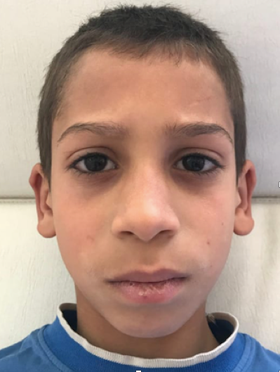
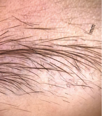
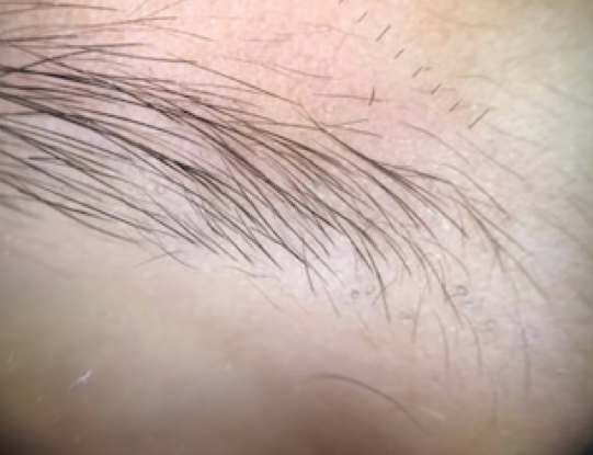
This case concerns an 8-year-old child, followed for spinulosic psoriasis under topical dermocorticoids, in whom a rarefaction of the density of the head of the eyebrows was noticed during his routine consultation, which was mistakenly taken by the mother as being an eyebrow involvement of his psoriasis. Dermatological examination found partial depilation of the heads of both eyebrows without the presence of crusts or scales (Figure 1). The traction sign was positive. In addition, the patient did not report any functional signs, especially no itching. Trichoscopic examination revealed short broken hairs, corkscrew hairs, zigzag hairs, comma hairs and @ hairs (Figure 2). These different dermosocpic aspects made us evoke a Tinea Capitis of the eyebrows in the child. Mycological sampling confirmed the diagnosis with the presence of trichophyton. The child was treated with oral griseofulvin 20mg/Kg/day for 6 weeks with good improvement. In fact, the trichoscopic signs of Tinea Capitis have completely regressed (Figure 3).
The eyebrow location of Tinea Capitis seems to be exceptional, even in children, and can be confused with other hair diseases. Trichoscopy is a good tool for the clinical diagnosis of ringworm, helping the clinician to speed up the therapeutic management. Trichoscopy is considered positive when at least one specific sign is found on at least 2 hairs Image Article [2]. Among those described in the literature are: comma hair, corkscrew hair, zigzag hair, morse hair, whitish sheath. Mycological confirmation is mandatory before starting an oral antimycotic treatment [2, 3].
Consent
The examination of the patient was conducted according to the principles of the Declaration of Helsinki. The authors certify that they have obtained all appropriate patient consent forms, in which the patients gave their consent for images and other clinical information to be included in the journal. The patients understand that their names and initials will not be published and due effort will be made to conceal their identity, but that anonymity cannot be guaranteed.
References
-
Waśkiel-Burnat A, Rakowska A, Sikora M, Ciechanowicz P, Olszewska M, et al. (2020) Trichoscopy of Tinea Capitis: A Systematic Review. Dermatol Ther (Heidelb) 10(1): 43-52.
-
Dhaille F, Dillies AS, Dessirier F, Reygagne P, Diouf M, et al. (2019) A single typical trichoscopic feature is predictive of tinea capitis: a prospective multicentre study. Br J Dermatol 181(5): 1046-1051.
-
Dhaille F, Dillies AS, Dessirier F, Reygagne P, Lombart F, et al. (2018) Évaluation de la trichoscopie dans le diagnostic de teigne, étude prospective multicentrique sur 2 ans, à propos de 100 patients. Annales de Dermatologie et de Vénéréologie 14(20): S80-S81.
- Epithelioid Granuloma; 3cases with Different Clinical Features
- Advancing Representation in Dermatology Clinical Trials: Ethical, Scientific, and Regulatory Imperatives for Inclusion Across all Fitzpatrick Skin Types
- A Case of Atopic Dermatitis with Concurrent Psoriasis Vulgaris: Successful Treatment with Upadacitinib
- Innovation Lifting Eyeshadow: A Synthesis of Makeup and Optical Illusion
- Distinguishing Superficial Actinic Porokeratosis from Actinic Keratosis with UVF Dermoscopy: A Case Report
- High Mobility Group Box 1 (HMGB1) in Cutaneous Inflammation: An Immune Modulator Bridging Cellular Stress, Ferroptosis and Danger Signaling