Cyclops Lesion Without ACL Reconstruction: A Rare Case in a Patient with Intact Anterior Cruciate Ligament and Tibial Plateau Fracture
Cyclops lesions are most commonly seen after anterior cruciate ligament (ACL) reconstruction and are known to cause anterior knee pain and mechanical loss of extension. However, they are rarely reported in patients with ACL injuries that were not surgically reconstructed, and exceptionally rare in knees with intact ACLs. We present the case of a 42-year-old male with post-traumatic knee stiffness and pain, who was found to have a cyclops lesion despite no history of ACL reconstruction or complete tear. This case adds to the extremely limited literature on non-operative, intact-ACL-associated cyclops lesions.
Introduction
Cyclops lesions, or localized anterior arthrofibrosis, are fibroproliferative nodules commonly found in the intercondylar notch following ACL reconstruction [1, 2, 3, 4, 5]. These lesions can lead to mechanical block and loss of full knee extension. While they are frequently linked to graft impingement after surgery, there have been occasional reports in non-reconstructed ACL injuries [1, 2]. Their occurrence in knees with an intact ACL is exceedingly rare, with fewer than ten cases reported in the literature [3, 4].
Case Presentation
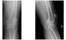
A 42-year-old medically free male presented with chronic left knee pain and limited extension, persisting following a road traffic accident (RTA) 16 years ago. He had no prior surgical intervention involving the ACL. On examination, he had a limited and painful range of motion in the left knee, particularly with terminal extension. Quadriceps strength was reduced. A left knee radiograph was performed and showed an old tibial plateau fracture (Figure 1).
Subsequent MRI of the left knee (Figure 2) revealed evidence of tibial plateau internal fixation, a focal nodular soft tissue mass of intermediate to low signal intensity projecting into the posterior aspect of Hoffa’s fat pad, consistent with a cyclops lesion and no cruciate ligaments or meniscal tears were observed.
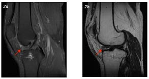
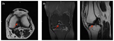
Figure 2: MRI of the left knee. Sagittal proton density fat saturation (2a), sagittal T2 non-fat saturation (2b) demonstrate the intermediate to low signal intensity soft tissue nodule near the tibial insertion of the anterior cruciate ligament. The patient was managed conservatively with medical treatment and physiotherapy aimed at improving range of motion and muscle strength (Figure 3).
Discussion
Cyclops lesions are composed of dense fibrous and granulation tissue and most commonly arise after ACL reconstruction, with an estimated incidence between 1% and 10% [5]. However, they have also been described in patients with no history of ligament reconstruction [6]. Early reports have documented cyclops lesions occurring in partially torn ACLs without surgical intervention [1, 2]. One case involved blocked extension due to a cyclops lesion in a completely torn ACL with no prior reconstruction [3]. Another case, published in 2022, represented one of the most recent examples of this rare presentation [4].
The development of a cyclops lesion in a knee with an intact ACL, as seen in this patient, may be associated with chronic inflammation, micro-tears, or trauma-induced fibrosis imitating classic postoperative mechanisms. MRI remains the most valuable imaging tool for diagnosis, especially when clinical examination reveals persistent loss of knee extension in the absence of a complete ligament tear [7]. MRI is highly effective for detecting cyclops lesions: it shows ~85% sensitivity and specificity, and over 90% accuracy for nodules >1 cm in patients with extension loss [8]. Treatment is mainly arthroscopic excision of symptomatic lesions which typically restore full extension and relieves pain with low recurrence rates; physiotherapy alone is often insufficient [9].
This case adds valuable documentation to the very limited number of such cases in the literature and underscores the importance of considering this entity when evaluating patients with chronic anterior knee pain and mechanical extension deficits.
Conclusion
This case illustrates a rare presentation of a cyclops lesion in a knee with an intact ACL and no surgical history. Radiologists and clinicians should include this entity in differential diagnoses for patients with chronic anterior knee pain and extension loss, even when no ACL rupture or reconstruction is documented. Early MRI assessment can facilitate conservative management and avoid unnecessary interventions.
Patient Consent
Written informed consent was obtained from the patient for publication of this case report.
Funding
This case report received no specific grant from any funding agency in the public, commercial, or not-for-profit sectors.
Conflict of Interest
The author declares no conflict of interest.
References
-
Runyan BR, Bancroft LW, Peterson JJ, Kransdorf MJ, Berquist TH, et al. (2007) Cyclops lesions that occur in the absence of prior anterior cruciate ligament reconstruction. Radiographics 27(6): e26.
-
Veselko M, Rotter A, Tonin M (2000) Cyclops syndrome occurring after partial rupture of the anterior cruciate ligament not treated by surgical reconstruction. Arthroscopy 16(3): 328-331.
-
Andronic O, Aslam Joiya S, Barbarosie C (2017) Blocked knee extension due to a cyclops lesion in a totally torn ACL with no prior reconstruction. Med-Surg J (Iasi) 121(3): 2017.
-
Awad M, Naqvi G, Khan A (2022) A rare case of cyclops lesion without ACL reconstruction. J Orthop Sports Med 4(2):100055.
-
Diermeier T, Rothrauff BB, Engebretsen L, Lynch A, Svantesson E, et al. (2021) The pathophysiology of cyclops lesions after ACL reconstruction. Knee Surg Sports Traumatol Arthrosc 2021.
-
Recht MP, Piraino DW, Cohen MA, Parker RD, Bergfeld JA (2013) Localized anterior arthrofibrosis (cyclops lesion) after reconstruction of the anterior cruciate ligament: MR imaging findings. AJR Am J Roentgenol 165(2).
-
Kambhampati SB, Lenhart RL, Patel NM (2020) Cyclops lesions of the knee: A narrative review of the literature. Orthop J Sports Med 8(8): 2325967120945671.
-
Bradley DM, Bergman AG, Dillingham MF (2012) MR imaging of cyclops lesions: sensitivity & specificity. AJR Am J Roentgenol 174(3): 719-726.
-
Abderraouf BF, Marouen S, Ameni J, Ammar I, Saida J (2022) Localized Anterior Arthrofibrosis of the Knee that Occurs after Meniscal Surgery. Int J Radiol Imaging Technol 8: 090.
- Ultrasound Guided Therapeutic Nerve Blocks
- Dosimetric Comparison between Two Dose Calculation Algorithms in SBRT Treatment of Lung Cancer in Ring-based and C-arm Radiation Therapy Equipment
- Adolescent Testicular Adrenal Rest Tumors: A Case Report and Review of the Literature
- Giant Intrathoracic Lipoma: A Rare Presentation
- Image of a Right Renal Angiomyolipoma Complicated by Hemorrhage
- Effect of Contrast Agents on Pregnant Women