Dried Umbilical Cord and Cord Blood Sample: Reliable Non Invasive Method of Sample Collection than Usual Invasive Method from Neonates for Forensic Identification
Forensic DNA profiling has an important role on the society by providing reliable evidence whether a man is the biological father of an individual or not. To do that biological samples are required for DNA analysis. It is very easy to collect blood samples from the adults but at the same time quite difficult to draw blood from neonates. It requires lot of skill as the process is invasive and many of the time collected amount is very few. To forbid that issue we have successfully isolated genomic DNA from a new born in a non-invasive way, by collecting the dried umbilical cord and the cord blood sample. Matching or exclusions between alleged parents and the child were established with the help of STR typing.
Soma Roy*, Ishani Mitra and Dinesh D
Central Forensic Science Laboratory, India *Corresponding author: Dr. Soma Roy, Central Forensic Science Laboratory, 30, Gorachand Road, Kolkata-700014, West Bengal, India, Email: soma.cfsl@gmail.com
Introduction
Forensic DNA fingerprinting can be defined as the comparison of the DNA in a person’s nucleated cells with that identified in biological matter found at the scene of a crime or with the DNA of another person for the purpose of identification or exclusion [1]. It is a useful tool for verifying a stated biological relationship like paternity or maternity [2]. It is the most accurate and widely acceptable technology to establish the biological relationship among two or more individual. Till date the preferred forensic specimen collection technique for DNA testing is blood sample either in liquid form or stain forms [3]. For collection of intra venous blood samples from the accused or the victim either direct liquid blood is obtained or bloodstains are made using lancet on FTA cards [4]. But cases related with exhibit sample collection from newborn babies, the methods are always difficult and painful. Venipuncture is the method of choice for blood sampling in term of neonates; however, it is complex and requires an experienced, knowledgeable and trained phlebotomist [5]. On the other hand umbilical cord blood is the blood left over in the placenta and in the umbilical cord after the birth of the baby. This cord blood is composed of all the elements found in whole blood. It contains red blood cells, white blood cells, plasma, platelets and some other hematopoietic stem cells [6, 7, 8]. During prenatal development, the umbilical cord is physiologically and genetically part of the foetus and, (in humans), normally contains two arteries (the umbilical arteries) and one vein (the umbilical vein), buried within Wharton’s jelly [9, 10, 11]. The umbilical vein supplies the foetus with oxygenated, nutrient-rich blood from the placenta. Conversely, the foetal heart pumps deoxygenated, nutrient-depleted blood through the umbilical arteries back to the placenta [12, 13].
The objective of this study was to solve the disputed paternity cases by using the DNA yield from dried umbilical cord or cord blood samples and other blood/bone samples to regularize the noninvasive sampling methods for neonates in forensic routine sample collection techniques.
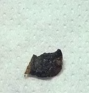
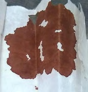
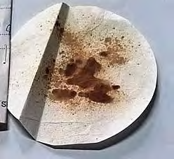
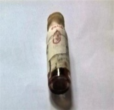
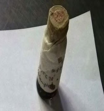
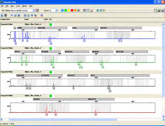
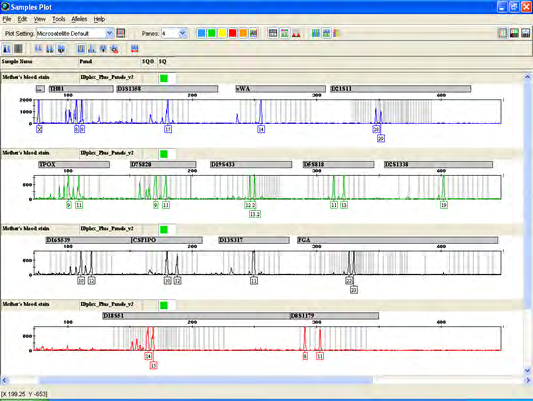
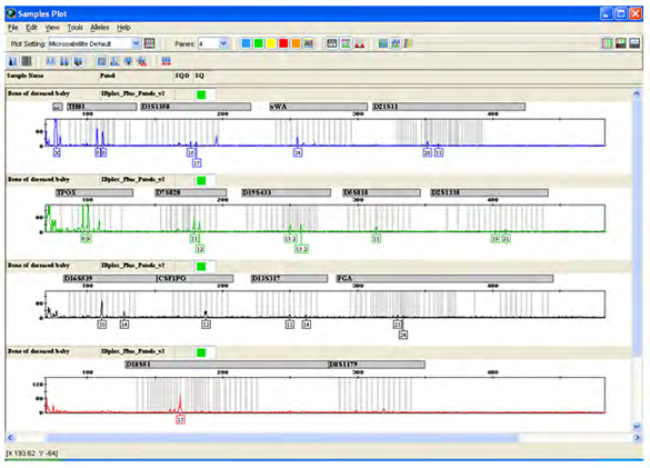
Case Study 1: A person was arrested and recommended jail custody for the allegation of a rape. A baby was delivered by the lady. Unfortunately the baby was expired within a few days and the body was buried under the soil. Police can only recover one small bone piece after few days. One small dry umbilical cord tissue of the baby was also recovered from the lady’s house. DNA profiling is done to identify the real biological father of the deceased baby.
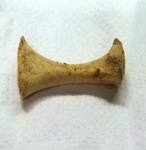
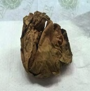
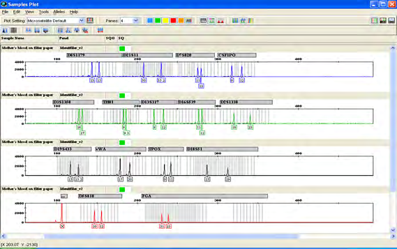
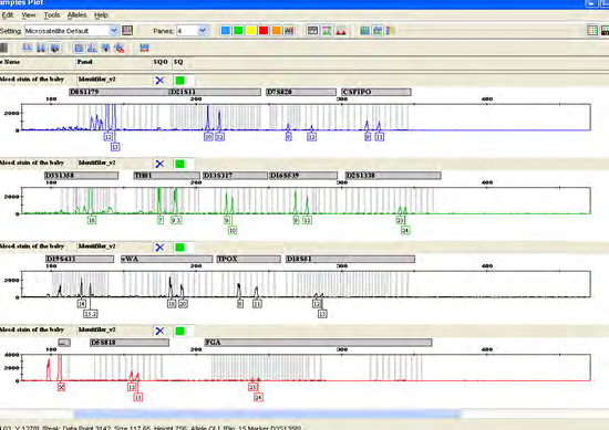
Exhibit samples of Case Study 2: The image of each case exhibits from case study 2 are given below in Figures 5-7.
Case Study 2: A person was arrested by police for allegation of a rape. The cord blood sample of the stillborn baby of the victim was collected and preserved by the doctor. DNA profiling is done to identify the actual biological father of the stillborn baby.
Materials and Methods
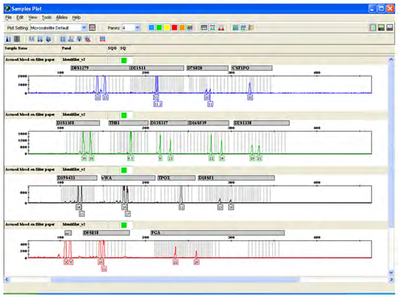
Exhibit samples of Case Study 1: The image of each case exhibits from case study 1 are given below in Figures 1-4.
Procedure for Isolation of Total DNA from Dried Umbilical Cord
Genomic DNA was isolated from dried umbilical cord tissue using Stain Extraction Buffer (SEB) followed by Amicon purification. The amount of dried umbilical cord was merely 1 inch in length. To isolate genomic DNA from it, at first cut a small part from the dried cord and then wash the cord with DNase free water to rehydrate it. When it becomes soften to cut in more small pieces take the rehydrated cord and cut them small, try to choose darker part of the cord and put in a 1.5ml eppendorf tube [14, 15]. Now, with the help of a pipette just smash the pieces lightly and add 400 µl stain extraction buffer into it followed by 20 µl of Proteinase K. Incubate at 56°over night.
From liquid blood and cord blood stain DNA was extracted using Phenol-Chloroform method followed by ethanol precipitation [16] and from femur bone using organic method and Amicon purification.
Analysis of Isolated DNA
Quality and quantity of isolated DNA was checked through 1% TAE gel electrophoresis.
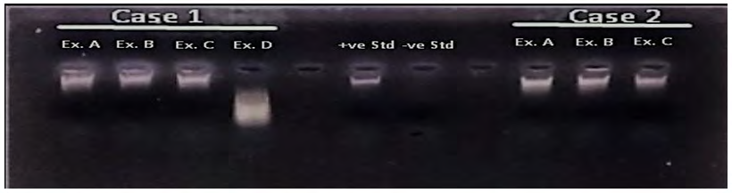
Figure 8:- The 1% Agarose gel electrophoresis results of isolated DNA Case-1 (Ex-A- Post mortem blood stain of suspected father, Ex-B- Dried umbilical cord of deceased baby, Ex-C- Blood stain of mother, Ex-D- Femur bone of deceased baby); “+ve” Std=20ng/l; “-ve” Std; Case-2 (Ex-A- Liquid blood of mother, Ex-B- Liquid blood of suspected father, Ex-C- Cord blood stain of the still born baby).
PCR Amplification and Sequencing Analysis
For Case 1 the isolated DNA PCR amplifications was performed using Investigator® IDplex Plus kit [17] and for Case 2 it was performed using AmpFlSTR®Identifiler®PCR Amplification Kit [18] which co-amplifies 15 autosomal STRs and a sex determination marker amelogenin in GeneAmp® PCR System 9700 (Applied Biosystems, Fosterb City, CA, USA ) [19] with the following cycling parameters given in table 1. The PCR products were then sequenced in ABI PRISM 3100 Genetic Analyzer (Applied Biosystems, Fosterb City, CA, USA ) using GeneMapper ver 3.5 software [20, 21].
| PCR Cycle | Investigator®IDplex Plus Kit | AmpFlSTR®Identifiler®PCR Amplification Kit |
|---|---|---|
| Initial incubation step | 95°C for 5 min | 95°C for 11 minute |
| Denature | 96°C for 10sec (30 cycles) | 94°C for 20sec (29 cycles) |
| Anneal | 61°C for 2 min.(30 cycles) | 59°C for 3 min (29 cycles) |
| Extension | 72°C for 0 min. | 72°C for 0 min |
| Final extension | NA | 60°C for 10 min |
| Final hold | 10°C for ∞ | 4°C for ∞ |
Table 1: PCR Cycle Conditions.
Results and Discussions
The resultant allele distributions for the studied loci with both the case exhibits are shown in the table below and the electropherograms are given at the end.
Discussion of Case 1
The genetic profile of Exhibit: B (Dried umbilical cord of deceased baby) and Exhibit –D (Bone of Deceased Baby) are identical at all the amplified loci. The genetic profile of the Exhibit C (Blood stain) is consistent as the biological mother of the Exhibit B and D at all the amplified loci but the Exhibit A (Blood stain) is not the biological father of the Exhibit B and D.
| CASE 1 | CASE 2 | ||||||
|---|---|---|---|---|---|---|---|
| Markers | Post mortem blood stain of suspected father | Dried umbilical cord of deceased baby | Blood stain of mother | Bone of Deceased Baby | Liquid Blood of Mother | Liquid Blood of Suspected Father | Cord blood stain of the still born baby |
| Amelogenin | X, Y | X,X | X, X | X, X | X, X | X, Y | X, X |
| THO1 | 6, 9 | 8, 9 | 8, 9 | 8, 9 | 6,7 | 6 | 6,7 |
| D3S1358 | 15, 16 | 16, 17 | 17 | 16, 17 | 18, 19 | 14, 17 | 17, 18 |
| vWA | 17 | 14 | 14 | 14 | 14,16 | 18 | 14, 18 |
| D21S11 | 32.2 | 29, 31 | 28, 29 | 29, 31 | 29 | 28, 29 | 29 |
| TPOX | 8, 11 | 8, 9 | 9, 11 | 8, 9 | 11 | 8, 12 | 11, 12 |
| D7S820 | 11, 12 | 11, 12 | 9, 11 | 11, 12 | 7, 12 | 10, 11 | 7, 10 |
| D19S433 | 14, 15.2 | 13.2, 15.2 | 12.2, 13.2 | 13.2, 15.2 | 13, 15.2 | 13, 15 | 13, 15 |
| D5S818 | 11, 12 | 11, 12 | 11, 13 | 11 | 11, 12 | 11 | 11 |
| D2S1338 | 21 | 19, 21 | 19 | 19, 21 | 18, 21 | 18, 19 | 18 |
| D16S539 | 10, 12 | 10, 14 | 10, 12 | 10, 14 | 11, 13 | 12 | 12, 13 |
| CSF1PO | 11 | 12 | 10, 12 | 12 | 11, 12 | 11,12 | 11 |
| D13S317 | 8 | 11, 14 | 11 | 11, 14 | 11, 13 | 11,12 | 11 |
| FGA | 24, 25 | 23, 24 | 22, 23 | 23, 24 | 21, 22 | 22, 24 | 21, 22 |
| D18S51 | 14, 20 | 15 | 14, 15 | 15 | 13, 15 | 14,15 | 15 |
| D8S1179 | 10 | 11, 15 | 8, 11 | NA | 11, 15 | 12, 13 | 11,13 |
Table 2: Allele distributions for the studied loci of both the cases.
Discussion of Case 2
The genetic profile of Exhibit: C (Cord blood stain of the Baby) is consistent as the biological daughter of the Exhibit: A (Liquid blood of the mother) and Exhibit: B (Liquid blood of the suspected father) at all of the amplified loci.

Conclusion
In this study we found statistically significant similarities in both genetic profiles between the dried umbilical cord tissue, cord blood sample and bone sample. We also observed that the bone sample did not yield a suitable amount of DNA for a generation of complete profile. This study established that both dried umbilical cord and cord blood of the still born baby can be used as alternatives to whole blood or dry blood spots for genomic DNA extraction from premature infants. Dried umbilical cord or cord blood samples are also a suitable source for newborn biological relationship establishment. Where blood sample collection is not possible, dried umbilical cord or cord blood samples may be an acceptable and alternative non invasive procedure to obtain neonatal DNA. This sampling method must be regularized during routine forensic sample collection of neonates.
Conflicts of Interest
The authors declare no conflicts of interest.
References
-
Butler JM (2005) Forensic DNA Typing: biology, technology and genetics of STR markers. Elsevier Academic, USA.
-
Roewer L (2013) DNA fingerprinting in forensics: past, present, future. Investig Genet 4(1): 22.
-
Arenas M, Pereira F, Oliveira M, Pinto N, Lopes AM, et al. (2017) Forensic genetics and genomics: Much more than just a human affair. PLoS Genet 13(9): e1006960.
-
Norrgard K (2008) Forensics, DNA fingerprinting, and CODIS. Nature Education 1(1): 35.
-
World Health Organization (2010) WHO Guidelines on Drawing Blood: Best Practices in Phlebotomy.
-
Lehmann AS, Haas DM, Mccormick CL, Skaar TC, Renbarger JL (2011) Collection of human genomic DNA from neonates: A comparison between umbilical cord blood and buccal swabs. Am J Obset Gynecol. April 204(4): 362.e1-362.e6.
-
Crouch SJ, Rowell KR, Beiser SO (2007) Umbilical cord blood for new born DNA identification. JOGNN Clinical Research. JOGNN 36(4): 308-312.
-
Weiss ML, Troyer DL (2006) Stem cells in the umbilical cord. Stem Cell Rev 2(2): 155-162.
-
Kabra M, Arora S, Maria A, Aggarwal R (2003) Preserved umbilical cord facilitates antenatal diagnosis of spinal muscular atrophy. Indian Paediatrics 40(5): 415-418.
-
Pena M, Lisandro L, Lojo MM (2009) DNA recovery from a 44-year old umbilical cord. Forensic Science International: Genetics Supplement Series 2(1): 238- 239.
-
Hara M, Kido A, Yamamoto Y, Takada A, Yoshimuru K, et al. (2006) STR typing of 77-year-old umbilical cord in maternity test. International Congress Series 1288: 444- 446.
-
Scaradavou A, Carrier C, Mollen N, Stevens C, Rubinstein P (1996) Detection of maternal DNA in placental/ umbilical cord blood by locus-specific amplification of the non-inherited maternal HLA gene. Blood 88(4): 1494-1500.
-
Briz M, Regider C, Monteaguda D, Somolines N, Garaulet C, et al. (1998) Detection of maternal DNA in umbilical cord blood by polymerease chain reaction amplification of minisatellite sequences. Bone Marrow transplantation 21: 1097-1099.
-
Rajatileka S, Luyt K, Bokle MEl, Williams M, Kemp H, et al. (2013) Isolation of human genomis DNA for genetic analysis from premature neonates: a comparison between newborn dried blood spots, whole blood and umbilical cord tissue. BMC Genetics 14: 105.
-
Sambrook J, Russell DW (2001) Molecular Cloning: A Laboratory Manual. CSH Laboratory Press, NY.
-
Investigator®IDplex Plus Kit. User’s Manual.
-
AmpFlSTR®Identifiler®PCR Amplification Kit. User’s Manual.
-
GeneAmp® PCR System 9700 - Applied Biosystems. User’s Manual.
-
ABI Prism 3100 Genetic Analyzer. Applied Biosystems. User’s Manual.
-
GeneMapperID v 3.2 software.
- Forensic Implications of Adverse Drug Reactions in Schizophrenia A Case Series
- Narcotics and Digital Forensics: Bridging Crimes in the Digital Age
- Ethics in Forensic Psychiatry: Principles, Dilemmas, and Human Rights
- Impact of Acute Stress on Attentional Orienting to Social Cues
- Head Injury and Intracranial Hemorrhage in Western Region of Libya
- A Forensic Study on Handedness: Examination of Handwriting Features in Right and Left Handed Writers