Spectrophotometric Analysis of Nitrite in Gunshot Recentness Examinations from Non Toxic Ammunitions: Expanding the Frontiers of Forensic Chemistry
Nitrite ions consist on an important chemical clue related to gunshot examination, concerning to estimation of the time since discharge. Current forensic laboratory methods for nitrite analysis in gunshot residues are well established for investigating conventional ammunition residues. However, the use of these methods to analyze the so-called ecological or “non-toxic” ammunition has been less studied. Here, we have developed a spectrophotometric flow injection analysis method to test nitrite in samples of “non-toxic” ammunition residues. The ammunition used in these experiments was the NTA (non-toxic ammunition) model, .38 caliber, We also investigated the kinetics of nitrite decomposition for this type of ammunition over time. Sulfanilic acid and 1-naphthylethylenediamine in acetic acid were used as colorimetric reagents. The linear dependence of the spectrophotometric signal on the nitrite concentration was as follows: A = 2.07 x 10⁴ μmol-1 L [NO2-] + 6.78 x 10⁻³ with a correlation coefficient (r) of 0.9994. The standard deviation (SD) of the analytical curve was 3.17 x 10⁻³ resulting in a limit of detection (LOD) = 0.46 μmol L-1 and a limit of quantification (LOQ) = 1.56 μmol L-1. The proposed instrumental method was more sensitive than conventional colorimetric tests and permitted the detection window to be extended to 32 days while ensuring the safety and reliability required for forensic analysis, in comparison to 7-10 days usually proposed for conventional ammunition. Therefore, the proposed method can be safely employed in forensic laboratories for routine analysis of nitrite ions in GSR samples from ecological or “non-toxic” ammunition.
Introduction
A Brief Overview of Gunshot Recentness Tests
Gunshot episodes produce a series of organic and inorganic chemical substances that can deposit on the target, the firearm, and the shooter’s hands and body after the gases from the explosion are propagated. These substances can be used to detect a potential shooter, as long as to determine time since the discharge, and the distances between the shooter and the target [1, 2, 3, 4, 5, 6, 7, 8, 9, 10, 11, 12, 13, 14, 15, 16, 17, 18, 19, 20, 21, 22, 23, 24].
One of the utilities of chemical substances produced by gunshot episodes consists on the determination of time since the discharge [25, 26, 27, 28, 29]. Generally organic compounds such as naphthalene, 2,6-dinitrotoluene, 2,4-dinitrotoluene, diphenylamine, and dibutyl phthalate for example, can be detected in these residues by separation techniques such as gas chromatography or capillary electrophoresis even after 30 days, depending on the effects of environmental factors such as occasional wind blow and direct sunlight on the estimation of time after spent cartridges were discharged [25, 29]. These instrumental methodologies are well developed for forensic analysis of GSR from conventional ammunition.
Gunshot residues can also produce inorganic compounds. Main inorganic salts identified in GSR of conventional ammunition consist basically on carbonate, sulphide, sulphate, nitrite, and thiocyanate [29].
On the other hand, modern ammunition based on smokeless powder (ecological ammunintion) mainly releases salts of nitrate and nitrite, which are formed after combustion of nitro-compounds based propellants [29].
In this case, an interesting approach to investigate whether a particular firearm has been recently fired by analyzing the presence of nitrite ions remaining from the firing, once this species is present in both conventional and ecological ammunition. The variety of organic nitrites present on conventional and ecological ammunition including nitrocellulose and nitroglycerine as well as a trace amounts of nitrate salts provides the production of nitrite ions after the explosion. Usually, the residue from the exothermic reaction of the primer for conventional ammunition can remain inside the weapon barrel for 7–10 days, depending on the packaging conditions. After this period, nitrite ions are naturally converted to nitrate through the action of water and oxygen present in atmospheric air [1].
In this context, the colorimetric Griess test has been widely employed to investigate nitrite in firearms since last century [1]. This test originally used sulfanilamide and 1-naphthylamine in acidic medium as colorimetric reagents. Currently, for reasons of lower toxicity [1], sulfanilamide and 1-naphthylamine have been replaced with sulfanilic acid and 1-naphthyl-ethylenediamine, respectively, as shown in (Figure 1).
![Figure 1: Adaptation of the Griess Test for Colorimetric Nitrite Ion Detection (Adapted From [1]).](/fulltextimages/10660/fig_1.png)
In addition to the Griess test, other methodologies that are relevant to forensic nitrite ion analysis have been reported [5, 6, 7, 8, 9, 10, 11, 12, 13]. Among optical methods, which present lower operational costs and easier manipulation in relation to separation methods such as chromatography and electrophoresis, UV-Vis spectrophotometric techniques have been used for detecting nitrite in weapons and for estimating the firing distance [5]. Fluorescence [6] has also been employed to analyze nitrite at concentrations ranging between 0 and 60 mmol L-1, boasting a limit of detection of 0.07 mmol L-1.
Classic colorimetric methods that use iodide [7] or modifications to the Griess test [8, 9, 10] have also been reported. Moreover, for samples of forensic interest, separation methods such as capillary electrophoresis can be used to detect and to distinguish between nitrite and nitrate [11, 21, 22, 23]; the analysis time is around 4 minutes, and the limits of detection for nitrate and nitrite are 6.7 and 4.3 mmol L-1, respectively.
Among the instrumental methods of forensic analysis, electrochemical techniques like differential pulse voltammetry [12] and amperometry [13] have been employed to detect and to quantify nitrite. The practicality of electrochemical sensors allows them to be coupled to flow injection analysis (FIA) systems, causing the analytical frequency of the method to increase considerably. Promsuwan et al. [13] described a glassy carbon electrode chemically modified with palladium for detecting nitrite in an FIA system. These authors obtained a linear response ranging from 0.10 to 4.0 mmol L-1 and limits of detection and quantification limits of 0.030 and 0.11 mmol L-1, respectively.
Non-Toxic Ammunition
A new class of ammunition for firearms, known as non- toxic or lead free ammunition, is currently available in the market [2, 14, 15, 16, 17, 18]. This type of ammunition may have the same chemical composition as gunpowder, but the primer composition is different. Instead of using standard primer mixes that contain heavy metals such as lead, barium, and antimony (lead styphnate, barium nitrate, antimony sulfide, etc.), the non-toxic primers employ diazole, strontium nitrate, nitrocellulose, potassium nitrate, aluminum powder, and tetrazene, which prevents the SEM/EDX based detection of fusion microspheres containing lead, barium, and antimony, commonly present in conventional ammunition residues [2, 24]. The absence of these characteristic particles in gunshot residue from lead free primers has hampered forensic work.
Literature papers on non-toxic ammunition have focused on analyzing gunshot residues collected from the shooter’s hands, to establish the shooting authorship, but information on recentness exams is lacking.
Despite the forensic importance of nitrite detection, studies related to non-toxic ammunitions are scarce. In this context, here we have developed a spectrophotometric flow analysis method based on a modified Griess test for examining gunshot recentness in samples of residues from non-toxic ammunitions. We have also investigated the nitrite decomposition kinetics along time for this type of ammunition.
Experimental
Reagents and Solutions
Sulfanilic acid (p-aminobenzenesulfonic acid), glacial acetic acid, and sodium nitrite were purchased from Synth. Naphthyledylenediamine (n-(1-naphthyl) ethylenediamine dihydrochloride) was obtained from Sigma-Aldrich. Sodium nitrate was acquired from ACS. All the reagents were analytical grade. Deionized water was used as solvent in the preparation of all the solutions.
A 40% acetic acid solution in water (m/m) was prepared and used as solvent for the standard sulfanilic acid (1.0 x 10⁻² mol L-1), naphthyledylenediamine (1.0 x 10⁻² mol L-1), sodium nitrite (1.0 x 10⁻³ mol L-1), and sodium nitrate (1.0 x 10⁻³ mol L-1) solutions. Carrying out the experiments in acetic acid medium allows nitrite to be converted to nitrous acid, which is the intermediate accounting for the reaction that produces the purple-colored species of colorimetric interest.
The standard nitrite solutions used to obtain the analytical curve were prepared as follows. First, 10 test tubes were labelled from 1 to 10. Then, with a micropipette, 2.0 mL of sulfanilic acid solution (1.0 x 10⁻² mol L-1) was added to each tube. Subsequently, different volumes of sodium nitrite solution (1.0 x 10⁻³ mol L-1) were added to tubes 1 to 10, namely 0, 10, 30, 50, 70, 100, 130, 150, 170, and 200 µL, respectively. Each tube was gently shaken. Next, with a micropipette, 4.0 mL of 40% acetic acid solution was added to each tube, and the solutions were slightly homogenized. Finally, by using a micropipette, 2.0 mL of naphthyledylenediamine solution (1.0 x 10⁻² mol L-1) was added to each tube, and the solutions were slightly homogenized.
Shotgun residue sample solutions were obtained by adding 20.0 mL of 40% acetic acid solution directly into the barrel of the gun. The bottom of the barrel was closed with a latex cover in order to avoid leakage. The inner surface of the barrel was then quickly washed with this 20.0 mL solution. Posteriorly, the sample solution was collected in a 50 mL beaker. Subsequently, 2.0 mL of sulfanilic acid solution (1.0 x 10⁻² mol L-1) and 2.0 mL of naphthyledylenediamine solution (1.0 x 10⁻² mol L-1) were added to each sample solution, in this order. After that, all the sample solutions were slightly homogenized.
Equipment
Shot residue samples for recentness testing were obtained by using a Taurus revolver, model RT 608, with .357 caliber and 6.5” barrel, as illustrated in (Figure 2a). The ammunition used in these experiments was the NTA (non- toxic ammunition) model, .38 caliber, CBC brand, flat tip model containing about 300 mg of propellant material per cartridge, jacketed, as illustrated in (Figure 2b). The choice of .38 caliber ammunition consists on the fact that most part of crimes by firearm involve use of short barrel guns like revolvers and pistols. In this case, .38 caliber is still a very popular kind of gun.
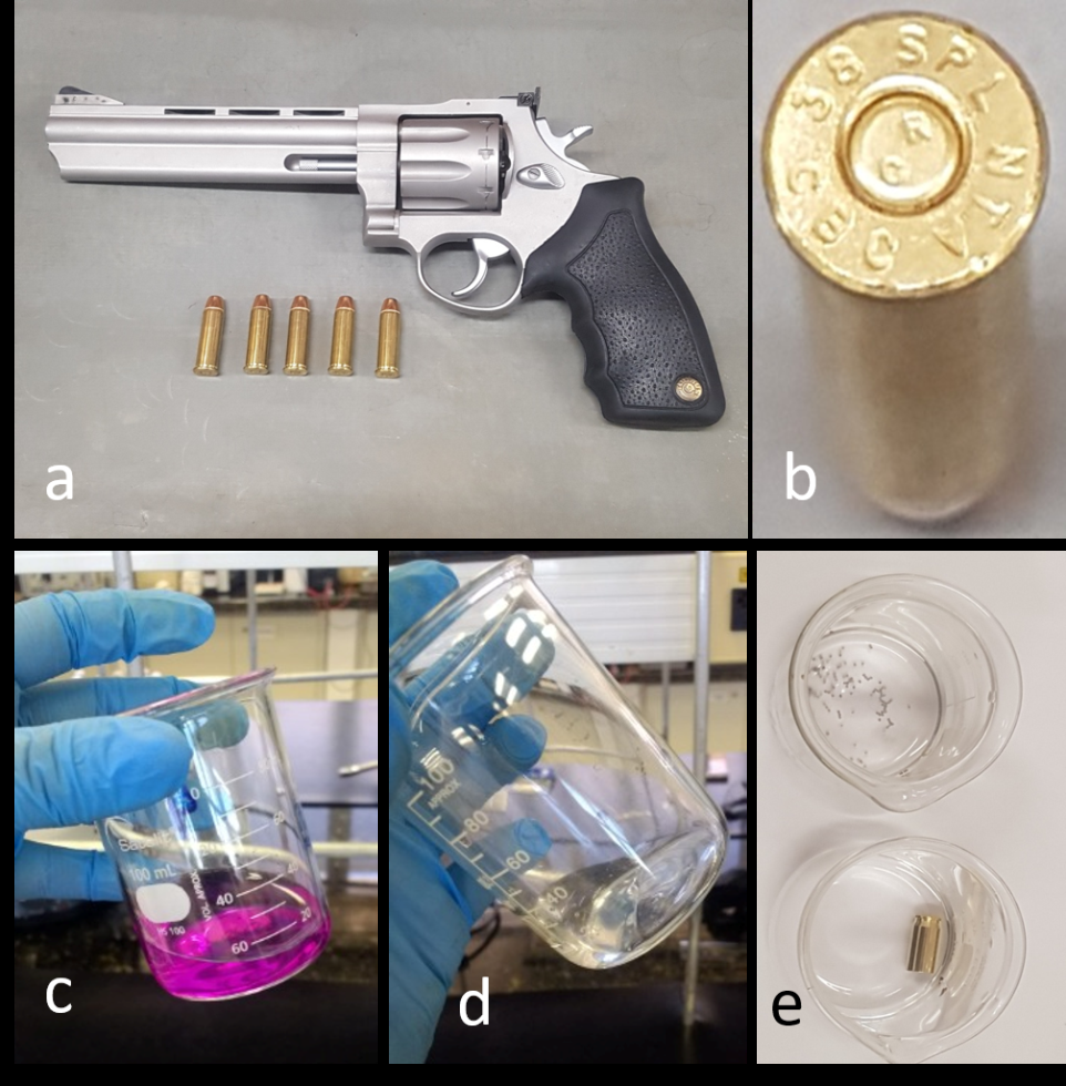
Figure 2: Description of the Revolver, Ammunition, and Initial Colorimetric Results Obtained with the Proposed Methodology. A) A Taurus RT 608 Revolver was Used for the Shooting; B) .38 Caliber NTA Ammunition was Used; C) Typical Purple Color Obtained During Colorimetric Detection of Nitrite Ions by Using Reagents Adapted from the Griess Test; D) Absence of Colorimetric Response for Nitrate Ions; E) Absence of Colorimetric Response for Nitrite Ions in the Chemical Components of the Powder and the Cartridge Before Firing.
Spectrophotometric analyses were conducted on a UV- Vis spectrophotometer, Marte model Spectro 560, coupled to a microcomputer. Flow injection analyses were performed by using a 4-channel peristaltic pump, Marte model MPV- 500, equipped with a portable handheld injector/switch and sampling loop consisting of a 150-µL Teflon tube. A flow cuvette with path length of 1.000 cm was used. The carrier solution consisted of 40% acetic acid solution in water at a flow rate of 1.4 mL min-1.
Experimental Procedure
Firearm shots were produced at a conventional shooting stand. Appropriate personal protective equipment, such as a simple filter mask, goggles, and a noise muffler, were used along all the experiments in the shooting stand.
To study the nitrite decomposition kinetics over time, nitrite residue produced by firing of a single ammunition was collected after a specific conditioning time interval; the weapon was placed in a conventional office drawer, protected from sunlight. Time intervals of 7, 9, 12, 15, and 32 days were used.
By using a standard sodium nitrite solution (1.2 x 10⁻⁵ mol L-1) previously treated with the colorimetric reagents, the maximum absorption wavelength (λmax) for this species was determined from 300 to 700 nm in the spectrophotometer. After λmax was determined, it was used in the spectrophotometric measurements in the proposed FIA system.
Results and Discussion
Colorimetric Analysis of Nitrite
The proposed methodology allowed colorimetric detection of nitrite even before the instrumental equipment is used. We observed a typical purple color when we added the reagents proposed in this methodology to the solutions containing nitrite (Figure 2c). In this case, the previous obtainment of a fast colorimetric positive result for nitrite allows this method to possible presumptive forensic tests.
Nitrate ions did not show colorimetric interference in any concentration range, as can be seen in (Figure 2d), where an aliquot of 500 mg of solid sodium nitrate was submitted to the colorimetric test. Additionally, the chemical components of the gunpowder and the fuse did not present free nitrite ions in their composition, given that we observed no purple coloration in the colorimetric tests (Figure 2e). In this case, it is important to mention that the proposed method is completely free of possible false-positive results from contamination of unburned gunpowder or propellants in general, that could perfectly occur in separation methods, such as gas chromatography or electrophoresis.
Spectrophotometric Analysis of Nitrite by the FIA System
The purple-colored product we obtained during the modified Griess test displayed a single absorption band in the visible region of the electromagnetic spectrum, with an absorbance peak located at 530 nm.
The duplicate injection of the standard nitrite solutions in the FIA system allowed us to obtain a fiagram with a baseline free of carryover effects (Figure 3a). The retention (tr) and washing (tw) times obtained for the FIA system when we injected more concentrated nitrite solutions were 53s and 34 s, respectively. The experimental results reported in (Figure 3a) indicated an analytical frequency of 20 h-1, which is consistent with the typical robustness of spectrophotometric FIA systems. As comparison, separation techniques usually present analytical frequencies ranging from 2 to 5 h-1, not mentioning higher consumption of reagents and solvents.
We used the average values of absorbance we obtained for each nitrite concentration to construct the analytical curve shown in (Figure 3b). The linear dependence of the spectrophotometric signal on the concentration of nitrite is given by equation 1:
4 1 3 2 2.07 10 6.78 10 µ − − − + = × A mol L NO x
Equation 1
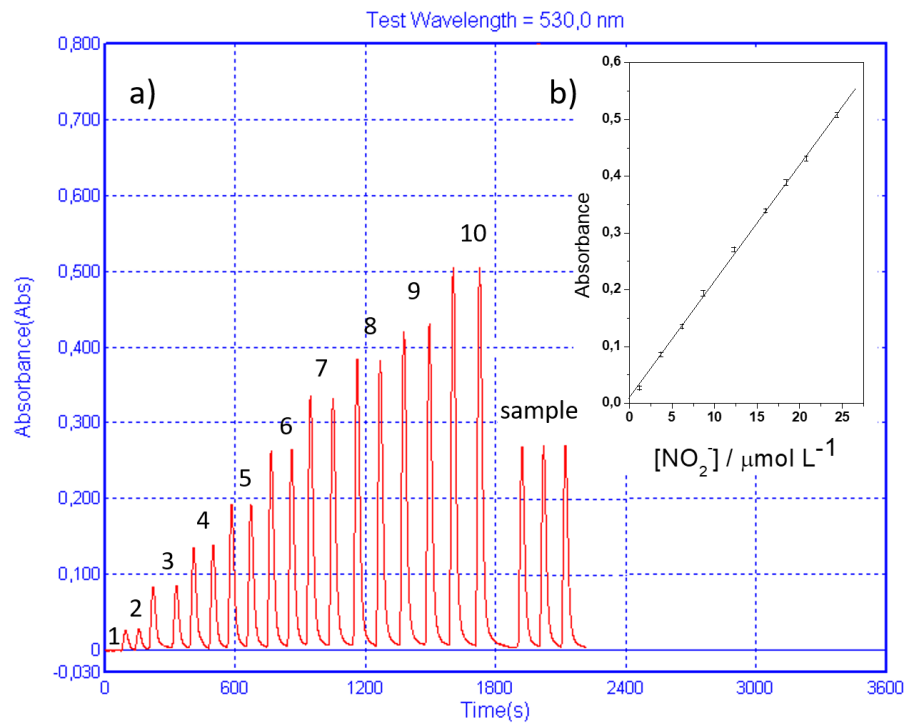
Linear regression of the analytical curve gave a linear correlation coefficient (r) of 0.9994. The standard deviation (SD) of the analytical curve was 3.17 x 10⁻³. The limits of detection (LOD = 0.46 µmol L-1) and quantification (LOQ = 1.56 µmol L-1) were calculated as 3 SD/m and 10 SD/m, respectively, where m is the analytical curve spectrophotometric sensitivity (2.07 x 10⁴ µmol-1 L). These results agreed with the results obtained by spectrophotometric methods regarding nitrite detection in samples of forensic interest (Table 1).
| Reagents | m (µmol L-1) | work range (µmol L-1) | LOD (µmol L-1) | LOQ (µmol L-1) | Ref. |
|---|---|---|---|---|---|
| sulfanilic acid+ 1-naphthylamine | 3.1 x 10⁴ | 2.1–42 | 0.58 | 1.93 | [5] |
| potassium iodide+ hydrochloric acid | 1.66 x 10⁴ | 1.31–84 | 0.52 | 1.73 | [7] |
| sulfanilic acid+ 3-aminophenol | 1.64 x 10³ | 105–525 | 31 | 95 | [8] |
| sulfanilic acid +naphthylethylenediamine | 2.07 x 10⁴ | 1.2–24.4 | 0.46 | 1.56 | This work |
Table 1: Comparison between the Analytical Parameters Reported in the Literature for Spectrophotometric Nitrite Detection in Samp
Investigation of Gunshot Recentness for Non- Toxic Ammunitions
The literature usually describes the effectiveness of nitrite colorimetric tests for examining gunshot recentness between 7 and 10 days [1–3] for conventional ammunitions. After this period, nitrite ions are commonly converted to nitrate, so there is no colorimetric response to the Griess test. In our study of the decomposition kinetics of nitrite from non-toxic ammunition, we verified that a single shot produced by using this ammunition generated persisting nitrite residues for at least 32 days in the weapon barrel (Figure 4).
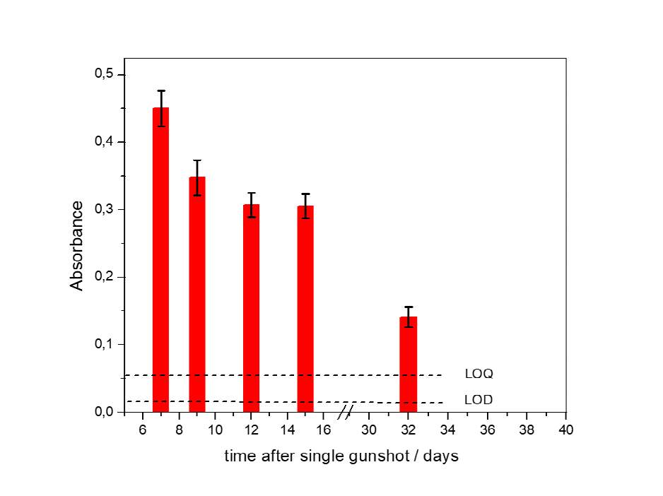
The persistence of nitrite ions in the barrel for a period higher than the maximum 10 days normally obtained in conventional ammunition can be explained by the stability of nitrite ions in the organic residues from the original components of non toxic ammunitions. It is important to remember that unburned residues of nitrocompounds such as nitrocellulose, nitroglycerine, and nitrotoluenes (2,6-dinitrotoluene, 2,4-dinitrotoluene) can encapsulate nitrite ions from the contact to air and water, decreasing its oxidation to the nitrate form, which presents no forensic interest for determination of time since the discharge.
Another interesting information about the nitrite ions present in the inner wall of the gun barrel consists on the fact that they do not present an accumulative characteristic, such as the metallic species in the hands of the shooter, for example [30]. When a single shot is produced, que amount of residues deposited in the barrel tends to a maximum value. This means that, if a subsequent shot is produced, the new gas flow tends to sweep the original amount of residues deposited and to produce a similar amount of new residues, with increases ranging from 1 to 3% for each additional shot. So, all experiments were carried out using single shots.
The deviation bars observed in the (Figure 4) in the absorbance measurements recorded in duplicate in this experimental stage were greater than the deviations we observed when we constructed the analytical curve. In fact, the analytical curve was obtained on the basis of intra- day measurements, whereas the recentness tests were based on inter-day measurements and therefore subject to greater influence of the environment, such as variations in temperature and relative humidity.
The results reported in the (Figure 4) also indicate that the detection and quantification of nitrite ions in GSR samples still possibilities the analysis of this forensic clue even for periods of time higher than 32 days, once the nitrite concentrations observed for this maximum 32 days-time limit presented higher than the LOD and LOQ concentration values [31].
Conclusions
Modifying the Griess test for detecting nitrite ions with sulfanilic acid and 1-naphthylethylenediamine in acetic acid medium promoted a considerable gain in spectrophotometric sensitivity in the visible spectrum region. The use of the proposed FIA system allowed us to obtain limit of detection in the order of 0.46 µmol L-1, which is the lowest value reported for this technique in forensic analysis to date. Additionally, the analytical frequency of 20 h-1 demonstrated excellent robustness in relation to conventional separation techniques as chromatography or electrophoresis, not mentioning the lower operational costs.
The considerably higher sensitivity of the proposed instrumental method in relation to the conventional colorimetric test for nitrite detection allowed the recentness exam time window to be expanded to 32 days, while providing the safety and reliability required for forensic analysis.
Conflicts of Interest
The corresponding author, in the name of all authors, declares that there is no conflict of interests in the proposed article.
Acknowledgements
The authors thank Dr. Cynthia Maria de Campos Prado Manso, who revised and edited this text.
Financial Support
The authors are thankful to CAPES (CAPES, PROCAD- SPCF – File number process 88887.613955/2021-00), FAPESP (File number 2022/12189-0), and CNPq (File number 302742/2022-0) for financial support.
References
-
Marinho PA, Guimarães LC, Velho JA (2015) Firearm shooting residue analysis, in: De Martinis BS, de Oliveira MF (Eds.), Experimental Forensic Chemistry, Cengage Learning, São Paulo, pp: 62-76.
-
de Oliveira MF, Ferreira B, Eleotherio IC. Velho JA (2019) Analysis of firearm shooting residues, in: Bruni AT, Velho JA, de Oliveira MF (Eds.), Fundamentals of Forensic Chemistry – 2nd edition, Millennium Publishers, Campinas, pp: 245-262.
-
de Oliveira MF, Bruni AT, Velho JA (2021) Química Forense, in: Velho JA, Geiser GC, Espíndula A (Eds.), Ciências Forenses – 4th edition, Millennium Editora, Campinas, pp: 187-210.
-
Jorge Gerardo FP, Ramírez Hernández GM, Rodríguez Méndez GC, Diaz EH, Murillo Leal JC, et al. (2021) The influence of contaminants in a shotgun barrel on the results of a griess test (a and b) for detecting Gunshot Residues. Revista Criminalidad 63(1): 61-76.
-
Petraco N, Yander M, Sardone J (1981) A method for the quantitative determination of nitrites in gunshot residue cases. Forensic Science International 18(1): 85-92.
-
Chaiendoo K, Ngamdee K, Limbut W, Saiyasombat C, Busayaporn W, et al. (2021) Gold nanoparticle-based cascade reaction-triggered fluorogenicity for highly selective nitrite ion detection in forensic samples. Microchem J 168: 106470.
-
Thipwimonmas Y, Jaidam J, Samoson K, Khunseeraksa V, Phonchai A, et al. (2021) A simple and rapid spectrophotometric method for nitrite detection in small sample volumes. Chemosensors 9(7): 161.
-
Jaluddin SN, Zain ZM, Abdul Halim MI, Safian MF, Abdul Rani MA, et al. (2021) Preliminary Evaluation of Gunshot Residue (GSR) Using 3-Aminophenol as a Substitute in Modified Griess Test. Indones J Chem 21(6): 1550-1559.
-
Berger J, Upton C, Springer E (2019) Evaluation of Total Nitrite Pattern Visualization as an Improved Method for Gunshot Residue Detection and its Application to Casework Samples. J Forensic Sci 64(1): 218-222.
-
Bailey JA, Casanova RS, Bufkin K (2006) A method for enhancing gunshot residue patterns on dark and multicolored fabrics compared with the modified Griess test. J Forensic Sci 51(4): 812-814.
-
Erol OO, Erdogan BY, Onar AN (2017) Nitrate and nitrite determination in gunshot residue samples by capillary electrophoresis in acidic run buffer. J Forensic Sci 62(2): 423-427.
-
Bohannan EW, Vangalen DA (1991) A sensitive electrochemical method for the analysis of nitrite ion and metals in gunshot residue. J Forensic Sci 36(3): 886- 892.
-
Promsuwan K, Kanatharana P, Thavarungkul P, Limbut W (2020) Nitrite amperometric sensor for gunshot residue screening. Electrochimica Acta 331(20): 135309.
-
Costa RA, Motta LC, Destefani CA, Rodrigues RRT, Santo KSE, et al. (2016) Gunshot residues (GSR) analysis of clean range ammunition using SEM/EDX, colorimetric test and ICP-MS: A comparative approach between the analytical techniques. Microchemical Journal 129: 339- 347.
-
Arouca AM, Vieira ML, Talhavini M, Weber IT (2022) Chromatographic analysis of byproducts from a non- toxic ammunition and a marked ammunition: an assessment of toxicity. Brazilian Journal of Analytical Chemistry 9(34): 138-161.
-
Cromie R, Newth J, Strong E (2019) Transitioning to Non- Toxic Ammunition: Making Change Happen. Ambio 48: 1079-1096.
-
Duarte A, Silva LM, Stori EM, Niekraszewicz LAB, Amaral L, et al. (2018) Characterization of Brazilian Ammunitions and their Respective Gunshot Residues with Ion Beam Techniques. Forensic Chem 7: 94-102.
-
Romano S, De Giorgio F, Onofrio CD, Gravina L, Abate S, et al. (2020) Characterisation of Gunshot Residues from Non-Toxic Ammunition and their Persistence on the Shooter’s Hands. Int J Legal Med 134: 1083-1094.
-
Blakey LS, Sharples GP, Chana K, Birkett JW (2018) Fate and Behavior of Gunshot Residue-A Review. J Forensic Sci 63(1): 9-19.
-
Goudsmits E, Sharples GP, Birkett JW (2015) Recent Trends in Organic Gunshot Residue Analysis-a Review. Trends in Anal Chem 74: 46-57.
-
McCord BR, Hargadon K, Hall K, Burmeister S (1994) Forensic Analysis of Explosives using Ion Chromatographic Methods. Anal Chim Acta 288(1-2): 43-56.
-
Hargadon KA, McCord BR (1992) Explosive Residue Analysis by Capillary Electrophoresis and Ion Chromatography J l Chrom 602(1-2): 241-247.
-
Northrop DM, Mac Crehan WA (1992) Sample Collection, Preparation, and Quantitation in the Micellar Electrokinetic Capillary Electrophoresis of Gunshot Residues. J Liquid Chrom & Related Technol 15(6-7): 1041-1062.
-
Oommen Z, Pierce SM, (2006) Lead-Free Primer Residues: A Qualitative Characterization of Winchester WinCleanTM, Remington/UMC LeadLessTM, Federal BallistiCleanTM, and Speer Lawman Clean Fire TM Handgun Ammunition. J Forensic Sci 51(3): 509-519.
-
Chang KH, Yew CH, Abdullah AFL (2015) Study of the Behaviors of Gunshot Residuesfrom Spent Cartridges by HeadspaceSolid-Phase Microextraction–Gas Chromatographic Techniques. J. Forensic Sci 60(4): 869- 877.
-
Meng HH, Caddy B (1997) Gunshot Residue Analysis–a Review. J Forensic Sci 42(4): 553-570.
-
Dalby O, Butler D, Birkett JW (2010) Analysis of Gunshot Residue and Associ-Ated Materials–a review. J Forensic Sci 55(4): 924-943.
-
Andrasko J, Stahling S (2003) Time Since Discharge of Pistols and Revolvers. J Forensic Sci 48(2): 1-5.
-
Gallidabino MD, Weyermann C (2020) Time Since Last Discharge of Firearms and Spent Ammunition Elements: State of the Art and Perspectives. Forensic Sci Int 311: 110290.
-
De Donato A, Gutz IGR (2005) Fast Mapping of Gunshot Residues by Batch Injection Analysis With Anodic Stripping Voltammetry of Lead at the Hanging Mercury Drop Electrode. Electroanalysis 17(2): 105-112.
- Forensic Implications of Adverse Drug Reactions in Schizophrenia A Case Series
- Narcotics and Digital Forensics: Bridging Crimes in the Digital Age
- Ethics in Forensic Psychiatry: Principles, Dilemmas, and Human Rights
- Impact of Acute Stress on Attentional Orienting to Social Cues
- Head Injury and Intracranial Hemorrhage in Western Region of Libya
- A Forensic Study on Handedness: Examination of Handwriting Features in Right and Left Handed Writers