Visualizing Cellular Material on Touched Surfaces
Touch deposits are comprised of oils, proteins, free-DNA molecules, nucleated cells, and cells undergoing programmed cell death. Previous studies have researched the use of fluorescent nuclear staining dyes, such as Diamond™ Nucleic Acid Dye (DD), to visualize DNA on an object, but not many studies have been conducted using other dyes, such as Nuclear Fast Red (NFR) and Trypan Blue (TB), on porous and non-porous substrates in comparison. The purpose of this research was to investigate and compare the effectiveness of the chemical enhancement reagents Diamond™ Nucleic Acid Dye, Nuclear Fast Red, and Trypan Blue for the visualization of DNA and cells. This research study shows that Diamond™ Nucleic Acid Dye and Nuclear Fast Red are effective at visualizing DNA on simulated forensic evidence. Trypan Blue was effective at visualizing apoptotic or necrotic cells with disrupted membranes. All three staining methods could be performed easily and are useful tools for forensic examiners to better understand the nature of the cellular material left behind after a single touch to the surface. Diamond™ Nucleic Acid Dye worked best on nonporous dark or clear surfaces while Nuclear Fast Red and Trypan Blue dyes worked well on all surfaces with high contrast-colors.
Abbreviations
NFR: Nuclear Fast Red; TB: Trypan Blue; IRB: Institutional Review Board; PI: Propidium Iodide.
Introduction
Touch DNA, or trace DNA, is DNA that has been transferred to a surface when epithelial cells, or skin cells, have been shed or deposited. This can occur when there is physical contact between the donor and the object being handled. In the case of touch deposits, DNA may originate from the donor as shed corneocytes (non-living cells) or nucleated cells (keratinocytes) from the hands, cell types that have been transferred to the donor’s hands, or it may exist in the form of residue from fragmented cells (cell-free DNA) [1]. All these forms of DNA and cells are not visible to the naked eye. Histological staining is used in pathology and cell biology to highlight different areas of the cell for study. This concept was applied to forensic evidence processing techniques to document mock evidence biological material adherent to porous and nonporous surfaces prior to potential downstream DNA profiling techniques.
In forensic science, DNA profiling encompasses techniques utilized by scientists to obtain an individual’s genetic information that may hold evidentiary value or reference information for the case [2]. When an item is submitted to a forensic lab for DNA testing, it is up to the scientist to determine intuitively where the trace DNA may be located on the item of evidence. Current methods of DNA collection for trace DNA may result in partial or no DNA profiles due in part to the lack of visualization of the evidence as it is microscopic and latent [3]. Pre-screening an item for touch DNA with dyes may provide a higher probability that a successful DNA profile will be obtained, since an area where DNA has been visualized can be targeted for collection.
Diamond™ Nucleic Acid Dye can interact with genomic DNA and cause fluorescence when visualized with a 480 nm excitation source and 510 nm emission filter [4]. Diamond™ Nucleic Acid Dye has been effective at locating trace DNA on select substrates under controlled conditions and had no effect on downstream DNA analysis, but issues with background fluorescence, non-specific staining, and absorbance of light on black surfaces hinder its ability to be incorporated into routine forensic casework [5]. Nuclear Fast Red dye stains nuclei containing cells a pink color. It has already been established as a commonly used stain in forensic science to test for the presence of spermatozoa in screening samples for sexual assault casework. Trypan Blue stains dead cells a blue color by binding proteins in membrane-compromised cells. It has previously been used for the quantification and evaluation of cell viability since it is a dye that is excluded from living cells. Diamond™ Nucleic Acid Dye has been used to visualize DNA in agarose gel electrophoresis methods but only recently explored for examination of cellular material on forensic surfaces. The selection of these three dyes was to establish the effectiveness of dye-based imaging on mock evidence surfaces, both porous and nonporous, and to have choices based on surface type and for color contrast like the traditional approach used to enhance latent fingerprints with powders in crime scene imaging.
Studies on the Use of Diamond™ Nucleic Acid Dye
In a study conducted by Champion et al., the exploration of Diamond™ Nucleic Acid Dye (DD) solution visualization on eight types of substrates was conducted [1]. This study used three non-absorbent substrates (glass, plastic bank note, tooth) and five absorbent substrates (postage stamp, cigarette, black cotton fabric, paper bank note, matchstick). The study had donors deposit their DNA onto each of the substrates and the samples were visualized using the Dino- Lite EDGE Am4115T-GFBW digital fluorescence microscope [1]. The study found that DD solutions prepared with 75% EtOH were more effective on non-absorbent substrates, while DD solutions prepared with water were more effective on absorbent substrates.
A study conducted by Cook R, et al. [6] used blood, semen, and saliva, and other samples from volunteers, as well as stock DD in 75% ethanol solutions or 0.1% / 0.01% Triton X-100 solutions [6]. Fluorescence imaging was followed by DNA testing on the samples. DD worked well visualizing trace DNA under controlled conditions, but when applied to operational casework, issues with fluorescence and absorption arose [6].
A study conducted by Hughes DA, et al. [7] applied varying ethanol DD concentration solutions via a micropipette and a spray bottle to non-porous substrates, including aluminum foil, glass microscope slides, clear polypropylene, clear acrylic, white PVC material, and black crystalline silicon after DNA deposition. The substrates were examined under the portable Dino-Lite fluorescence microscope and a Zeiss fluorescence microscope. The spray DD application was the best method for maximum surface coverage and identification of DNA [6]. Researchers noted that while the Zeiss fluorescence microscope did a great job at visualization, it was not a portable microscope. They found that while the Dino-Lite microscope is portable, it was not able to be used for larger surface area imaging but was nonetheless effective with locating the fluorescence of DNA [7].
Research conducted by Kanokwongnuwut P, et al. [8] measured how well DD performed when visualizing cellular material deposited from different positions on the hand [8]. Researchers used participants who were high, low, and medium DNA shedders and had them deposit their DNA on sheets of plastic, which were then sprayed with a 75% ethanol DD solution. The handprints were visualized using the Dino-Lite microscope and DinoXcope software for MacOS [8]. The fingertips demonstrated the highest amount of transferred cellular material.
Studies on the Use of Trypan Blue
Trypan Blue (TB) is a common cell staining technique often used in the medical field. In research conducted by Kerschbaum HH, et al. [9] TB was used to identify how the dye was incorporated into nonviable cells [9]. Researchers studied this phenomenon by applying Trypan Blue to the cells to create labelled cell structures and proteins. The researchers found that dead cells were able to show even and intense Trypan Blue staining [9]. They were also able to observe even, but faint, Trypan Blue labelling in the nucleus and cytoplasm of viable, or live, cells [9].
In another study, Chan LL, et al. [10] explored the formation of dim and diffuse objects in Jurkat cell samples that have been stained with TB [10]. The study used 0.4%, 0.2%, and 0.1% working concentrations of Trypan Blue to stain the Jurkat cells, and used propidium iodide (PI), a fluorescent staining dye, for comparison. The study found that live cells were not stained, while the dead cells were stained by Trypan Blue and appeared blue, dark, and compact [10].
Studies on the Use of Nuclear Fast Red
Nuclear Fast Red dye has often been used in the forensic community for sperm cell staining. Researchers use the technique of Christmas tree staining to microscopically examine each semen sample by applying Nuclear Fast Red followed by picroindigocarmine (PICS) [11]. The Nuclear Fast Red dye caused the nuclei of the sperm cells in the samples to be stained red in color, while the picroindigocarmine stained the cytoplasm and tails of the cells green.
Another study conducted by Allery JP, et al. [12] determined which type of cytological staining for spermatozoa detection was the most effective [12]. The study compared hematoxylin- eosin, Christmas tree, and alkaline fuchsin staining. Researchers found that when compared with alkaline fuchsin staining, Christmas tree and hematoxylin-eosin staining were better methods for microscopic sperm detection [13, 14].
Studies on Factors that Affect Trace DNA Collection
The amount of trace DNA that can be collected from a surface depends on the type of substrate that it is deposited on. In a study conducted by Bonsu DOM, et al. [13] the persistence and recovery of DNA on metal surfaces was explored when collecting DNA samples [13]. Due to an attraction between the ions in the metal surfaces and the negatively charged DNA molecules, it can cause an ionic bond to form, which can make it difficult to recover DNA [13]. The study emphasized that rough surfaces have a better DNA recovery rate than smooth surfaces.
Another study conducted by Alketbi SK, et al. [14] investigated how surface type affected the amount of trace DNA recovered. The study had participants deposit DNA with their index, middle, and ring fingers of both hands on a variety of non-porous and porous surfaces, including glass, wood, banana skin, copier paper, and textured plastic [14]. The DNA was recovered 30 minutes after deposition and profiled. The study found that the surface DNA deposited on had a great impact on the amount of DNA collected. Non-porous surfaces yielded the highest amount of DNA recovered, while the porous surfaces yielded the lowest amount of DNA [14].
Methods
Approval from the University of New Haven’s Institutional Review Board (IRB) was obtained prior to the initiation of the human subject research for this study. Two experiments were conducted in this study. Experiment 1 surfaces were selected to simulate commonly handled types of bags and purses that could be relevant evidence in purse snatching scenarios where the perpetrator’s DNA would be highly desirable as recovered DNA evidence from the straps or bag itself. Experiment 2 surfaces were selected to simulate common ligature, binding and restraint materials that would be used to bind a victim during the commission of a crime and would have touch DNA from the perpetrator on the surfaces. These surfaces are commonly ropes, cords and large conjoined zip-ties but have large surface areas that are nonspecifically swabbed to collect DNA and the dyes on these surfaces could be useful to focus DNA collection efforts. Focused biological evidence collection yields a concentrated DNA sample for improved profile recovery.
Nuclear Fast Red and Trypan Blue Staining- Experiment 1
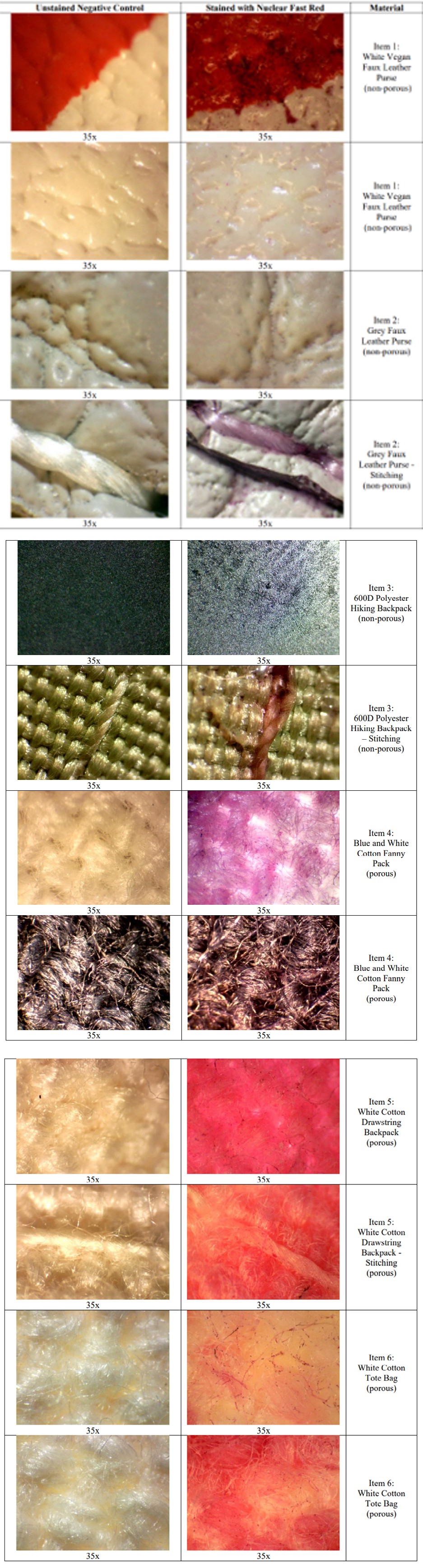
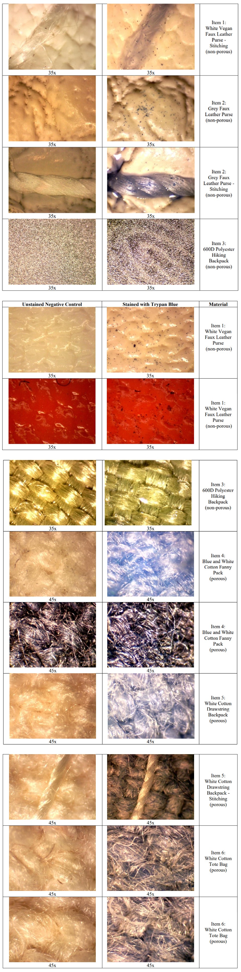
Substrate Selection: Three non-porous items and three porous items were selected for use to simulate evidence from a purse or bag theft case. The three non-porous items were (1) a white, vegan faux leather purse, (2) a grey faux leather purse, and (3) a yellow green 600D polyester hiking backpack. The three porous items were (1) a blue and white colored cotton fanny pack, (2) a white, cotton drawstring backpack, and (3) a white, canvas tote bag. Prior to their use, portions of each bag were designated for the application of each dye using a marker. The application areas were chosen for locations that would be considered common points of contact on the item. Prior to DNA deposition, each substrate was placed into a UV Crosslinker (Spectrolinker XL-1500, 120 μJ/cm2) for thirty minutes to remove any exogenous DNA that may have been on the items from the manufacturing process.
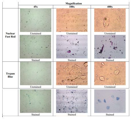
Controls: One positive control on a glass slide was performed for each of the dyes. Each control was analyzed under a compound light microscope at 45x, 100x, and 400x magnification to compare the unstained and stained appearance of the cells. Each of the substrates were examined and imaged under the binocular stereomicroscope (VWR) prior to DNA deposition to serve as a negative control. The substrates were imaged using the Dino-Eye Eyepiece Camera (AM7025X) and associated DinoXcope software for Mac.
Dye Preparation: The Nuclear Fast Red (G-Biosciences®) and 0.4% Trypan Blue (Thermo Fisher Scientific) reagents were purchased as premade solutions.
Trace DNA Deposition: One female donor was selected and used throughout the study to deposit DNA onto each substrate. Prior to each DNA deposition, the donor was asked to wash her hands for thirty seconds using soap and warm water. Immediately after washing and drying the hands, the donor was asked to move the thumb, index, and middle fingers of the left hand across her face to collect the DNA. She was then asked to place each of those fingers at the designated locations on the item for ten seconds before lifting them off the surface. The stain was immediately applied to each of the fresh touch deposits. Each finger deposit was considered representative and treated as three individual fingerprints and examined on each surface.
Nuclear Fast Red and Trypan Blue Staining and Visualization
Nuclear Fast Red or Trypan Blue dye was applied to each touch deposit via a 1000 uL micropipette. Enough NFR or TB was applied to completely cover the touch deposit. The stain was allowed to sit for ten minutes before it was de-stained with molecular biology grade water in a sink. Each area was de-stained until the rinse water residue was clear. Each touch deposit was visualized under the stereomicroscope (Vista Vision, WF10X/22 eyepiece lens), starting at a magnification of 35x. If cells were hard to visualize the magnification was increased to 45x, 100x or 400x. Using the Dino-Lite USB Eyepiece Camera (AM7025X) and associated DinoXcope software for Mac, images were captured and analyzed for cell morphology and adherence. Cell counts were made visually by counting the number of cells in microscopic fields of view in triplicate and averaged per fingerprint per surface.
Diamond Dye Staining and Visualization- Experiment 2
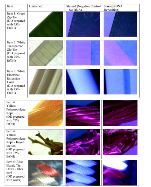
Substrate Selection: There were four non-porous items and one porous item that were chosen to be used in this study to simulate items of evidence that could be used as bindings and/or ligatures seen in forensic casework. The four non- porous items were a 19.5cm long green plastic zip tie, a 10cm long white transparent plastic zip tie, yellow polypropylene rope, and a white electrical extension cord. The one porous item was a “Haul Master” elastic tie down or bungee cord. All the items were sterilized by spraying with DNA ExitusPlus followed with 70% ethanol solution to remove any exogenous DNA that may have been deposited on the items from the manufacturing or handling of the items. The application areas were chosen based upon where a perpetrator may touch the item when tying it around a victim as a binding or ligature. These items were selected for screening with the newer fluorescent dye due to the difficulty in observing TB on dark surfaces and to address imaging issues with shiny and clear or white surfaces with NFR.
Controls: One positive control and one negative control were performed for each of the DD solutions. A fingerprint was deposited on a glass slide for the two positive controls and was then sprayed with either DD and EtOH or DD and water solutions. Each control was examined and imaged at 20x magnification under a stereomicroscope with a Dino- Eye Microscope Piece Camera (AM7025X) and the associated DinoXcope software for macOS (data not shown).
Dye Preparation: Two DD solutions were prepared for this study. The DD (Promega Corporation) was diluted from 10,000X stock concentration to a 20X working solution in a dark room. The solution for the nonporous substrates was prepared by diluting 5µl of DD in 9.5ml of 75% molecular- grade EtOH. The solution for the porous substrates was prepared by diluting 5µl of DD in 9.5ml of molecular-grade water. Both solutions were put into misting travel spray bottles (Elanor) and were labeled accordingly. The body of both bottles were wrapped entirely in aluminum foil to prevent light exposure to the DD solutions.
Touch DNA Deposition: One female donor was selected for the study to deposit DNA onto each substrate to ensure consistency of DNA deposition. The donor was asked to place her thumb, index finger, and middle finger at the designated locations on the item for 10 seconds before lifting off. The appropriate DD solution was applied to that designated area of DNA deposition as well as the designated area for the negative control on the item. Each finger deposit was considered representative and treated as three individual fingerprints and examined on each surface.
Diamond Dye Staining and Visualization: The DD solution was applied to each item in a darkened room immediately after DNA deposition. A plastic pump spray bottle was used to apply the DD solution and was held about 6 inches away from the application area on the item. Each designated area for DNA deposition and designated area for the negative control were visualized at 20x magnification under the stereomicroscope using a Nightsea SFA Light Base as an alternate light source. Images were captured using the Dino- Eye Piece Camera (AM7025X) and the associated DinoXcope software for macOS.
Hazards and Safety Precautions: Diamond Dye is cytotoxic and genotoxic as a concentrated stock but nontoxic when diluted (1:10,000) as recommended. Trypan Blue may be a carcinogen. Nuclear Fast Red may cause eye damage and skin burns. Eye and face protection and gloves are recommended for use with all three dyes.
Results
For the negative and positive control slides, ridge marks from oils and secretions of the unstained fingerprints were apparent at all magnifications. The nuclei inside the cells were visible for NFR magnification and for TB, the cytoplasm was stained blue (Figure 1).
For the white leather purse (Item 1), deposited DNA could clearly be observed with NFR staining (Figure 2). The contrast of the stained cells with the background color of the purse was better for the white colored surfaces of the purse. For the red colored surfaces of the purse, cells were still able to be visualized, but the contrast was lower. For the grey leather purse (Item 2), trace DNA could also be clearly observed (Figure 2). Much of the cellular material was visualized in the indentations of the material, rather than directly on the surface. For the 600D polyester hiking backpack (Item 3), trace DNA could not be observed for the black colored surfaces (Figure 2). Ridge patterns from the fingerprint deposit on the dark colored surface were visible with oblique lighting, but no cells were able to be identified. For the yellow green surfaces of the backpack, trace DNA could be clearly observed, but the number of cells that could be identified were limited (Figure 2). While some cells could be identified on the material pattern, most of the cells were found on the stitching. There was a good contrast between the stained cells and the background due to the lighter color of those regions of the backpack. For the blue and white cotton fanny pack (Item 4), trace DNA was clearly observed, but mainly for the white colored material (Figure 2). For the white colored parts of the item, the cells could be observed on the individual fibers, and there was a good contrast between the stained cells and the background. For the white cotton drawstring backpack (Item 5), trace DNA was clearly observed (Figure 2). There was a high contrast between the stained cells and the background of the substrate. Cells were also able to be observed on the stitching of the backpack (Figure 2). For the white cotton tote bag (Item 6), trace DNA was also clearly observed (Figure 2). Like the drawstring backpack, there was high contrast between the cells and the background. Cells could be seen on individual fibers.
Trypan Blue
Trypan Blue performed very well with high cell counts and high contrasts for the non-porous substrates. For the white faux leather purse (Item 1), the cells were clearly observed on both the white and red colored surfaces (Figure 3). Although the white surface had better contrast, the red surface did provide high contrast that allowed the blue stained cells to be clearly identified. For the grey faux leather purse (Item 2), cells were clearly visualized, even for the indented areas of the substrate (Figure 3). Likewise, cells were observed on the stitching of the purse, including both the stitch itself and the surrounding area (Figure 3). For the 600D polyester hiking backpack (Item 3) the black colored parts of the item provided little cell visualization. Although the fingerprint ridge marks were visible under the microscope, due to the dark color of the material, cells were not able to be visualized because of a lack of contrast with the dye (Figure 3). The yellow green surfaces of the backpack had clear cell visualization due to high contrast with the dye, but the number of visualized cells were limited. For the blue and white cotton fanny pack (Item 4), cells were visualized on both the blue and white colored surfaces, but due to the porous nature of the surface, de-staining of the dye was difficult, and some of the surface backgrounds blended with the color of the dyed cells (Figure 3). On the white colored portions of the fanny pack, while cells were visualized, they were more difficult to discern because the background had absorbed the dye. On the dark blue colored portions of the fanny pack, cells were also visualized, but they were difficult to see (Figure 3).
For the drawstring backpack (Item 5), cells were observed on both the backpack material and the stitching of the straps (Figure 3). There was greater cell visualization on the stitching fibers than the material of the main body of the bag. For the canvas tote bag (Item 6), there was high contrast between the light color and the darkness of the bag surface (Figure 3).
Diamond™ Nucleic Acid Dye
Touch DNA visualization with DD prepared with 75% EtOH was shown to be successful for all the nonporous items (Figure 4). An abundance of fluorescent cells was visualized for both zip ties (Items 1 and 2) in the designated areas for touch DNA deposition. There appeared to be some minimal auto-fluorescence for the white transparent zip tie (Item 2) but that did not affect the visualization of the fluorescent cells. For the green zip tie (Item 1), the cells fluoresced a yellow color. For the white transparent zip tie (Item 2), the cells fluoresced into a white color. Visualization of cells deposited on the white electrical extension cord (Item 3) was more challenging due to the fluorescence of the white cord itself. Additionally, the indentations on the cord and the bumpy texture of the surface of the outlet box at the end of the cord most likely caused reflectance from the light source, further contributing to the difficulty of visualizing the fluorescent cells. However, there were still a small number of fluorescent cells that were able to be seen on both the cord and outlet box, and they fluoresced a white color. For the yellow polypropylene rope (Item 4), visualization of the fluorescent cells was successful due to the high contrast between the substrate and fluorescing cells. The yellow polypropylene rope (Item 4) appeared as a reddish-purple color under the alternate light source and the cells fluoresced a bright yellow color, making visualization easy. The fraying of fibers on the rope posed no challenges as the fluorescent cells were able to be as easily visualized. The elastic tie down (Item 5) had both nonporous and porous surfaces which were sprayed with the appropriate DD solutions. The nonporous metal hooks for both tie downs were sprayed with the DD prepared with 75% EtOH solution while the porous vinyl fiber cord was sprayed with the DD prepared with water solution. Fluorescent cells were visible on the hooks for both the blue and yellow elastic tie downs (data not shown). There was high contrast between the black colored hooks and the yellow-colored fluorescing cells, which made it easy to observe the fluorescent cells. For the blue elastic tie down (Item 5), very few fluorescent cells were observed on the vinyl fiber cord. This may have been due to the background fluorescence of the white colored fibers within the cord as the cells also fluoresced in a white color or lack of cell adherence.
Discussion and Conclusion
For the microscopic visualization of cells and DNA on simulated evidence, this study illustrates the concepts of: (1) dye incorporation into shed epithelial cells and nuclei, (2) a substrate preference for different dyes based on contrast, (3) the differential adherence of cells to porous and nonporous surfaces, and (4) reflectance, absorbance and fluorescence concepts with use of an alternate light sources and a cellular dye method. Overall, these three cellular stains are relevant and effective in visualizing trace DNA on simulated mock crime scene evidence from a latent fingerprint and can be used for focusing an analyst’s efforts to collect from areas suspected to have cellular material and DNA. In this survey of three cellular stains, all were successful on light colored backgrounds. Nuclear Fast Red dye compared to Trypan Blue dye was superior for visualization of cells on both porous and nonporous surfaces (Table 1).
| Substrate | Ave. Cell Count (cells/mm) | ||
|---|---|---|---|
| NFR | TB | Surface Type | |
| White leather purse | 119 | 165 | nonporous |
| Grey leather purse | 35 | 113 | nonporous |
| 600D polyester hiking backpack | 11 | 13 | nonporous |
| Blue and white cotton fanny pack | 30 | 7 | porous |
| White cotton drawstring backpack | 60 | 8 | porous |
| White cotton tote bag | 70 | 7 | porous |
Table 1: Estimated Cell Counts for Nuclear Fast Red and Trypan Blue Dyes and Surfaces.
Trypan Blue dye was most effective on light-colored nonporous surfaces due to the background fabric substrates absorbing some of the dark dye and a resultant reduction of contrast between surface and dye on porous surfaces. Diamond Dye worked well on both light and dark surfaces, but the reflectance and auto-fluorescence of some surfaces made it challenging to capture some of the microscopic images. Diamond Dye was most efficient for visualization of cellular material on the simulated mock evidence for dark and nonporous surfaces, providing high contrast images, but due to the brightness of the dye fluorescence, it does allow for visualization and imaging touch deposits on even white- and yellow-colored substrates.
One limitation to this study includes the lack of data on the impact of dye on the quality of the STR profile after the DNA extraction, quantitation, PCR amplification and capillary electrophoresis steps. There are references to support no or little impact on the ability to generate the DNA profile after the dye has been used to visualize cells on slides but little or no published validation studies on the PCR aspects such as stutter, allele drop-out and degradation [5, 15, 16]. Another limitation deals with environmental factors such as humidity, light, time and temperature that also have not been extensively studied to determine the impact on the use of these dyes for the visualization of cells on evidence substrates for dye performance except for post-blast IED devices and water submerged surfaces with Diamond™ Nucleic Acid Dye [17, 18]. The shedder status (genetic predisposition to leave trace cells behind on the surface) of the two donors used in this study was unknown but held as a constant for each experiment to reduce variability. However, variability in cell counts and results could occur when these methods are applied to authentic casework samples with unknown donors and varying shedder status. Additional replicates with a greater number of donors with defined shedder status and more surfaces are goals of future studies.
The advantage of each dye includes the relative ease of use with both NFR and TB being provided as pre-made ready-to-use solutions and stored at room temperature. The disadvantage to both Nuclear Fast Red and Trypan Blue dyes is that a destaining process is required that may result in some loss of cellular material from the surface prior to imaging. Diamond™ Nucleic Acid Dye is provided frozen and must be diluted with either ethanol or water prior to use and is effective for up to one week in solution if stored at -20C. No destaining is required for this dye before imaging so this method takes less time than the NFR and TB dye methods. There is also a small, estimated cost difference when purchasing these dyes for use [Trypan Blue ($30USD/100mL); Nuclear Fast Red ($80USD/100mL); Diamond™ Nucleic Acid Dye ($20USD/100mL of ethanol or water)]. Since this is a microscopy-based approach, there is little additional cost for forensic laboratories that already have stereomicroscopes for evidence examination, however, the cost of the Dino-Eye Eyepiece Camera (AM7025X) is approximately $650 USD, and this includes a free downloadable software package for capturing images. The Nightsea SFA Light Base (estimated $1600USD) is important to use as an alternate light source in conjunction with the Diamond™ Nucleic Acid Dye to visualize the fluorescence of the dye. Overall, these cellular dyes and equipment are effective for imaging residual cells that remain on porous and nonporous surfaces that are likely to be encountered as forensic biology evidence from touch deposits.
Acknowledgements
Thank you to the University of New Haven for funding this research.
Conflicts of Interest
The authors declare no conflicts of interest.
References
-
Champion J, Kanokwongnuwut P, Van Oorschot RAH, Taylor D, Linacre A (2021) Evaluation of a fluorescent dye to visualize transfer DNA on various substrates. Journal of Forensic Sciences 66(4): 1435-1442.
-
Burrill J, Daniel B, Frascione N (2019) A review of trace “touch DNA” deposits: Variability factors and an exploration of cellular composition. Forensic Science International: Genetics 39: 8-18.
-
David Tan WC, Stasi A, Dhar BK (2022) Forensic DNA profiling in the southern border provinces of Thailand: Ethical and regulatory issues. Forensic Science International 336: 111322.
-
Mapes AA, Kloosterman AD, Poot CJ (2015) DNA in the criminal justice system: The DNA success story in perspective. Journal of forensic sciences 60(4): 851-856.
-
Haines AM, Tobe SS, Linacre A (2016) Optimization of Diamond Nucleic Acid Dye for quantitative PCR. BioTechniques 61(4): 183-189.
-
Cook R, Mitchell N, Henry J (2021) Assessment of Diamond™ Nucleic Acid Dye for the identification and targeted sampling of latent DNA in operational casework. Forensic Science International: Genetics 55: 102579.
-
Hughes DA, Szkuta B, van Oorschot RAH, Conlan XA (2022) “Technical Note:” Optimization of Diamond Nucleic Acid Dye preparation, application, and visualization for latent DNA detection. Forensic Sci Int 330: 111096.
-
Kanokwongnuwut P, Kirkbride KP, Lincare A (2019) Detection of cellular material within handprints. Forensic Science International: Genetics Supplement Series 7(1): 194-196.
-
Kerschbaum HH, Altug B, Schurz M, Oberascher K, Bresgen N (2021) Trypan Blue – Adapting a dye used for labelling dead cells to visualize pinocytosis in viable cells. Cellular Physiology and Biochemistry 55(S1): 171- 184.
-
Chan LL, Rice WL, Qiu J (2020) Observation and quantification of the morphological effect of trypan blue rupturing dead or dying cells. PLoS ONE 15(1): e0227950.
-
Leubitz SS, Savage RA (1984) Sensitivity of picroindigocarmine/nuclear fast red (PIC/NF) stain for detection of spermatozoa: a serial dilution study of human ejaculate. American journal of clinical pathology 81(1): 90-93.
-
Allery JP, Telmon N, Mieusset R, Blanc A, Rougé D (2001) Cytological detection of spermatozoa: comparison of three staining methods. Journal of forensic sciences 46(2): 349-351.
-
Bonsu DOM, Higgins D, Austin JJ (2020) Forensic touch DNA recovery from metal surfaces-A review. Science & Justice 60(3): 206-215.
-
Alketbi SK, Goodwin W (2019) The effect of surface type, collection, and extraction methods on touch DNA. Forensic Science International: Genetics Supplement Series 7(1): 704-706.
-
Di Martino D, Giuffrè G, Staiti N, Simone A, Le Donne M, et al. (2004) Single sperm cell isolation by laser microdissection. Forensic Sci Int 2(146) Suppl: S151-S153.
-
Strober W (2015) Trypan Blue Exclusion Test of Cell Viability. Curr Protoc Immunol 2(111): 1-3.
-
Nolan M, Handt O, Linacre A (2023) Persistence of cellular material after exposure to water. Journal of Forensic Sciences 68(6): 2128-2137.
-
Martin B, Kanokwongnuwut P, Taylor D, Kirkbride KP, Armitt D, et al. (2020) Successful STR amplification of post-blast IED samples by fluorescent visualisation and direct PCR. Forensic Science International: Genetics 46: 102256.
- Forensic Implications of Adverse Drug Reactions in Schizophrenia A Case Series
- Narcotics and Digital Forensics: Bridging Crimes in the Digital Age
- Ethics in Forensic Psychiatry: Principles, Dilemmas, and Human Rights
- Impact of Acute Stress on Attentional Orienting to Social Cues
- Head Injury and Intracranial Hemorrhage in Western Region of Libya
- A Forensic Study on Handedness: Examination of Handwriting Features in Right and Left Handed Writers