Preliminary Study on Human Dose of X and Γ Flaw Detection Accident Based on FLUKA
Introduction
X and γ-ray flaw detection technology are widely used in industry field, which can effectively ensure the quality and safety of products and other equipment of active nuclear facilities [1, 2]. However, radiation is a double-edged sword, and X, γ-ray flaw detection also cause incidents or accidents from time to time [3, 4, 5]. Once the occurrence of a radiation accident, the estimation of human exposure dose was the first problem that doctors must face in the rescue. In order to solve this problem, FLUKA simulation software was employed to study the energy deposition and dose of each organ under the typical conditions assumed that human body were irradiated by X or γ rays. Preliminary research on the dose distribution has been conducted, and the results have important reference significance for the dose assessment of accident-exposed personnel.
Objects and Methods
Mock object
The simulation was based on the Chinese male reference human body model developed by Tsinghua University [6, 7]. The phantom, which was 1.76 meters tall and 60 kg in weight, contained 103 tissues or organs. This phantom had been also applied widely in the field of radiation protection [8, 9], in order to study on the energy deposition and dose distribution of various tissues and organs of the human body who was irradiated by X-ray, Cs-137 and Co-60 radiation sour.
Calculation Method
In this study, we firstly converted the reference human body model into the voxel structure files that could be recognized by FLUKA [6, 7, 10], and then the simulated space structure and human body materials were constructed using the flair program of the FLUKA software [11]. When modeling, circular radiation fields of 200 keV and 300 keV X-ray were set to irradiate the human chest with a radiation field diameter of 10 cm. Besides, the distance between the Cs-137 or Co-60 radiation source and the human body was set to be 1 cm, and the surrounding space was set as an air sphere with a diameter of 1000 m. The number of simulated particles was 1×10⁸.
Quality control
After the Fluka-based model was successfully debugged, 0.10, 0.15, 0.20, 0.30, 0.40, 0.50, 0.60, 0.80, 1.00, 2.00, 4.00, 6.00, and 10.00 MeV X-ray was simulated to isotropically irradiate human body model. The simulation results were compared with that of reported work based on GEANT4 [4] and the deviation was less than 5%.
Results
Dose distribution
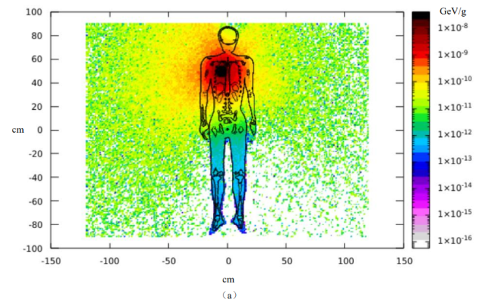
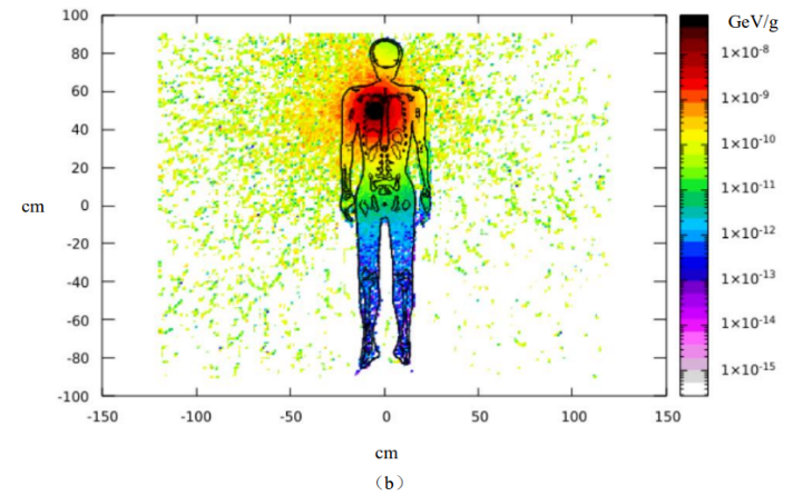
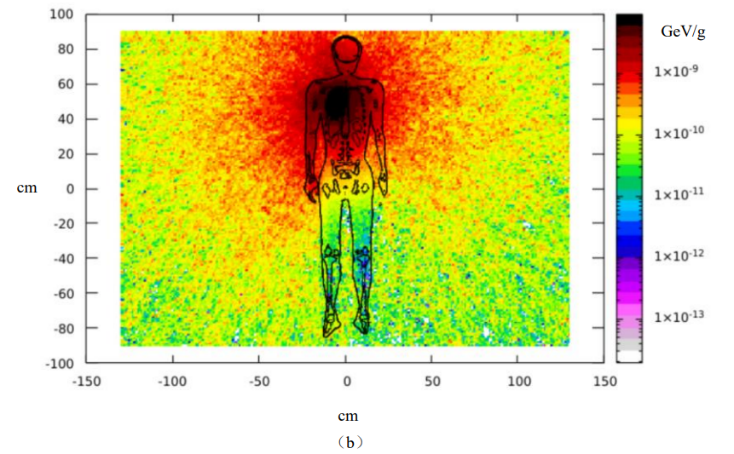
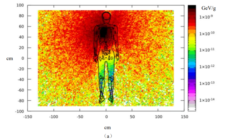
The 200 keV X-ray, 300 keV X-ray, Cs-137 and Co-60 radiation sources were respectively simulated to directly irradiate the human body. Then, the spatial distributions of absorbed dose for human body could be got, which were shown in Figure 1 and Figure 2, respectively. When 200 keV and 300 keV X-rays irradiated human chest, the energy deposition was high in the tissues and organs of the chest region. Among them, the absorbed dose of the lung was the highest, and the area in the irradiation field reached the maximum, followed by the heart and liver that were closer to the lungs. When Cs-137 and Co-60 radiation sources were simulated to irradiate the human body at close range, the spatial distribution of the absorbed dose of the human body was generally the same as that of 200 keV and 300 keV X-rays irradiation.
Energy Deposition and Absorbed Dose in Major Organs
The simulated results of energy deposition in 103 tissues and organs of the human body were statistically analyzed. The top 5 organs of energy deposition were lung, heart, muscle, soft tissue, liver, and skin. The energy deposition of the five organs and the whole body was listed in Table 1, whose unit was GeV/photon. The top 5 organs of energy deposition at each energy point of X ray were the same. The masses of lung, heart, muscle, soft tissue, liver, skin and whole body used in simulation calculation were given in Table 2.
| Ray types | Lung | Heart | muscle &soft tissue | Liver | Skin | Whole body |
|---|---|---|---|---|---|---|
| 200keV X ray | 5.07E-05 | 5.33E-05 | 3.75E-05 | 1.58E-06 | 1.99E-06 | 1.07E-04 |
| 300keV X ray | 8.20E-05 | 7.79E-06 | 5.76E-05 | 2.50E-06 | 3.08E-06 | 1.67E-04 |
| Cs-137 γ ray | 2.21E-05 | 5.78E-06 | 4.65E-05 | 5.09E-06 | 3.60E-06 | 9.66E-04 |
| Co-60 γ ray | 3.74E-05 | 1.01E-05 | 8.55E-05 | 9.13E-06 | 5.37E-06 | 1.74E-05 |
Table 1: Simulation results of energy deposition in the body and main organs (GeV/photon).
| Organ | Lung | Heart | Muscle and soft tissue | Liver | Skin | Whole body |
|---|---|---|---|---|---|---|
| Mass | 1250g | 744g | 40524g | 1460g | 2401g | 60000g |
Table 2: The masses of the whole body and main organs of the human body.
To obtain the absorbed dose of organ in unit of Gy during single photon irradiation, the energy deposition per unit mass of the organ should be obtained firstly, that is, divide the organ energy deposition data in Table 1 by its mass, and then multiply it by 1.602176462×10⁻⁷, the absorbed dose of each organ could be obtained [10]. The absorbed doses of lung, heart, muscle and soft tissue, liver and skin were shown in Table 3. Among them, the lung had the largest absorbed dose, followed by the heart and liver, while the skin, muscle and soft tissues had the third highest absorbed dose.
| Ray types | Lung | Heart | Liver | Muscle & soft tissue | Skin |
|---|---|---|---|---|---|
| 200keV X ray | 6.50E-15 | 1.12E-15 | 1.74E-16 | 1.48E-16 | 1.33E-16 |
| 300keV X ray | 1.05E-14 | 1.68E-15 | 2.09E-16 | 2.28E-16 | 2.05E-16 |
| Cs-137 γ ray | 4.79E-15 | 2.18E-15 | 1.00E-15 | 3.38E-16 | 3.58E-16 |
| Co-60 γ ray | 2.72E-15 | 1.24E-15 | 5.59E-16 | 1.84E-16 | 2.40E-16 |
Table 3: Absorbed dose of the top 5 organs for energy deposition, Gy/photon.
Discussion
In this article, the authors preliminarily studied energy deposition and absorbed dose in various tissues and organs when the human body was irradiated at close range by the typical energy of X-ray (200 keV and 300 keV X-ray) and γ-ray (Co-60 and Cs-137 radiation sources). Chinese reference human body model based on FLUKA was feasible for simulating the absorbed dose of human body in industrial flaw detection radiation accidents. The obtained basic data could be used to quickly calculate the organ equivalent dose, whole body absorbed dose, and local absorbed doses in areas of interest under the same similar accidents. These calculation results also could provide critical dose data for the treatment of exposed persons.
It should be pointed out that although muscle and soft tissue had higher energy deposits in this study, this was due to their large mass distribution in the human body. The overall absorbed dose of muscle and soft tissue was low, but the absorbed dose in the irradiation field was still high.
The calculation methods and calculation results used in this work have important reference value and reference significance for the dose calculation of accidental exposure in Co-60 and Cs-137 radiation field. However, the simulation results for X-ray exposure of 200 keV and 300 keV have certain limitations, because the X-ray machines generate X-ray with continuous energy spectrum when working, and only the X-ray with single energy spectrum was used for simulation calculation. Therefore, there may be certain deviations from the actual results, but it can be used for conservative evaluation in accidental exposure.
References
-
Luo L (2017) Radiation Shielding of Industrial X-Ray Radiography Room. Chin J Radiol Health 26(5): 588-590.
-
Zheng LZ (2020) Study on the Radiation Shielding Calculation Method for Industrial X-ray Radiography Room. Ind Saf Environ Prot 46(11): 89-92.
-
Sun L, Liu YL, Guo KL, Wang YY, Li XY, et al. (2016) Physical Dose Estimation for the Patient in Early Stage of “5.7” 192Ir Source Accident in Nanjing. Chin J Radiol Med Prot 36(5): 340-344.
-
Lu W, Wu Z, Qiu R, Li C, Yang B, et al. (2017) Physical Dosimetric Reconstruction of a Radiological Accident at Nanjing (China) for Clinical Treatment Using Thudose. Health Phys 113(5): 327-334.
-
Li XH, Wang YN, Wang JH, Li J, Deng BH, et al. (2020) Overview and Analysis of 192Ir Industrial Crack Detection Radiological Accident in Peru in 2014. Nucl Saf 19(3): 34-52.
-
Liu H, Qiu R, Pan YX, Li JL (2016) Development of lymphatic nodes in the Chinese reference male voxel model(CRAM) with applications to radionuclide therapy. J Tsinghua Univ (Sci&Technol) 56(12): 1290-1296.
-
Chen Y, Qiu R, Li C, Wu Z, Li J (2016) Construction of Chinese Adult Male Phantom Library and Its Application in the Virtual Calibration of in Vivo Measurement. Phys Med Biol 61(5): 2124-2144.
-
Pan YX, Qiu R, Liu LY, Ren L, Zhu HJ, et al. (2014) Chinese Reference Human Voxel Phantoms for Radiation Protection: Development,Application and Recent Progress. Radiat Prot 34(4): 199-205.
-
Lu W, Wu Z, Qiu R, Li C, Yang B, et al. (2016) Dose Coefficient Calculation of External Exposure of Radionuclides Based on Chinese Reference Voxel Phantom. Chin J Comput Phys 33(05): 613-624.
-
Ferrari A, Sala PR, Fasso A, Ranft J (2005) FLUKA: A Multi- Particle Transport Code. Stanford Linear Accelerator Center, Stanford University.
-
ICRU (1984) Stopping Powers for Electrons and Positrons, ICRU Report 37, Oxford University Press.
- Contribution of 18FDG PET in Atypical HORTON Disease
- Living Conditions, Healthy Practice and State of Households of a Town Rural in Colombia
- Background to the Health and Safety Regulation at Work in Colombia
- Risk Factors Psychology Workers Sena (Center for the Petrochemical Industry) Regional Bolívar, Colombia
- Diffuse Intense Pleural FDG Uptake with Smooth Thickening: A MARKER of Tuberculosis in Isolated Pleural Effusion
- Hypermetabolic Splenomegaly with Infarct in FDG PET/CT: A Clue to Scrub Typhus in PUO