Giant Trichobezoar: A Rare Cause of Gastric Outlet Obstruction
Bezoars are uncommon conglomerates made up of foreign bodies like vegetable fibers, hair, inspissated/formula milk, semi-fluid medications or pills etc. in the alimentary tract of humans and certain animals. These elements remain undigested by the gastric fluids resulting in their accumulation within the gastrointestinal tract, most commonly the stomach and proximal portions of the small bowel. Presented here is an unusual case of giant Trichobezoar due to Trichophagia in the absence of trichotillomania in a 16-year-old girl who presented with a palpable epigastric mass along with symptoms of gastric outlet obstruction.
Introduction
Bezoars are a rare entity which is characterized by accumulation of organic or non-biological substances inside the gastrointestinal system [1]. They are found in less than 0.5% of patients undergoing upper gastrointestinal endoscopies [2]. Bezoars have different compositions, comprising from agglomeration of vegetable fibers (phytobezoar), hair (trichobezoar), milk (lactobezoars), stones, plastic, cotton, medicines (pharmacobezoars) and resin fibers [3]. Amongst all the bezoars, the chief type is phytobezoars, which accounts for 40% of all the bezoars and are more prevalent in adults [4]. Trichobezoars, which are concretions of mostly swallowed hair, form the next most common group. They are generally found in children and adolescent females with psychiatric and neurological disorders such as trichotillomania (urge to pull out one’s hair), trichophagia (urge to eat one’s hair), mental retardation, emotional disturbances and/or pica [5]. Trichobezoars occur in 20- 30% patients with trichotillomania who also engage in trichophagia [6] however, trichophagia and trichobezoars are rarely described in the absence of trichotillomania [7, 8, 9]. We report an unusual case of a giant trichobezoar associated with trichophagia in the absence of trichotillomania in an adolescent girl.
Case Report
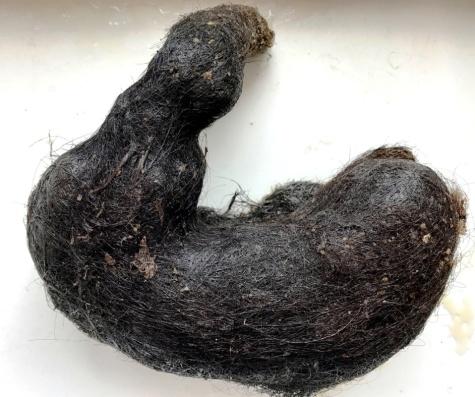
A 16-year-old illiterate female from a rural background came to the surgical outpatient department with the complaints of intermittent pain and fullness in the upper abdominal region since last 4 months. The pain had been increasing slowly in intensity over the last month and was now accompanied by nausea, vomiting, decrease in appetite and weight loss. Her personal, family, past medical and surgical, birth, developmental histories were all noncontributory. On her general examination, marked pallor and cachexia was evident. On abdomen examination, a large, firm, non-tender mass in the epigastrium was palpated. Laboratory results revealed iron deficiency anemia with following parameters: hemoglobin- 5.9 g/dL, white blood cell count- 11,500/cumm, platelet count- 4,80,000/μl, MCV: 60 fl, RDW: 27%, ferritin: 20 ng/mL (normal range:30-400 ng/mL), and iron: 18 μ/dL (normal range: 33-193 μ/dl). The peripheral blood smear showed microcytic hypochromic red blood cells. Rest of her hematological, biochemical and microbiological tests were within normal limits. An abdominal x-ray exhibited a large gastric mass and foci of intraabdominal free air. A computed tomogram of the abdomen revealed free air and a heterogeneous mass completely filling and distending the gastric cavity. The patient refused for an endoscopy and underwent an exploratory laparotomy with a transverse incision in the epigastric region. An anterior gastrostomy was done between two stay sutures. During the procedure, a large trichobezoar measuring 20 x 18 x 8 cm in size was found in the stomach (Figure 1). There was no intraoperative spillage or extension of hair particles into the distal intestine. Postoperative recovery was uneventful. A psychiatry consultation was sought and the patient was found to be suffering from pica since early childhood, as she had an urge to eat hairs which were thrown on the ground and stuck onto the clothes after combing the head hair, especially under stressful conditions. However, the patient denied pulling out her own hairs. Physical examination of scalp and other body parts did not reveal any evidence of alopecia or pulling of hair/short hair. Past history did not reveal any evidence of depressive disorders, obsessive compulsive disorder, other anxiety disorders, eating disorders, emotional disturbances, mental sub-normality and other impulse control disorders. Her family history of any mental illness was non-contributory. However, an appropriate counseling was done for both the patient as well as her family members. On one year follow-up, the patient is doing well and did not reveal any behavior suggestive of pica.
Discussion
The word bezoar comes from either the Arabic "badzehr” or Persian “padzehr" or Hebrian “beluzaar”, all of them means an antidote or counter poison [10]. Historically, bezoars from the intestine of animals were originally worn as charms. They were also incorporated into potions for use as antidotes and acted as remedies to prevent disease. The first exposition on the bezoar was made by Imad ul oia [11] and the earliest references on bezoar were made by Sushruta in India which dates back to 12th century BC [12]. Trichobezoar is formed as a result of hair ingestion either self-hair or from others and is an uncommon disorder in humans. It was first reported in 1779 by Baudamant [13] and the first surgical removal was done by Schonhorn in 1883 [14]. It was once the commonest types of bezoar and accounted for more than half of the 303 cases collected by DeBakey and Ochsner in 1938 [15]. Nevertheless, since then several case reports and small series have been documented in the literature on this rare enigmatic entity [16, 17]. Trichobezoar has several clinical and pathological features which warrant the attention from treating physicians. Its occurrence is predominantly observed in adolescents; nevertheless its incidence in children less than 10 years of age is on a rise [18, 19]. Females with long hairs and psychiatric disturbances are more prone to have trichobezoars than the males [8, 20, 21]. Several factors are associated with trichobezoar formation like child neglect, abuse, mental retardation, pica, obsessive compulsive disorder, depression and anorexia nervosa which in turn leads to trichotillomania and trichophagia [5, 22, 23, 24]. Trichotillomania usually starts in adolescent age and results in highly variable patterns of hair loss, ranging from small undetectable patches of hair loss to total baldness [25]. It has been estimated that 20% of patients with trichotillomania also have trichophagia and 30% of them develop trichobezoar [26]. Nevertheless, few authors have also documented that sometimes there is no evidence of trichotillomania in patients presenting with trichophagia and trichobezoar [7, 8]. Trichobezoar have also reported in co-occurrence with pica of infancy and childhood, which is a clinical condition without trichotillomania, as seen in our case. A person with pica often eats non-nutritive materials like soil, dirt, hairballs, ice, paint and sand, etc. It is usually seen in the setting of low intelligence; however, it can also occur in individuals with normal intelligence [9]. The pathogenesis of trichobezoars is still debatable. However, most of the researchers have attributed it to the enzyme-resistant properties and smooth, slippery surface of the human hair which makes it impervious to digestion and peristalsis in the gastrointestinal system. Subsequently, retention and accumulation of the eaten hairs in between the gastric mucosal folds occur, which result in the formation of a hair ball along with food and mucus [27]. The mass is usually black because of the acidic nature of the stomach that denatures the proteins. There is decomposition and fermentation of fat resulting in a foul smelling breath odour [28]. Trichobezoars are mainly confined to the stomach however, as the hair ball gets too large to leave the stomach, gastric atony may result. The growing mass effect of the ball can sometimes be sufficient to cause chronic malnutrition and iron deficiency anemia, like in our case [29]. Rarely there is extension of the hair ball from the stomach into the small intestine and colon. This condition is named Rapunzel syndrome, which was first described in 1968 by Vaughan ED, et al. [30] since then fewer than hundred cases of Rapunzel syndrome have been reported in the literature [31, 32]. The clinical presentation of trichobezoar is usually late and there are no pathognomonic signs or symptoms pertaining to it. Most of the patients remain asymptomatic for many years and remain so until the bezoar increases in size. Initially the patients may have anorexia, nausea, vomiting, early satiety, weight loss, epigastric pain, sensation of fullness and abdominal discomfort related to meals. Later patients may develop features of gastric outlet obstruction with passage of hair fragments in vomitus or in stools [33, 34, 35].
Proper physical examination reveals hair loss, halitosis or a well defined palpable epigastric mass which is mobile in all directions. Crepitus over the lump and indentability (Lamerton’s sign) are specific signs of trichobezoars and provide important diagnostic clues [36]. It has been estimated that only 1% of patients with trichophagia develop a trichobezoar [37]. Researchers have also mentioned that if the history of the patient fails to provide sufficient findings revealing trichophagia and trichotillomania, then this epigastric mass can be misdiagnosed as a tumor or splenomegaly and trichobezoar springs up on the surgical table as a surprise. Therefore, a high index of suspicion is required for detecting trichobezoars [5, 27]. In our case, the patient had an epigastric mass and other symptom of gastric outlet obstruction. However, there was no evidence of alopecia and she didn’t reveal any history related to trichotillomania or trichophagia initially. Other helpful indicators for detecting trichobezoars are laboratory investigations which usually reveal microcytic hypochromic anemia due to iron deficiency, slight leucocytosis and a positive test for occult blood. Radiological examination also aids in clinching the diagnosis. Plain abdominal films may reveal amorphous, granular or whirlpool-like configurations of solid and gaseous material within the stomach [38]. The ultrasonography (USG) has a limited role because the high echogenicity of hair and the presence of multiple acoustic interfaces created by trapped air and food create a hindrance [26, 39], however, USG shows a typical curvilinear trichobezoar with bright echogenic band. Computed tomography (CT) is one of the best tool in the diagnosing trichobezoars. The image is quite characteristic, consisting of a well-defined, non-enhanced mass filling the stomach, with a spotted/ mesh like pattern due to air being trapped between hairs. CT scans facilitate evaluation of its size, location, and potential complications [40], on the other hand a mass with signal density similar to air is seen on magnetic resonance imaging (MRI) [41]. On barium meal studies, an intragastric mass with barium retained on its honeycomb surface is observed. Nevertheless, a definitive diagnosis of gastric bezoars is usually established by endoscopy. Furthermore, endoscopy of the duodenum is essential to rule out the possibility of Rapunzel syndrome as well as it plays a pivotal therapeutic role [28, 42, 43, 44, 45]. In our case X- ray as well as CT scan aided in diagnosing trichobezoar. Endoscopy was not done in our case as the patient didn’t give consent for it. The treatment approach depends on the size, location and the complications of the trichobezoar followed by post treatment prophylaxis against further bezoar formation by the patient. Many complications are associated with a large eroding or obstructing trichobezoar which includes gastric ulceration /perforation /emphysema, obstructive jaundice /cholangitis, acute pancreatitis, intestinal intussusception, appendicitis, nephrotic syndrome and other malabsorption related complications like protein-losing enteropathy, iron deficiency, and megaloblastic anemia [46, 47, 48, 49]. Small bezoars may be removed by endoscopic techniques, chemical dissolution or fragmentation [50] while large bezoars are surgically resected. Surgery can be open or by laparoscopy. Open surgery and extraction is the usual mode of removing the trichobezoar as it is technically easy, have low rates of complications, 100% success rate and enables complete assessment of small bowel for other areas of bezoar [20, 27]. Laparoscopic approach has its own advantages and disadvantages, though it improves the cosmetic appearance, has fewer postoperative complications, and reduce the hospital stay of the patient [27] but at the same time the laparoscopic removal of an entire bezoar is difficult without spillage of hairs into the peritoneal cavity [51], the procedure is complex with risk of contamination and overall less success rates have been observed [5]. Bezoars have a tendency to recur [52], therefore these patients should be regularly followed up [53, 54] and the underlying predisposing condition for its occurrence should be dealt properly with psycho education, counseling, behavior therapy and teaching adaptive coping skills to prevent its relapse [55].
Conclusion
Trichobezoar is a rare and a fascinating entity as it can create a diagnostic conundrum and therapeutic delay leading to life-threatening complications in an individual. It should be considered as an important differential diagnosis of anemia, abdominal mass and other symptoms of a gastric outlet obstruction in young psychiatric female patients. A detailed medical history with proper clinical examination are mandatory in such patients and the presence of trichotillomania should not be considered as an essential prerequisite for its diagnosis as trichophagia can occur without trichotillomania. An appropriate psychiatric assessment and care along with a long-term follow-up are recommended as a regular part of treatment to prevent its recurrence.
References
-
Kement M, Ozlem N, Colak E, Kesmer S, Gezen C, et al. (2012) Synergistic effect of multiple predisposing risk factors on the development of bezoars. World J Gastroenterol 18(9): 960-964.
-
Yau KK, Siu WT, Law BK, Cheung HY, Ha JP, et al. (2005) Laparoscopic approach compared with conventional open approach for bezoar-induced small-bowel obstruction. Arch Surg 140(10): 972- 975.
-
Ruiz HD, Palermo M, Ritondale O, Pest E, Pest P, et al. (2005) Tricobezoares gastroduodenales: una causa poco frecuente de obstrucción del tracto de salida. Acta Gastroenterol Latinoam 35(1): 24-27.
-
Eng K, Kay M (2012) Gastrointestinal bezoars: history and current treatment paradigms. Gastroenterol Hepatol (NY) 8(11): 776-778.
-
Gonuguntla V, Joshi DD (2009) Rapunzel syndrome: a comprehensive review of an unusual case of trichobezoar. Clin Med Res 7(3): 99-102.
-
Grant JE, Odlaug BL (2008) Clinical characteristics of trichotillomania with trichophagia. Compr Psychiatry 49(6): 579-584.
-
Tiago S, Nuno M, João A, Carla V, Gonçalo M, et al. (2012) Trichophagia and trichobezoar: Case report. Clin Pract Epidemiol Ment Health 8: 43-45.
-
Islek A, Sayar E, Yılmaz A, Boneval C, Artan R (2014) A rare outcome of iron deficiency and pica: Rapunzel syndrome in a 5-year-old child. Turk J Gastroenterol 25: 100-102.
-
Mehra A, Avasthi A, Gupta V, Grover S (2014) Trichophagia along with trichobezoar in the absence of trichotillomania. J Neurosci Rural Pract 5(1): 55- 57.
-
Goldstein SS, Lewis JM, Rothstein R (1984) Intestinal obstruction due to bezoars. J Gastroenterol 79(4): 313-318.
-
Eigood C (1935) A treatise on the bezoar stone. Ann Med History 7: 73-80.
-
Lall MM, Ohall Jc (1975) Trichobezoar: a colleclin analysis of 39 cases from India with a case report. Ind Paed 12: 351-353.
-
Malpani A, Ramani SK, Wolverson MK (1988) Role of sonography in trichobezoars. J Ultrasound Med 7(12): 661-663.
-
Rees M (1984) Intussusception caused by multiple trichobezoars: a surgical trap for the unwary. Br J Surg 71(9): 721.
-
DeBakey M, Ochsner A (1938) Bezoars and concretions. Surgery 4(6): 934-963.
-
Parshad R, Prabhu S, Kumar GVR, Mukherjee A, Bhamrah D (2002) Trichobezoars: Case Reports and Review of Literature. JK Science 4(4): 202-205.
-
Anipindi NP (2016) Trichobezoar with gastric outlet obstruction: A case report. J Evid Based Med Healthc 3(39): 1951-1954.
-
Talaiezadeh AH, Javaherizadeh H (2011) An unusual trichobezoar in a non-psychiatric nine-year-old girl. Przegląd Gastroenterologiczny 6(6): 409-410.
-
Cintolo J, Telem DA, Divino CM, Chin EH, Midulla P (2009) Laparoscopic removal of a large gastric trichobezoar in a 4-year-old girl. JSLS 13(4): 608-611.
-
Jain M, Solanki SL, Bhatnagar A, Jain PK (2011) An unusual case report of Rapunzel Syndrome trichobezoar in a 3-year-old boy. Int J Trichol 3(2): 102-104.
-
Joda AE, Salih WM, Al-Nassrawi RM, Nawzat H (2018) Rapunzel Syndrome Trichobezoar in a 4-Year Old Boy: An Unusual Case Report with Review of Literature. SM J Pediatr Surg 4(1): 1057.
-
Naik S, Gupta V, Naik S, Rangole A, Chaudhary AK, et al. (2007) Rapunzel syndrome reviewed and redefined. Dig Surg 24(3): 157-161.
-
Ventura DE, Herbella FAM, Schettini ST, Delmonte C (2005) Rapunzel syndrome with a fatal outcome in a neglected child. J Pediatr Surg 40: 1665-1667.
-
Armstrong JH, Holtzmuller KC, Barcia PJ (2001) Gastric trichobezoar as a manifestation of child abuse. Curr Surg 58(2): 202-204.
-
Morales HL, Catalán CH, Demetrio RA, Rivas ME, Parraguez NC, et al. (2014) Gastric trichobezoar associated with perforated peptic ulcer and Candida glabrata infection. World J Clin Cases 2(12): 918-923.
-
Wolski M, Gawłowska-Sawosz M, Gogolewski M, Wolańczyk T, Albrecht P, et al. (2016) Trichotillomania, trichophagia, trichobezoar- summary of three cases. Endoscopic follow up scheme in trichotillomania. Psychiatr Pol 50(1): 145- 152.
-
Gorter RR, Kneepkens CM, Mattens EC, Aronson DC, Heij HA (2010) Management of trichobezoar: case report and literature review. Pediatr Surg Int 26(5): 457-463.
-
Middleton E, Macksey LF, Phillips JD (2012) Rapunzel syndrome in a pediatric patient: A Case Report. AANA Journal 80(2): 115-119.
-
Demirel AH, Akbal E, Köklü S, Kuru S, Cakal B, et al. (2011) An unusual cause of iron deficiency anemia and abdominal pain in a young female. Am Surg 77(1): 127-128.
-
Vaughan ED, Sawyers JL, Scott HW (1968) The Rapunzel syndrome: an unusual complication of intestinal bezoar. Surgery 63(2): 339-343.
-
Hamid M, Chaoui Y, Mountasser M, Sabbah F, Raiss M, et al. (2017) Giant gastric trichobezoar in a young female with Rapunzel syndrome: case report. Pan Afr Med J 27: 252.
-
Kwong WT, Kalmaz D (2014) A modern form of Rapunzel syndrome: trichobezoar composed of synthetic hair extensions. Clinical gastroenterology and hepatology 12(5): A33-A34.
-
Rutkoski JD, Gittes GK (2015) Bezoar-related stercoral perforation in an 11-year old child: Has anyone seen my other sock? Journal of Pediatric Surgery Case Reports 3(5): 219-222.
-
Sinha AK, Vaghela MM, Kumar B, Kumar P (2017) Pediatric gastric trichobezoars with acute life threatening and undifferentiated elective bipolar clinical presentations. Journal of Pediatric Surgery Case Reports 16: 5-7.
-
Sharma V (1991) Gastrointestinal Bezoars. J Indian Med Assoc 89: 338-339.
-
Lamerton AJ (1984) Trichobezoar: Two case reports; A new physical sign. Am J Gastroenterol 71(5 ): 354- 356.
-
Irving PM, Kadirkamanathan SS, Priston AV, Blanshard C (2007) Education and imaging. Gastrointestinal: Rapunzel syndrome. J Gastroenterol Hepatol 22(12): 2361.
-
Wadlinglon WB, Rose M, Holcomb OW (1992) Complications of trichobezoars: a 30-year experience. South Med J 85(10): 1020-1022.
-
Hoovera K, Piotrowskib J, Pierreb K, Katzc A, Goldsteinb AM (2006) Simultaneous gastric and small intestinal trichobezoars: A hairy problem. J Pediatric Surg 41(8): 1495-1497.
-
Billaud Y, Pilleul F, Valette PJ (2002) Mechanical small bowel obstruction due to bezoars: Correlation between CT and surgical findings. J Radiol 83(5): 641- 646.
-
Sinzig M, Werner H, Hasselbach H, Illing P (1998) Gastric trichobezoar with gastic ulcer: MR findings. Paediatr Radiol 28(5): 296.
-
Shadwan A, Mohammad A (2000) Small bowel obstruction due to trichobezoar: Role of upper endoscopy in diagnosis. Gastrointest Endoscop 52(6): 784-786.
-
Andrus CH, Ponsky JL (1998) Bezoars: cIassification, pathophysiology, and treatment. Am J Gastroentrol 83(5): 476-478.
-
Ibuowo AA, Saad A, Okonkwo T (2008) Giant gastric trichobezoar in a young female. Int J Surg 6(6): 4-6.
-
Jiledar, Singh G, Mitra SK (1996) Gastric perforation secondary to recurrent trichobezoar. Indian J Pediatr 63(5): 689-691.
-
Klipfel AA, Kessler E, Schein M (2003) Rapunzel syndrome causing gastric emphysema and small bowel obstruction. Surgery 133(1): 120-121.
-
Malhotra Gupta G, Janowski C, Sidlow R (2017) Gastric perforation secondary to a trichobezoar: A case report and review of the literature. Journal of Pediatric Surgery Case Reports 26: 11-14.
-
Nwankwo E, Daniele E, Woller E, Fitzwater J, McGill T, et al. (2017) Trichobezoar presenting as a gastric outlet obstruction: A case report. Int J Surg Case Rep 34: 123-125.
-
Soufi M, Benamr S, Belhassan M, Massrouri R, Ouazzani H, et al. (2010) Giant trichobezoar of duodenojejunal flexure: a rare entity. Saudi J Gastroenterol 16(3): 215-217.
-
Fraser JD, Leys CM, Shawn D (2009) Laparoscopic removal of a gastric trichobezoar in a pediatric patient. Laparoendosc 19(6): 835-837.
-
Ripollés T, García-Aguayo J, Martínez MJ, Gil P (2001) Gastrointestinal Bezoars. Am J Roentgenol 177: 65- 69.
-
Gurzu S, Jung I (2013) Gastric trichowool bezoar in an 18-year-old girl. S Afr J Surg 51(1): 33-34.
-
Kırpınar I, Kocacenk T, Koçer E, Memmi N (2013) Recurrent trichobezoar due to trichophagia: A case report. Gen Hosp Psychiatry 35(4): 439-441.
-
Hisamuddin K, Brandt CP (2008) Hairball in the Stomach: A case of gastric trichobezoar. Clin Gastroenterol Hepatol 6(3): A28-A28.
-
Albal M, Sadriwala Q, Bansod P (2017) Trichobezoar in Paediatrics: An Uncommon Cause of Gastric Outlet Obstruction. JCR 7: 165-168.
- Management of Chronic Insertional Achilles Tendinopathy Using Flexor Hallucis Longus Tendon Transfer in Patients Over 50 Years of Age: A Four-Case Series Following the CARE Guidelines
- Application of Induced Pluripotent Stem Cells in Bone Tissue Engineering: Current Status and Prospects
- Surgical Management of Upper Thoracic Esophageal Squamous Cell Carcinoma with Concomitant Hypersplenism: Integration of Chai's Supra-Thoracic Apex Technique with Laparoscopic Splenectomy - A Technical Innovation Case Study with Systematic Review
- Evaluation of Masticatory Functional Efficiency of Stomatognathic System in Patients Undergoing Open Reduction Internal Fixation for Treatment of Pan-Facial Trauma: A Prospective Study
- Hepatic Abscess Secondary to Appendiceal Phlegmon an Unusual Complication of Appendiceal Phlegmon
- Report of Lumboperitoneal (LP) Shunt Procedure in Over Decades Experiences, Systematic Narrative Review