Talar Extrusion Injury: Anatomy, Diagnosis, Treatment, and Outcomes
Talar extrusion is a very rare injury and is defined as talar dislocation from surrounding tibiotalar, subtalar, and talonavicular joints. It often results from high energy trauma. These injuries often have poor outcomes and serious complications including avascular necrosis, infection, and osteoarthritis. There are limited case reports and case series in the literature regarding talar extrusion injuries. In this review, we discuss the complex anatomy, diagnosis, treatment options, complications, and outcomes for patients with talar extrusion injuries.
Introduction
The talus is the second-largest tarsal bone in the foot and is essential to the function of the foot and ankle [1]. Talus dislocations and fractures are rare injuries accounting for less than 1% of all fractures; however, it is the second most common fractured tarsal bone, after the calcaneus [2, 3, 4]. Total traumatic extrusion of the talus accounts for 2% of all talar injuries, and it is estimated that this phenomenon only occurs in 0.06% of all dislocations [5, 6]. Most talus injuries occur after high energy trauma but can also occur from low energy trauma [7]. The mechanism of talar neck fractures and dislocations is forced dorsiflexion of the foot, driving the talus against the anterior tibial plafond, which was first described by Anderson due to pilot’s exerted pressure on the rudder pedal as they crashed to the ground [8]. If the dorsiflexion continues, the talus will rotate around the intact deltoid ligament and subluxate between the posterior aspect of the medial malleolus and the Achilles tendon [8, 9]. In rare cases, disruption of the intact deltoid ligament can lead to complete dislocations, and extrusion of the talus can occur [10].
Talar extrusions are defined as dislocation of the talus from the tibiotalar, talonavicular, and subtalar joints [11]. In about 25% of these injuries there is supination of the foot, which causes medial neck comminution and an associated medial malleolus fracture [8, 9]. Treatment of talar extrusion injuries, also known as total talus dislocation, can be either conservative or surgical. Conservative management often has a limited role for these injuries and is reserved for small nondisplaced fractures. Therefore, surgical management is often undertaken for these injuries. Due to the tenuous blood supply of the talus, one major complication after talus injury is avascular necrosis (AVN). AVN of the talus often occurs in the talar neck or body and is seen on radiographs within 6 to 8 months from time of injury resembling an opacity of the involved bone [12, 13]. This can lead to collapse of the talus and posttraumatic arthritis.
There seems to be paucity in the literature regarding management and outcomes when treating talar extrusion injuries. Published reports on this topic are mostly limited to case reports and small case series. Multiple surgical techniques, including primary talectomy with tibiocalcaneal arthrodesis and immediate reduction with fixation have been discussed in the literature [14]. Therefore, the purpose of this study is to review and determine the efficacy of the management and treatment of talar extrusion injuries, with additional literature review on talar anatomy and overall outcomes regarding this rare injury.
Anatomy
The talus is the second largest bone in the hindfoot just behind the calcaneus and plays a vital role in the stability of the ankle joint [15]. The talus articulates with the tibia, fibula, calcaneus and navicular [16]. The anatomy of the talus consists of the head, neck, body, lateral and posterior processes [17]. The body of the talus has a trapezoid shape with a convex superior dome with the wider anterior portion allowing for increased stability in dorsiflexion of the ankle [17]. The talus does not have any muscular or tendinous attachments, and is covered in up to 70% of cartilage [18]. Without any musculotendinous attachments, this increases the vulnerability for dislocation [19].
The talus has an extraosseous blood supply with contributions from all three major vessels in the lower extremity [18]. Gelberman, et al. [20] found that the peroneal artery provides two branches, the posterior tibial artery provides three branches, and the anterior tibial artery provides six branches, which anastomose to form a complex network of vessels surrounding the talus. The main blood supply to the talus is the artery of the tarsal canal. This is a branch of the posterior tibial artery and supplies nearly two- thirds of the body [20]. Miller, et al. [21] demonstrated that the posterior tibial artery supplies approximately 47% of the total blood supply to the talus [21].
Presentation and Diagnosis
The talus is the only bone in the lower extremity with no muscular attachment, making it susceptible to complete dislocation. This injury is mostly involved with high energy trauma, falls from significant heights, and snowboarding when an axial load is applied to a plantar flexed foot [22]. The exact position of the foot relative to the talus determines the direction of dislocation, including anterior, posterior, medial, lateral, and complete dislocations [23]. A systematic review conducted by Weston, et al. [14] compared 86 cases of total talar dislocation [14]. They found that a majority of talar extrusion injuries are open compared to closed, with 73 injuries being open compared to 13 being closed [14]. In addition, isolated total talar dislocations, indicating no associated fractures, were present for 29 of the 86 patients, with 72% of these patients having open injuries [14].
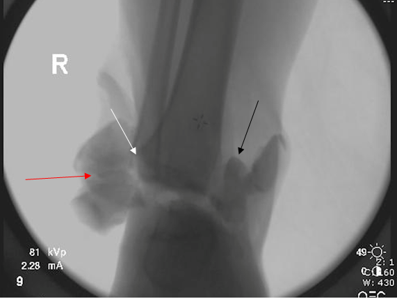
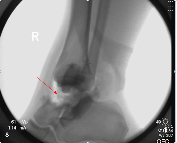
Upon presentation, patients often have significant pain, ecchymosis, swelling, and decreased range of motion of the foot and ankle. An obvious deformity is commonly appreciated, but may be masked by substantial amounts of swelling. Complete talar dislocations are frequently open injuries, but there is a rare possibility of this injury being closed. Due to the high risk of associated injuries, including medial and lateral malleoli, calcaneus, navicular, cuneiforms, cuboid, and metatarsals, multiple radiographs and additional imaging need to be obtained [23]. Anteroposterior (AP), lateral, and oblique radiographs of the foot and ankle are the first steps in diagnosis, with additional Canale or Harris views to evaluate talar neck fractures or talocalcaneal joint disruption, respectively [24]. Associated fractures and intraarticular fractures are often difficult to assess with plain radiographs, therefore additional imaging is often required [25]. A computed tomography (CT) scan can assist in diagnosing talar injuries, whether initial radiographs are negative or further information needs to be obtained. It provides excellent visualization of the bony anatomy and gives the provider more information of the injury and can assist with preoperative planning. Additionally, magnetic resonance imaging (MRI) is an option to detect talar revascularization, early development of AVN, and additional soft tissue injury at subsequent follow up appointments [6].
Treatment and Outcomes
Initial treatment and evaluation of talar fractures involves reduction of dislocations as well as urgent assessment of open injuries. Management of open fractures applies to the initial management of the extruded talus with timely antibiotics and tetanus, and proper irrigation of the wound [26]. While an attempt to standardize treatment of the extruded talus has been made, there is still controversy over proper treatment. Historical treatment of talar extrusion consisted of primary talectomy, but recent literature proposes reimplantation of the talus as the mainstay of treatment [27]. Due to the severity of this injury, urgent surgical management is often necessary as closed reduction is often not adequate. A case review of 25 subtalar dislocations demonstrated that open reduction was required in 32% of subtalar dislocations alone [26]. In a retrospective review investigating results of reimplantation for extruded talus, Smith, et. al. [27] 19 patients diagnosed with extruded talus were identified and treated with reimplantation [27]. The method of initial reimplantation varied depending on the degree of soft tissue disruption. If no further soft tissue attachments were identified, the extruded talus was irrigated with sterile saline, wrapped in sterile bacitracin and reimplanted in the operating room [27]. A case report by Fleming and Hurley included antibiotic polymethylmethacrylate beads in the wound with delayed wound closure [28]. Results and fixations of remaining fractures following reimplantation were treated with open reduction and internal fixation utilizing screws and plates [29].
A similar alternative was proposed, which suggested talar reimplantation supplemented by tibiotalocalcaneal arthrodesis [28]. Due to the proposed and common complication of avascular necrosis, Mohammad et. al. utilized initial treatment of the extruded talus with tibiotalocalcaneal arthrodesis [28]. This was thought to decrease the risk of secondary procedures and provide added stability to the subtalar, tibiotalar, and talocalcaneal joints [28]. Regardless of the decision to proceed with a tibiotalocalcaneal arthrodesis, most current literature continues to recommend reimplantation as opposed to complete talectomy.
Following talar reimplantation, it has been recommended that patients remain non-weight bearing in a cast for 6 weeks if the wound is amenable for closure. If there is significant soft tissue disruption, immobilization with an external fixator was recommended for approximately 6 weeks [27]. Following removal of either cast or external fixator, patients were non-weight bearing for an additional 6 weeks [26, 27, 29]. Current literature suggests that talar reimplantation remains the primary treatment method for the extruded talus, with overall good outcomes and preserved ankle mobility [30]. Although infection and avascular necrosis tend to be the most commonly associated complications with these injuries, Smith, et. al. saw no acute infections postoperatively with talar reimplantation in all twenty-seven of their patients [27]. Talar reimplantation also appears to have no significant impact on ankle range of motion in the post-operative course [29].
Complications
Common complications with fractures of the talar body and neck are subsequent subtalar arthritis as well as AVN. In the setting of talar neck fractures, subtalar arthritis has been reported to occur at varying rates, with some studies reporting 100% occurrence [31]. In regard to AVN, the rates have been reported to be correlated to the severity of the injury. In the setting of talar neck fractures rates have been reported to be as high as 48-53% in Hawkin’s III and IV patterns [31]. Due to the rarity of the injury, rates of subtalar arthritis and AVN have not been as clearly defined with talar extrusion.
In a systematic review of total talar dislocations involving the subtalar, tibiotalar, and talonavicular joints by Weston, et al. [14] AVN was found to be the most common complication and present in 25% of the cases [14]. Radiographic evidence of arthritis was only present in 22% of cases; however, the mean follow-up time in this review was only 32 months. There were no correlations of outcomes to treatment methods in this study; however, 26% of patients did require a second surgery such as an arthrodesis after failed fixation [14]. Of note, 26% of patients had a primary arthrodesis after the injury, showing that in the limited time period of this study, over 50% of patients ended up with a fusion [14].
Infection is also a concern when discussing talar extrusion injuries. A study by Smith, et al. investigated infection risk with reimplantation of the talus after talar extrusion [27]. In 19 patients with a minimum of 1 year follow up, they found the infection risk to be relatively low, occurring in only two of their patients. They concluded that reimplantation was a relatively safe procedure with the benefits outweighing the risks [27].
The study by Smith, et al. [27] also describes complications that occur when there is a talar fracture associated with a talar extrusion [27]. There were 8 patients that had an associated talar neck or body fracture with a talar extrusion injury that reached 1 year of clinical and radiographic follow up. All 8 patients had evidence of talar collapse, osteonecrosis, or subtalar arthritis at the 1–2-year mark. There were 5 patients with extrusion injury without fracture that met the same criteria of 1 year of clinical and radiographic follow up. Three of these patients had no significant abnormalities at the 1-year mark. Overall, there were 17 secondary surgeries performed in 7 out of the 19 patients. Four of the 7 that required secondary surgery did have an associated fracture [27].
Conclusion
Fractures and dislocations of the talus are difficult to treat in the acute and long-term setting. They can occur due to a variety of different mechanisms, but often occur during an axial load while the foot is in a dorsiflexed position. Talar fracture-dislocations will commonly present as an open fracture and subsequent extrusion, as there are no muscle or tendinous attachments to hold it in place. With talar extrusion injuries, the risk of long-term arthritis increases as the subtalar, tibiotalar, and talonavicular joints are disrupted. High rates of avascular necrosis are appreciated due to complete cut off of blood supply. For talar extrusions, previous literature supported management with primary talectomy. However, recent studies discuss reimplantation of the talus as the mainstay of treatment. If reimplantation is possible, the talus needs to be thoroughly cleaned and debrided to decrease the risk of infection. Regardless of treatment decision, these patients should be taken to the operating room due to the nature of an open fracture- dislocation.
References
-
Caracchini G, Pietragalla M, De Renzis A, Galluzzo M, Carbone M, et al. (2018) Talar fractures: radiological and CT evaluation and classification systems. Acta Biomed 89(S1): 151-165.
-
Lin S, Hak DJ (2011) Management of talar neck fractures. Orthopedics 34(9): 715-721.
-
Vallier HA, Nork SE, Benirschke SK, Sangeorzan BJ (2003) Surgical treatment of talar body fractures. J Bone Joint Surg Am 85(9): 1716-1724.
-
Vosoughi AR, Fereidooni R, Shirzadi S, Zomorodian SA, Hoveidaei AH (2021) Different patterns and characteristics of Talar injuries at two main orthopedic trauma centers in Shiraz, south of Iran. BMC Musculoskeletal Discord 22(1): 609.
-
Karampinas PK, Kavroudakis E, Polyzois V, Vlamis J, Pneumaticos S (2014) Open talar dislocations without associated fractures. Foot Ankle Surg 20(2): 100-104.
-
AlMaeen BN, ElMaghrby IS, AlNour MK, Alrefeidi TA, Abu Adas SM (2020) Complete Revascularization of Reimplanted Talus After Isolated Total Talar Extrusion: A Case Report. Cureus 12(5): e7947.
-
Vallier HA (2015) Fractures of the Talus: State of the Art. J Orthop Trauma 29(9): 385-392.
-
Anderson HG (1919) The Medical and Surgical Aspects of Aviation. London: Henry Frowde, Oxford University Press. pp: 255.
-
J R B, Ma A, W K, A OG (2017) The Diagnosis, Management and Complications Associated with Fractures of the Talus. Open Orthop J 11: 460-466.
-
Elgafy H, Ebraheim NA, Tile M, Stephen D, Kase J (2000) Fractures of the talus: experience of two level 1 trauma centers. Foot Ankle Int 21(12): 1023.
-
Ortiz-Cruz JR, Ojeda Boscana IL (2019) Talar Extrusion, A Very Rare Sequela of Trauma: A Case Report. Am J Case Rep 20: 575-579.
-
Hawkins LG (1970) Fractures of the neck of the talus. J Bone Joint Surg Am 52(5): 991-1002.
-
Fortin PT, Balazsy JE (2001) Talus fractures: evaluation and treatment. J Am Acad Orthop Surg. 9(2): 114-127.
-
Weston JT, Liu X, Wandtke ME, Liu J, Ebraheim NE (2015) A systematic review of total dislocation of the talus. Orthop Surg 7(2): 97-101.
-
Khan IA, Varacallo M (2023) Anatomy, Bony Pelvis and Lower Limb, Foot Talus. Treasure Island (FL): StatPearls Publishing.
-
Hegazy MA, Khairy HM, Hegazy AA, Sebaei, MA, Sadek SI (2023) Talus bone: Normal anatomy, anatomical variations and clinical correlations. Anatomical Science International 98(3): 391-406.
-
Al-Jabri T, Muthian S, Wong K, Charalambides C (2021) Talus Fractures: All I need to know. Injury 52(11): 3192- 3199.
-
Parekh SG, Kadakia RJ (2021) Avascular Necrosis of the Talus. J Am Acad Orthop Surg 29(6): e267-e278.
-
Van Opstal N, Vandeputte G (2009) Traumatic talus extrusion: case reports and literature review. Acta Orthop Belg 75(5): 699-704.
-
Gelberman RH, Mortensen WW (1983) The arterial anatomy of the talus. Foot Ankle 4(2): 64-72.
-
Miller AN, Prasarn ML, Dyke JP, Helfet Dl, Lorich DG (2011) Quantitative assessment of the vascularity of the talus with gadolinium-enhanced magnetic resonance imaging. J Bone Joint Surg Am 93(12): 1116-1121.
-
Kramer IF, Brouwers L, Brink PR, Poeze M (2014) Snowboarders’ ankle. BMJ Case Rep. 2014: bcr2014204220.
-
Cheruvu MS, Narayana Murthy S, Siddiqui RS (2023) Subtalar dislocations: Mechanisms, clinical presentation and methods of reduction. World J Orthop 14(6): 379- 386.
-
Melenevsky Y, Mackey RA, Abrahams RB, Thomson NB (2015) Talar Fractures and Dislocations: A Radiologist’s Guide to Timely Diagnosis and Classification. Radiographics. 35(3): 765-779.
-
Bibbo C, Anderson RB, Davis WH (2003) Injury characteristics and the clinical outcome of subtalar dislocations: a clinical and radiographic analysis of 25 cases. Foot Ankle Int 24(2): 158-163.
-
Mohammad HR, A’Court J, Pillai A (2017) Extruded talus treated with reimplantation and primary tibiotalocalcaneal arthrodesis. Ann R Coll Surg Engl 99(4): e115-e117.
-
Smith CS, Nork SE, Sangeorzan BJ (2006) The extruded talus: results of reimplantation. J Bone Joint Surg Am 88(11): 2418-2424.
-
Fleming J, Hurley KK (2009) Total talar extrusion: a case report. J Foot Ankle Surg 48(6): 690.e19-23.
-
DeLee JC, Curtis R (1982) Subtalar dislocation of the foot. J Bone Joint Surg Am 64(3): 433-437.
-
Breccia M, Peruzzi M, Cerbarano L, Galli M (2014) Treatment and outcome of open dislocation of the ankle with complete talar extrusion: a case report. Foot (Edinb) 24(2): 89-93.
-
Dodd A, Lefaivre KA (2015) Outcomes of Talar Neck Fractures: A Systematic Review and Meta analysis. J Orthop Trauma 29(5): 210-215.
- Management of Chronic Insertional Achilles Tendinopathy Using Flexor Hallucis Longus Tendon Transfer in Patients Over 50 Years of Age: A Four-Case Series Following the CARE Guidelines
- Application of Induced Pluripotent Stem Cells in Bone Tissue Engineering: Current Status and Prospects
- Surgical Management of Upper Thoracic Esophageal Squamous Cell Carcinoma with Concomitant Hypersplenism: Integration of Chai's Supra-Thoracic Apex Technique with Laparoscopic Splenectomy - A Technical Innovation Case Study with Systematic Review
- Evaluation of Masticatory Functional Efficiency of Stomatognathic System in Patients Undergoing Open Reduction Internal Fixation for Treatment of Pan-Facial Trauma: A Prospective Study
- Hepatic Abscess Secondary to Appendiceal Phlegmon an Unusual Complication of Appendiceal Phlegmon
- Report of Lumboperitoneal (LP) Shunt Procedure in Over Decades Experiences, Systematic Narrative Review