A Comparison of the Anthropometrical Measurements of External Ear between Normal persons and Diabetic Patients in Iraq
The human ear is an important distinguishing organ of the human face. Defects on the auricle surface of the ear may allow a person to be identified. Since the auricle's structure is complex, a plastic surgeon needs to know its different auricular dimensions, which will be useful for designing an ear prosthesis that will aid in forensic recognition of the person. Objectives: The objective of the current study to compare the anthropometric measures of Pinna bilateral external ear in normal and diabetic patients in Iraq with sexual differences Subject and Methods: Research performed at the Al-Faiha General Hospital outpatient clinic and the Basra Teaching Hospital. Two groups were obtained from 95 patients with an average age of 40-60 years in normal as well as Type II diabetes mellitus. Vernier caliper measured anthropometric measurements of the outer ear, including total ear length (TEL), total ear width (TEW), total lobules length (TLL) and total lobules width (TLW), to the nearest 0.1mm. Results: Each group in our study consisted of 55 female patients (58%) and 40 male patients (42%) aged 40-60 (50±9.8). In the study, high measurements were found when comparing anthropometric measurements in male and female persons. It was observed, in each group, that the measurements of the right ear for males and females are larger than the left ear and there were significant differences in anthropometric measurements (P
Introduction
The ear would affect the appearance of people and provide to the human face an esthetic, so that the external ear was made up of auricle/pinna and external auditory meatus [1]. The pinna absorbed the air vibrations and consisted of the thin elastic cartilage plate coated with skin and intrinsic and extrinsic muscles innervated by facial nerve [2]. The auricle is usually associated with congenital anomalies as a malposition related to Down’s and Turner’s syndrome [3], and with injuries and pathological conditions as in cancer, the auricle has often acquired defects [4].
Correcting these conditions needed information on the usual auricular measurements, because recent anthropometric measurement studies from different populations show a lot of variability depending on age , sex and ethnic group, but there is a difference between right and left ears even in the same person [5]. The perception of human differences has an important role in plastic surgery, prosthetics [6] the form, size and orientation of each auricle are special fingerprints and allow the inference that the male has larger ears than the female [7] in addition to the fact that both the length and the width of the ear increase with age [8]. From birth to 99 years of age, the increase of both ear length and width in females was continuous while in males it stopped at around 50 and 70 years of age [9].
In detecting sounds that come from the front rather than those from the back, the pinna angle also helps to identify the sounds; the relation with the size of the head and the frequency of the audible sound this effect applies at higher frequencies, while in the middle frequencies the head casts the sound shadow itself and in lower frequencies the point of sound arrival between the ears helps to identify the sound [10]. So a huge challenge in face-plastic operations, particularly in the field of aurical reconstruction, lies in the discovery of the best treatment for patients with advanced biotechnologies as well as surgical techniques, such as costal cartilage grafts, prosthesis, alloplastic tools and advances in tissue engineering of tissue of cultural tissue of clinical interest [11].
The connective tissue and blood vessels changes in diabetes mellitus affects ear pinna [12] because the common risk factor for diabetes mellitus is a vascular condition which includes the pathological effect of advanced glycation accumulations, impaired nitrous oxide inhibition of vasodilatory reaction, and a smooth dysfunction in the muscles [13].
Diabetes mellitus characterized by insulin deficiency causing high blood glucose and also linked resistance to insulin. Over time such chronic hyperglycemia, particularly in genetically susceptible individuals, can cause tissue damage. The formation of fibrosis, involving the predominant extracellular matrix (ECM) accumulation, is a pathological response to tissue injury [14].
Invasive studies (tissue biopsy) are typically needed to accurately evaluate morphological changes in the target organs and the degree to which fibrotic changes influence their function. The ECM contains an insoluble network of collagen, structural glycoprotein, elastine, proteoglycan- hyaluron and integrin, which not only assist cells in their mechanical role but also mediate complex interactions between cells or between the cells and the extracelular matrix of vascular tissue [15]. The aim of the study to compare the anthropometric measurement of the external ear of right and left ears in normal persons and diabetic patients in Iraq with sexual differences in addition to determine anthropometric measurements as a method for external ear identification in normal persons and type II diabetes mellitus patients.
Subject and Methods
The research was conducted at the Al-Fayha General Hospital and the Basra Teaching Hospital outpatient clinic. The data was collected for the period from19th November of 2019 to the 20th of February of 2021. Both group with average age 40-60 a normal persons and Type II diabetes mellitus patients ; their HbA1c (8-12) for diabetes medication treatments. I choose the level of HbA1c (8-12) for the diabetic patients because they represent the majority of the diabetic patients in outpatients clinic, I expect that diabetes mellitus affected all parts of the body.
All subjects with congenital auricular malformation of the external ear (pinna) and all persons with previous history of trauma to the external ear (pinna) or surgery are not involved in study. The anthropometric tests for “Pinna” external ear include total ear length (TEL), total ear width (TEW), total lobular length (TLL) and total lobular width (TLW), calculated by a specific investigator by Vernier caliber (Mitutoyo Corp., Japan) to 0.1 mm (Figure 1).
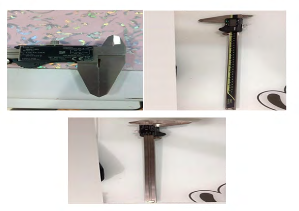
The questionnaire formula was developed and discussed with the experts for the purpose of our analysis following receiving guidance from the medical ethics committee. The data collection was performed after verbal consent from the patients. The anthropometric measurements of the “pinna” external ear included total ear length (TEL) measure the distance between the higher projection of the helix and the lower projected ear lobule. Correspondingly total width of ear (TEW) measures the distance between the posterior point and most anterior point of the auricle. The total length of the lobule (TLL) calculates the distance from the bottom end of the lobule to the base of the tragus notch. Total lobule width measured at midpoint of the lobular height is the horizontal width of the lobule as in (Figure 2).
Results
The sample size was 190 cases separately classified into two categories, each with 95 normal persons and type II mellitus patients. The females 55(58%) and males 40(42%) in each group; the patient’s age ranged from 40 to 60 years (50±9.8).The anthropometric comparision was high in males relative to women and, in both females and male patient groups, the right auricles were larger than left, with significant differences (P<0.05) in anthropometric measurements. Our diabetic type Ⅱ group results decrease in all parameters compared with normal group, but no major differences except in total ear width exist; as shown in (Tables 1-10).
| Gender | Male | Female | P value | ||
|---|---|---|---|---|---|
| Variables | Mean (cm) | SD | Mean (cm) | SD | |
| Total length of ear | 6.51 | 0.31 | 6.12 | 0.37 | 0.275 |
| Total width of ear | 3.28 | 0.34 | 2.56 | 0.33 | 0.046* |
| Total lobule length | 1.91 | 0.31 | 1.75 | 0.15 | 0.003* |
| Total lobule width | 1.98 | 0.14 | 1.59 | 0.15 | 0.017* |
Table 1: Comparism of left ear between male and female persons in normal group.
| Gender | Male | Female | P value | ||
|---|---|---|---|---|---|
| Variables | Mean (cm) | SD | Mean (cm) | SD | |
| Total length of ear | 6.76 | 0.36 | 6.16 | 0.38 | 0.991 |
| Total width of ear | 3.56 | 0.31 | 3.42 | 0.31 | 0.002* |
| Total lobule length | 2.02 | 0.15 | 1.77 | 0.15 | 0.00819* |
| Total lobule width | 2.03 | 0.15 | 1.78 | 0.15 | 0.00411* |
Table 2: Comparism of right ear between male and female persons in normal group.
| Gender | Male | Female | P value | ||
|---|---|---|---|---|---|
| Variables | Mean (cm) | SD | Mean (cm) | SD | |
| Total length of ear | 6.76 | 0.36 | 6.51 | 0.31 | 0.0081* |
| Total width of ear | 3.56 | 0.31 | 3.28 | 0.34 | 0.842 |
| Total lobule length | 2.02 | 0.15 | 1.91 | 0.31 | 0.0621 |
| Total lobule width | 2.03 | 0.15 | 1.98 | 0.14 | 0.0019* |
Table 3: Comparism the right and left ear of male persons in normal group.
| Gender | Male | Female | P value | ||
|---|---|---|---|---|---|
| Variables | Mean (cm) | SD | Mean (cm) | SD | |
| Total length of ear | 6.16 | 0.38 | 6.12 | 0.37 | 0.0041* |
| Total width of ear | 3.42 | 0.31 | 2.56 | 0.33 | 0.833 |
| Total lobule length | 1.77 | 0.15 | 1.75 | 0.15 | 0.0193* |
| Total lobule width | 1.78 | 0.15 | 1.59 | 0.15 | 0.0025* |
| Variables | Mean (cm) | SD | Mean (cm) | SD | |
| Total length of ear | 6.42 | 0.44 | 6.12 | 0.45 | 0.942 |
| Total width of ear | 2.89 | 0.49 | 2.37 | 0.26 | 0.00719* |
| Total lobule length | 1.9 | 0.31 | 1.7 | 0.23 | 0.0262* |
| Total lobule width | 1.94 | 0.18 | 1.57 | 0.17 | 0.0413* |
Table 4: Comparism the right and left ear of female persons in normal group.
| Gender | Male | Female | P value | ||
|---|---|---|---|---|---|
| Variables | Mean (cm) | SD | Mean (cm) | SD | |
| Total length of ear | 6.65 | 0.61 | 6.1 | 0.39 | 0.8701 |
| Total width of ear | 2.93 | 0.28 | 2.78 | 0.25 | 0.0095* |
| Total lobule length | 2.01 | 0.3 | 1.72 | 0.21 | 0.0162* |
| Total lobule width | 1.97 | 0.25 | 1.75 | 0.16 | 0.0022* |
Table 5: Comparism of right ear between male and female patients in diabetic type ΙΙ group.
| Gender | Male | Female | P value | ||
|---|---|---|---|---|---|
| Variables | Mean (cm) | SD | Mean (cm) | SD | |
| Total length of ear | 6.65 | 0.61 | 6.42 | 0.44 | 0.0001* |
| Total width of ear | 2.93 | 0.28 | 2.89 | 0.49 | 0.518 |
| Total lobule length | 2.01 | 0.3 | 1.9 | 0.31 | 0.185 |
| Total lobule width | 1.97 | 0.25 | 1.94 | 0.18 | 0.00251* |
Table 6: Comparism the right and left ear of male patients in diabetic type ΙΙ group.
| Gender | Male | Female | P value | ||
|---|---|---|---|---|---|
| Variables | Mean (cm) | SD | Mean (cm) | SD | |
| Total length of ear | 6.1 | 0.39 | 6.12 | 0.45 | 0.0016* |
| Total width of ear | 2.78 | 0.25 | 2.37 | 0.26 | 0.762* |
| Total lobule length | 1.72 | 0.21 | 1.7 | 0.23 | 0.0038 |
| Total lobule width | 1.75 | 0.16 | 1.57 | 0.17 | 0.0513* |
Table 7: Comparism the right and left ear of female patients in diabetic type ΙΙ group.
| Gender | Right Ear Variables | Normal Group | Diabetic Group | P value | ||
|---|---|---|---|---|---|---|
| Mean (cm) | SD | Mean (cm) | SD | |||
| Male | TEL | 6.76 | 0.36 | 6.65 | 0.61 | 0.10619 |
| TEW | 3.56 | 0.31 | 2.93 | 0.28 | 0.02260* | |
| TLL | 2.02 | 0.15 | 2.01 | 0.3 | 0.76258 | |
| TLW | 2.03 | 0.15 | 1.97 | 0.25 | 0.11905 | |
| Female | TEL | 6.16 | 0.38 | 6.1 | 0.39 | 0.3998 |
| TEW | 3.42 | 0.31 | 2.78 | 0.25 | 0.02398* | |
| TLL | 1.77 | 0.15 | 1.72 | 0.21 | 0.38037 | |
| TLW | 1.78 | 0.15 | 1.75 | 0.16 | 0.87532 | |
| Mean(cm) | SD | Mean(cm) | SD | |||
| Male | TEL | 6.51 | 0.31 | 6.42 | 0.44 | 0.27078 |
| TEW | 3.28 | 0.34 | 2.89 | 0.49 | 0.01635* | |
| TLL | 1.91 | 0.31 | 1.9 | 0.31 | 0.50047 | |
| TLW | 1.98 | 0.14 | 1.94 | 0.18 | 0.37649 | |
| Female | TEL | 6.12 | 0.37 | 6.12 | 0.45 | 0.9726 |
| TEW | 2.56 | 0.33 | 2.37 | 0.26 | 0.04173* | |
| TLL | 1.75 | 0.15 | 1.7 | 0.23 | 0.59112 | |
| TLW | 1.59 | 0.15 | 1.57 | 0.17 | 0.2371 |
Table 8: Comparism of right ear between normal persons and diabetic type II groups.
Discussion
The outer ear is an essential component of human facial complex. It has described the face and transmitted details about an individual’s age and sex [16]. For esthetic surgery the external ear shape and facial proportions are critical; this data helps plastic surgeons in correcting ear defects. It is important to be aware that there are no standardized ear morphology and differences in ethnic groups [17, 18]. Up to now all available researchers have focused on anthropometric external ear measurement, but none of these have been contrasted between normal persons and diabetic patients.
It was shown that sexual dimorphism occurred between females and males with higher values in males in auricular dimensions [19]. Bozkir MG, et al. [20] found sexual dimorphism in external ear dimensions; results indicate a considerable difference in ear height between Turkish and Japanese populations. In the same analysis, the total auricular height and width in the Turkish population were also seen to be longer in men. It has therefore been concluded that in males auricular measurements are substantially greater than in females [20]. These discrepancies can be related to the assertion that the expansion of auricle begins earlier with males than with females and continues to the older period [21]. Gender differences can also be affected by sexually differing genetic factors [9].
Our study results related to the anthropometrical measurements of right ear in male persons (TEL, TEW, TLL and TLW) respectively are 6.76±0.36, 3.56±0.31, 2.02 ±0.15, 2.03±0.15 but in the left ear are 6.51±0.31, 3.28 ±0.34, 1.91±0.31, 1.98±0.14 while in the female persons; the right ear anthropometrical measurements (TEL, TEW, TLL and TLW) respectively are 6.16 ±0.38, 3.42±0.31, 1.77±0.15, 1.78±0.15 but in the left ear are 6.12±0.37, 2.56±0.33, 1.75±0.15, 1.59±0.15. Our study shows all the anthropometric measurements in males are larger than females and the right auricle measurements is larger than left in both female and male persons of normal group and there is a significant differences between both sexes the differences in male and female may be related to a statement that an auricular growth started early in male than in female which is continuous up to old age [21]. When Compared with Gupta AK, et al. [22] who conducted his research with 134 Nepalese students and 66 Indian students, our male person’s measurements are larger than the results and our female persons are larger than the results of their study except for total ear width and lobular width. In Asai Y, et al. [23] who performed his analysis on 400 consecutive Japanese, the average total ear length is 64.1mm which is smaller than our results; however, our findings have been in agreements with Alexander K, et al. [24] in 2010, which conducted its study of 420 volunteers. ; The findings from the Caucasian are similar to our findings but are better than those from the African/ African Caribbean countries. Ear dimensions differ according to genetic and environmental factors; however, our ear size is similar to the Caucasian ethics community.
The anthropometric measurements of right ear in male diabetes mellitus patients; (TEL, TEW, TLL and TLW) respectively are 6.65±0.61, 2.93±0.28, 2.01±0.30, 1.97 ±0.25 but in the left ear are 6.42 ±0.44, 2.89±0.49, 1.90±0.31, 1.94±0.18 while in the female diabetes mellitus patients; the right ear (TEL, TEW, TLL and TLW) respectively are 6.10±0.39, 2.78±0.25, 1.72±0.21, 1.75±0.16 but in the left ear are 6.12±0.45, 2.37±0.26, 1.70±0.23, 1.57±0.17. Here still the measurements are larger in males than females and the right auricle measurements is larger than left in both female and male patients. No previous studies are performed in diabetic patients linked to anthropometric measurements, but if our findings of diabetics type Ⅱ are compared to the normal population, all parameters are decreased, but with no major differences except for total ear width(TEW). All these changes could be important because of connective tissue in the long- term Products of changes in the content and quantity of the extracellular matrix structural macromolecule the vessels and tissues [25]. Diabetes mellitus associated with several macro- and micro-molecules complications, as well as neuropathic changes, which may cause hearing loss when the risk increases with diabetes greater than without it because of inadequate glycemic controls [26]. The limitation of the study.1-limit age group 40-60 years .2-comparison of the ear dimensions with various population corresponding to the age group. 3- The measurements of the normal persons among known people to me.
Conclusion
For normal persons and diabetics, knowledge of ear parameters is vital for plastic and reconstructive surgeon in the hearing aid technology sector to produce acceptable ears. It is also beneficial for diabetics to focus on anthropometric measures due to the increased risk of hearing loss over time.
References
-
Japatti SR, Engineer PJ, Reddy BM, Tiwari AU, Siddegowda CY, et al. (2018) Anthropometric Assessment of the Normal Adult Human Ear. Ann Maxillofac Surg 8(1): 42- 50.
-
Snell R (2012) Clinical Anatomy by Regions. 9th (Edn.), Walters’s kluwere, Lippincott Williams and Wilkins, pp: 561.
-
Coward TJ, Watson RM, Scott BJ (1997) Laser Scanning for the Identification of Repeatable Landmarks of the Ears and Face. Br J Plast Surg 50(5): 308-314.
-
Pless J (1976) Carcinoma of the external ear. Scand J Plast Reconstr Surg 10(2): 147-151.
-
Purkait R, Singh P (2007) Anthropometry of the normal human auricle: A study of adult Indian men. Aesthetic Plast Surg 31(4): 372-379.
-
Deopa D, Thakkar HK, Prakash C, Niranjan R, Barua MP (2013) Anthropometric measurements of external ear of medical students in Uttarakhand region. Jour of the anat Society of India 62(1): 79-83.
-
Healthcote JA (1995) Why do old men have big ears?. BMJ 311(7021): 1668.
-
Meijerman L, van der Lugt C, Maat GJR (2007) Cross- sectional anthropometric study of the external ear. J Forensic Sci 52(2): 286-293.
-
Shireen S, Karadkhelkar VP (2015) Anthropometric Measurements of Human External Ear. J of Evolution of Med and Dent Sci 4(59): 10333-10338.
-
Alberti PW (2006) The Anatomy and Physiology of the Ear And Hearing. Medicine 2: 53-62.
-
Storck K, Staudenmaier R, Buchberger M, Strenger T, Kreutzer K, et al. (2014) Total reconstruction of the auricle: our experiences on indication and recent technique. Biomed Res Int.
-
Huntley AC (1989) Cutaneous Manifestations of Diabetes Mellitus. Dermatologic Clinics 7(3): 531-546.
-
Cade TW (2008) Diabetes-Related Microvascular and Macrovascular diseases in the physical therapy setting. Phys Ther 88(11): 1322-1335.
-
Ban CR, Twigg SM (2008) Fibrosis in diabetes complications: Pathogenic mechanisms and circulating and urinary markers. Vasc Health Risk Manag 4(3): 575- 596.
-
Hashimoto G, Inoki I, Fuji Y, Aoki T, Ikeda E, et al. (2002) Matrix metalloproteinases cleave connective tissue growth factor and reactivate angiogenic activity of vascular endothelial growth factor 165. J Biol Chem 277(39): 36288-36295.
-
Muteweye W, Muguti GI (2015) Prominent ears: Anthropometric study of the external ear of primary school children of Harare Zimbabwe. Ann Med Surg 4(3): 287-292.
-
Ali K, Meaike JD, Maricevich RS, Olshinka A (2017) The Protruding Ear: Cosmetic and Reconstruction. Semin Plast Surg 31(3): 152-160.
-
Farkas LG (1978) Ear morphology in Treacher Collins’, Apert’s and Crouzon’s syndromes. Arch Otorhinolaryngol 220(1-2): 153-157.
-
Brucker MJ, Patel J, Sullivan PK (2003) A morphometric study of the external ear: age and sex related differences. Plast Reconstr Surg 112(2): 647-652.
-
Bozkir MG, Karakas P, Yavuz M, Dere F (2006) Morphometry of the external ear in our adult population. Aesth Plast Surg 30(1): 81-85.
-
Taura M, Adamu L, Modibbo M (2013) External ear anthropometry among Hausas of Nigeria; the search of sexual dimorphism and correlations. World Journal of Medicine and Medical Science Research 1(5): 091-095.
-
Gupta AK, Ambekar MN (2017) Anthropometric study of external ear: comparative study in medical students of Nepalgunj medical college in Nepal. Journal of Nepalgunj Medical College 15(2): 49-52.
-
Asai Y, Yoshimura M, Nago N, Yamada T (1996) Why do old men have big ears? Correlation of ear length with age in Japan. BMJ 312(7030): 582.
-
Alexander KS, Stott DJ, Sivakumar B, Kang NA (2011) A morphometric study of the human ear. J Plast Reconstr Aesthet Surg 64(1): 41-47.
-
Rosenbloom AL, Silverstein JH (1996) Connective Tissue and Joint Disease in Diabetes Mellitus. Endocrinol Metab Clin North Am 25(2): 473-483.
-
Kim MB, Zhang Y, Chang Y, Ryu S, Choi Y, et al. (2017) Diabetes mellitus and the incidence of hearing loss: a cohort study. Int J Epidemiol 46(2): 717-726.
- Pattern of Breast Lesions in Ovu Inland, Delta State, South Southern Nigeria
- Morphometric Analysis of the Human Femur: Exploring Platymetric and Robusticity Indices Among the Nigerian Population
- Anatomical Variation of Arteria Lusoria: Clinical Implications for Dysphagia Lusoria and Surgical Risk
- Morphometric Study of the Vertebral Body and Pedicle of Typical Cervical Vertebrae Using Radiological Image
- Epigenetic Mechanisms Driving Human Evolutionary Changes
- Neuroprotective Effects of Ginkgo Biloba Extract on Bilateral Common Carotid Artery Ischaemic Stroke Induced in Wistar Rat