Assays for Anti-Cancer Screening of Medical Plants: A Review
Cancer is one of the most common causes of death around the world, and this is due to the weakness of the treatment strategies used, which may result from a delay in diagnosis and thus the ineffectiveness of treatment, or as a result of the bad side effects of cancer treatment drugs. The International Agency for Research on Cancer revealed that by 2030, cancer cases are expected to reach 26 million new cases and 17 million deaths. This led those in charge of the medical process and scientific research to search in the botanical pharmacy for plants that have a cytotoxic effect on cancer cells, and the advantages of these natural medicines are that they do not have any side effects on health in addition to their effective ability to treat cancer cells.
Introduction
There are different types of cancer found in the world. The forms of human cancer include:leukemia, stomach cancer, lung cancer, liver cancer, bone cancer, breast cancer, prostate cancer, kidney cancer, cervical cancer and so on. It affects about 20% of the population of the African continent [1]. Treatment of patients with fractures takes several days and strategies depending on the history and stage of the injury. Similarly, treatments of cancer using anti- cancer drugs take longer days with undesirable side effects. Therefore, research has made quick attempts to find some known plants capable of destroying the cytotoxic effect of cancer cells without causing many side effects [2].
To assess the toxic effect of substances on cells, the selectivity index (SI) must be calculated. If it is less than 1, means that the compound has a toxic effect on cells, greater than 1 while less than 10 indicate a weak toxic effect on cells. But if the SI is greater than 10, it means that the compound is safe and does not have much toxic effect on cells [3].
The use of natural products have always been an important matter in the field of medical care, as many plants have been used in the pharmaceutical industry, especially in the field of oncology, as the emergence of anti-cancer drugs relied on improving the phytochemical properties of medicinal plants or preparing synthetic compounds mimicking what is extracted from natural plants. As long as research continues in natural plants for compounds that have a toxic effect on cancer cells and have no role on normal cells, there will be greater break through [4].
In recent times, the idea of research in the development of anti-cancer compounds has begun. The presence of cells resistant to chemotherapy has caused the spread of disease and high death rates. The passion to carryout research on the use of anti-cancer compounds extracted from natural plants is worthwhile due to the low cost involved and it’s little or no toxicity level [5].
In a situation where natural plants contain phytochemical compounds from secondary metabolism, such compounds would have anti-cancer properties, for example flavonoids, which act as an anti-cancer by encouraging programmed cell death. This leads to toxic events in carcinogenic cells while the normal cells remain as they are and thus have been done [6].
Although approved anticancer drugs are available to day for the treatment of more than 200 different tumor entities, effective therapies for most of these tumours are lacking. Out of the 92 registered drugs, 17 are considered by oncologists to be more broadly applicable and 12 additional agents are perceived as having certain advantages in some clinical settings.
These are generally cytotoxic and function in a very restricted number of molecular mechanisms. In this manner, the requirement for novel medications to treat the malignant disease requiring systemic treatment is tireless [7]. Globally, the rate of use of medicinal plants with an anti- cancer effect ranges from 10-40% and up to 50% in the countries of the Asian continent. As for Europe, what is spent on these plants is 5 billion dollars every year [8].
Disclosure and improvement of anti-cancer specialists is the focal point of numerous pharmaceutical organizations and different associations, similar to the National Cancer Institute (NCI). Regardless of various endeavours, fruitful advancement of malignant growth treatment is disappointing by lacking comprehension of tumor-genesis. Anti-cancer medication advancement endeavours center around cytotoxic aggravates that reason tumor end. Late spurt in the comprehension of disease pharmacology has encouraged the improvement of objective centered medications that are intended to specifically repress atomic markers engaged with malignancy development and metastasis [9].
A pre selection called the screening process is therefore required. The aim of screening efforts is to identify products that will produce anti-tumor effects matching the activity criteria.
The objective of this paper is to review assays for anti- Cancer screening of medical plants.
Development of Multidrug Resistance in Patients
The phenomenon of multiple drug resistance refers to the resistance and development of cancer cells against anti-cancer drugs, which leads to treatment failure and consequently death. Multiple drug resistance to anticancer drugs remains a major challenge, as there is resistance to every anti-cancer drug and this is done through many strategies, either by reducing the drug dose, increasing the flow, reducing or preventing programmed cell death, repairing the DNA (DNA), and activating detoxification systems [10] (Figure 1).
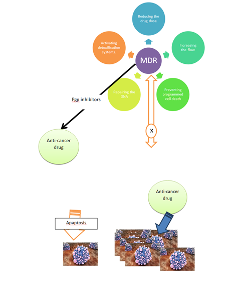
Selenium compounds, Pgp inhibitors, NSC23925, and Dexrazoxane, all of which inhibits the expression of ABCB1 gene and Pgp, are among the most important compounds that contribute to solving the problem of cellular drug resistance [11].
The increase in Pgp gene expression counteracts the effects of some anti-cancer drugs such as doxorubicin and vincristine [12].
As a result of the great variation in the extent of the patient’s response to treatment or resistance due to the dependence of this on genetic factors, but at the present time and with knowledge of the genomic structure and metabolism of each patient, scientists were able to develop a strategy for each patient that is commensurate with his health condition and the sick history of God to avoid the problem of drug resistance [13].
Modern strategies depend on the combination of two types of drugs, as using one type works to kill sensitive cells while letting the resistant cells grow and spread. Among the modern strategies also is to stop the supply of energy to cells, as cancer cells need more glucose than normal cells, and in the absence of them they will die [14].
When potential cytotoxic mixes are acquired by compound blend or extraction from regular sources, the resulting stages associated with medication advancement procedure are as per the following:
Preliminary invitro Screening
Invitro cell culture models are utilized to assess tumorigenic movement of new medication up-and-comers. Roughly 10,000 medications are tried in-vitro models on an annual premise to explore the degree and explicitness of hostile to malignant action [15]. In vitro Methods
- Tetrazolium salt assay
- Sulphorhodamine B assay
- H-Thymidine uptake
- Dye exclusion test
- Clonogenic test
- Cell counting assay
- Morphological assay
- Cell Counting Kit-8 In Vivo Methods
- Carcinogen induced models
- Viral infection models
- Transplantation Models
- Genetically Engineered Mouse Models
- In vivo hollow fibre assay
Clinical Development
Medication poisonous quality is tried in human volunteers to recognize greatest endured portion in stage I clinical preliminaries. To evaluate adequacy and to affirm sedate measurements, stage II analyses are led in patients of chosen tumor type pursued by gigantic stage III investigations. The entire method of medicine divulgence and improvement is innately time and resource eating up with particularly low accomplishment rates. Out of 10,000 medications screened routinely, only 100 are attempted in pre-clinical study, 5-10 are taken up for clinical preliminaries, at last only 1 or 2 mixes are supported by NDA as advanced sedation for treatment. The typical time starting from another composite association to get exhibit support is around 9 to 12 years with ordinary uses running from 0.5 to 2 billion US$ depending upon the infection.
Preclinical and clinical medication improvement has overwhelmingly relied upon creature frame works. The time and costs of medicine headway generally increase during pre- clinical invivo testing using creatures and besides incoming about clinical starters, it is fundamental to recognize assuring medication spearheads correctly at the outset times of drug improvement [16].
Productive assurance and progression of the most unique drug candidates requires reliable and amazing invitro test structures. Novel systems have been grasped for making dependable invitro models that duplicate invivo tumor lead. Complex correspondence between different cell types working inside the 3D tissue ECM microenvironment is outstandingly essential for tissue improvement, homeostasis and tumor pathogenesis. Quality verbalization, nosy direct of various human cancers are susceptible to ‘solid factor signs’ and demonstrations absolutely one of a kind in the closeness or nonattendance of stroma. Prevailinginvitro models join - 2D monolayer culture and 3D spheroid models [16].
InVitro Models for Tumours
2D Monolayer: Cells developed straightforwardly on 2D tissue culture plates vary generously, from those developed in 3D microenvironment, in the morphology, cell-cell cooperation, cell-network collaborations and separation. 2D models change from invivo tumor conditions in the going with ways [17]:
- Gene articulation profile in 2D frameworks regularly varies from in vivo conditions, as a result of2D cell association and restricted cell-cell connections.
- Impassive vehicle of supplement and excretory items is adequate in 2D framework, for upkeep of ordinary pathophysiology. In any case, such over-disentangled mass exchange does not remain constant in vivo conditions.
- As opposed to hypoxic condition of in vivo tumours, uniform sustenance and oxygenation is given to monolayer cells.
- Shortage of in vivo like thick stroma in 2D monolayer which shows little IC50level of medications relating to finish bioavailability of medication possibility to the objective cells. In any case, IC50 estimations of medications in vivo carcinomas are nearly greater. These distinctions show less invivo productivity from 2D cell culture frameworks.
3D Spheroids: Spheroids allude to invitro aggregate of cells set up both from a solitary cell type or from a blend of different cell types: cancer cells, insusceptible cells, epithelial cells, fibroblast cells, mesenchyme foundational microorganisms (MSCs) and endothelial cells. These totals are more successful than 2D frameworks since like tumours; spheroids ordinarily contain a blended populace of surface- uncovered and profoundly covered cells, multiplying and non-multiplying cells, well-oxygenated and hypoxic cells. In spite of the fact that it expands cell-cell connections, it intrinsically needs stromal mediation. The normal time required for a tumor spheroid to progress from a total of couple of cells to an expanded structure with suitability and proliferative angles ranges from 1 to about fourteen days. It is exceedingly testing to get spheroids of uniform size in a reproducible way. An evident answer for the above models is to use invivo creature models [18]. Creature Models: In spite of the fact that coherent characteristics and ethics of using creatures for research are perpetually being tended to, it is assessed that countless creatures are still being commonly used in wide scope of research studies. In any case, invivo models address a part of the repressions of invitro systems, moral concerns, long test term and non-human science addresses colossal downsides. Moreover, medications and strategies that gives off an impression of being empowering in creature models have normally shelled in on schedule or later times of clinical preliminaries. Other than the cost of drug improvement augments extensively during pre-clinical invivo creature testing. In this way, we need to Reduce, Replace and Refine the usage of creatures. A basic issue is low predictively from right currently used preclinical research models. For financial and good concerns, this prerequisites the headway of 3D malignancy models which overcome the space between standard 2D monolayer and creature models [19].
In vitro 3D Tumor Models: This is exceedingly fundamental to distinguish inadequate medicine up-and-comers in starting times of testing than afterwards, which set aside, would both time and money. To upgrade careful assurance of the highly unique medication particles from the massive and creating collection of mixes, test systems which eagerly replicate an invivo solid carcinoma are exceptionally basic. In this manner, it needs to make logically reasonable in vitro cell culture models which immovably recreate invivo microenvironment. Regardless, the affirmation of thick and practical illness counterpart’s invitro, it is some of the critical confronts in tissue planning [20].
A natural tissue isn’t just a blend of cells inside a load of dormant macromolecules; anyway it is genuinely a multifaceted combination of cell and cell-incorporated network coordinated by homeostatic concordance. Cells are coordinated and compelled by the ECM piece and organization, which, in this way, are organized, accumulated and reconstructed by cells. Tissue brokenness despite when risen up out of cell fragments impacts the extracellular space and the other way around. Henceforth, a dependable in vitro 3D malignancy model should replicate the entire tissue, consolidating the ECM assembling in order to dependably and reasonably reflect both the physiological and over the top status of invivo hazardous tissue. Invitro 3D malignant development models emulating normally significant constraints of invivo microenvironment, for example, cell− cell, cell−ECM connections will be a significant gadget to assess the restorable viability of anticancer medications [18].
The 3D models are logically been used to accurately imitate properties and conduct of human malignant tissues. Innovative frameworks are being used for improving strong in vitro models that better restate in vivo settings for testing the counter tumorigenic sufficiency of anticancer drugs. These models additionally can possibly improve and to streamline medicate conveyance frameworks for compelling chemotherapy. The essentialness of in vitro 3D tumor models has been comprehensively seen and their headway is ascending at a faster rate. Precious stone S. Shin adjusted hydrogel format to culture spheroids in a hydrogel framework comprising of miniaturized scale wells and consequently moved the spheroids to a microfluidic divert speaking to in vivo powerful smooth motion [20].
Hui-li Ma revealed the limit of HeLa cell-gathered spheroids to fill in as a diagnostic instrument for nanoparticle which goes about as movement vehicles for chemotherapeutics [21]. They revealed 3D imaging of nanoparticle entrance into HeLa spheroids using HeLa cells created in 2D culture as the control structure. Moutusi Mitra manufactured surface built, polymeric biodegradable micro- particles to be used as a stage for 3D advancement of Y79 cell line in order to survey the consequences of tumorigenic medications [21].
Qgyi He tissue built a pancreatic malignant growth model via pancreatic disease undifferentiated organisms conveyed from a well-defined electro spun structure of poly (glycolide- co-trim ethylene carbonate) and gelatine [22]. AgateNyga built up an innovative colorectal malignant growth model using HT29 cells entrenched in collagen type I which is epitomized in a non-thick collagen type I gel populated by a mix of fibroblasts and endothelial cells [23]. Jayme L Horning grew boson malignant growth model by creating MCF-7 cells on porous, biodegradable polymeric micro particles [24]. Chandraiah Godugu declared a 3D culture system for improvement of spheroids in Algi Matrix TM stages and coming about cytotoxicity appraisal of tumorigenic medication [25].
In Vivo Assays for Screening of Anti-Cancer Drugs
The use of animal models is indispensable in understanding cancer biology and the effect of anti-cancer drugs on cells.
Tetrazolium Salt Assay
- This assay is a delicate, quantifiable and dependable colorimetric assay that estimates feasibility, expansion and initiation of cells. The assay depends on the limit of mitochondrial dehydrogenase enzymes in living cells to convert the yellow water-dissolvable substrate 3-(4, 5-dimethylthiazole-2-yl) - 2, 5-diphenyl tetrazolium bromide (MTT) into a dark blue formazan product which is water insoluble.
- The quantity of formazan formed is directly proportional to the cell number in scope of cell lines.
- MTT Formazan Metabolically Active Cell Insoluble [26].
Method: It is performed to determine the enzymatic properties. Cells from particular cell lines in log phase of growth are trypsinised, It is counted in haemocytometer and adjusted multiwall plates (96 well plates). The cells are treated with a various concentration of drug for specified duration.
After MTT dye is added in each well and plates are incubated at 37° C for 4 hrs. in a CO2 incubator. The plates are taken out from the incubator and dark blue colored formazan crystals are thoroughly dissolved in DMSO at room temperature. The plates are then read on ELISA reader at 570nm to analyze the percent cell viability regarding control is calculated [26].
Haemocytometer Cell Counts
- The most common routine method for cell counting which is efficient and accurate is with the use of a haemocytometer.
- The haemocytometer is divided into 9 large squares size
1 mm x 1 mm, chamber size 0.1 mm x 1 mm x 1 mm.
Method: 15-20 μL of cell suspension is added to the haemocytometer, then the cap is placed, then the number of cells is counted in the large squares, as for the red blood cells, they are counted in the small squares.
% Cell Availability = (OD of treated cells/OD of control cells) ×100
The Sulphorhodamine B Assay
- TheaSulphorhodamineaBaassayaMeasuresawhole- cultureaproteinacontent, which should be comparative to the cell number [27].
- Cell cultures are stained with a proper staining dye, Sulphorhodamine B. SRB is a bright pink anionic dye that binds to basic amino acid of cell.
- Unbound dye is then removed by washing with acetic acid.
- During the dead cells either lyses or are lost during procedure, the measure of SRB binding is corresponding to the number of live cells left in a culture after drug exposure.
Morphological Assay
- Huge scale, morphological changes that happen at the cell surface, or in the cytoskeleton, can be pursued and identified with cell viability.
- Damage can be identified by large decreases in volume secondary to losses in protein and intracellular ions due to altered permeability to sodium or potassium.
- Necrotic cells: nuclear swelling, chromatin flocculation, loss of nuclear basophilia.
- Apoptotic cells: cell shrinkage, nuclear condensation, nuclear fragmentation. Example: Morphological feature (human skin keratinocyte) of normal human skin keratinocyte and differentiated human skin keratinocyte.
Dye Exclusion Test
- This assay is built on the structural integrity of the cells.
- Live cells possess intact cell membranes that exclude certain dyes, such as trepan blue, Eosin, or presidium, whereas dead cells would have lost membrane integrity.
- Hence they would take up the dyes while the live cells exclude it [28]. Method: Cell lines are counted, cultured and inoculated in 96 well plates as above. Cell lines were brooded with various centralizations of test mixes for 4 days. Number of cultured cells in various wells were counted utilizing haemocytometer after staining with suitable dyes.
% Cell Viability = Number of Viable Cells Total Number of Cells (Viable + Dead) × 100 [21]
Tryphan Blue Dye
Tryphan Blue is a dye solution that dyes the dead cells blue, and since the cells are selective, the dye does not pass through the membrane, so it does not stain blue, while in the dead cells the dye passes through the membrane and is stained in the blue color that we see under the microscope [29].
Cell Counting Kit-8(CCK8)
CCK8 method is very common now for in vitro anticancer evaluation for the below reasons:
- High sensitivity, reliable result and good reproducibility;
- Easy to operate and save much time;
- Lower cytotoxicity to cells;
- Suitable for high-throughput drug screening.
Method: The cancer cell lines were seeded in 96-well plates with 100μL DMEM supplemented with 10% foetal bovine serum, and cultured at 37oC in a humidified CO2 incubator (95% air, 5% CO2) for 24h. While the cell lines grew to 90% in logarithmic growth, the culture medium was removed from each well, and 100μL fresh DEME was added to each well. Then, 10μL drug solution was added into each well (every concentration was repeated for 5 times) and the plates were incubated for another 48h at 37oC. Subsequently, 10μL CCK8 was added to each well, and the plates were cultured at 37oC for another 4 hours. The optical density was measured at a wave-length of 450 nm on an ELISA micro plate reader. DMEM and DMSO solution (V/V: 10/1) was used as a negative control. The results were expressed as the inhibition calculated at the ratio {[1-(OD450 treated/ OD450 negative control)] ×100} [xx].
Advantages
- Reduce the usage of animals
- Less time consuming
- Cost effective
- Easy to manage
- Capable of processing large number of compounds hurriedly with least quantity
- Limit of concentrations use are analogous to that required for in vivo studies [30].
Disadvantages
- Trouble in maintenance of cultures
- Give negative results for the compounds which get stimulated after body metabolism and vice versa.
- Difficult to maintain Pharmacokinetics [31].
In vitro Assays for Screening of Anti-Cancer Drugs
With the emergence of new types of anti-carcinomas, it was necessary to affect these compounds on cancer cells before their use by humans. Therefore, these drugs were tested on cells in the laboratory to study the efficacy of drugs and their toxic effect on cancer cells.
Chemical Carcinogen Model
- DMBA induced mouse skin papilloma
- Two stage experimental carcinogenesis Initiator DMBA(dimethylbenz[a]anthracene),Promotora– TPA(12-O-tetradecanoyl-phorbol-13-aacetate)
- Mice: Single dose–2.5 µg of DMBA, 5 to 10 μg of TPA in 0.2 ml of acetone twice weekly.
- Papilloma begins to appear after 8 to 10 wks. –Tumor incidence and multiplicity of treatment group is compared with DMBA control group.
- Mice are topically applied a single dose of 2.5 µg in acetone, followed by 5-10 µg of TPA in 0.2 ml acetone twice weekly on the same site starting one week after DMBA application.
- Percent tumor incidence and multiplicity of treatment groups is compared with DMBA control group. Drug under test can be administered either topically or oral route.
- The tumor incidence in this model is usually about 100% DMBA controls.
- In repeated topical application of DMBA Alone has also been shown to induce carcinogenesis.
- Drug efficacy is measured as percent reduction in carcinoma incidence, compared with that of carcinogen control.
- Mouse skin papilloma RAT Mammary Gland CA [31].
Mouse Mammary Tumor Virus (MMTV)
- Mouse Mammary Tumor Virus (MMTV) was the first mouse virus, isolated at Jackson Lab as the “non- chromosomal factor” that caused mammary tumours in C3H strain of mice.
- Some viruses cause cancer via random integration in certain cells. Some viruses carry cellular oncogenes. Abelsonamurinealeukemia virus – Abl Moloney murine sarcoma virus–Raff
- Engineered viruses now used routinely in the laboratory to induce cancer [32].
Transplantation Model
• Tumor cells or tissues (mouse or human) transplanted into a host mouse.
- Ectopic: Implanted into a different organ than the original (typically subcutaneous or kidney capsule) **
- Orthotopic: Implanted aintoathe aanalogous aorganaof a thea original a tumor. Advantages**
- Typically cheap, fast and easy to use › Not covered by patents
- In vivo screening tool implemented in 1995 by NCI.
In Vivo Hollow Fibres Assay
- Human tumor cell lines (lung, breast, colon, melanoma, ovary, and glioma.
- Cells suspended into hollow polyvinylidene fluoride fibres implanted IPaor SCainalabamice
- After in vivo drug treatment, fibres are eradicated and investigated in vitro.
- Tumorigenic activity assessed. 33
Subcutaneous Hollow Fibre Implants
- 2D and 3D Cell-based Assays in Drug Screening
- Currently, pharmaceutical, firm suspend large amount of a money on the compound efficacy and cytotoxicity test
- There is still a 78% failure rate for all drugs, which may be devastating to developing companies
- Effective compounds in vitro may be non-effective in vivo for many reasons, including differences between in vitro and in vivo target biology, interrelated biochemical mechanism, metabolism, poor penetration into solid tissues, etc.
- Presently, almost all cell-based assays or biosensors are created in 2D culture frameworks, though conventional 2D cultures generally experience the ill effects of contact inhibition and loss of local cell morphology and functionality.
- In contrast with 2D cultures, 3D models make a progressively practical portrayal of genuine human tissues, which is basic to numerous significant cell functions, including morphogenesis, cell metabolism, gene expression, differentiation and cell-cell interaction [32].
Conclusion and Recommendations
Cancer is one of the diseases of the era that has spread in a terrifying way, so studies have tended to provide anticancer drugs to reduce the percentage of deaths, and the modern era has turned to the use of medicinal plants as they are rich in phytochemical compounds antioxidant that work to eliminate cancer cells through programmed death of cells while preserving Healthy cells. However, in some cases, the so-called multi-drug resistance appeared, due to which the death rate increased, but the use of a combination of more than one drug contributed to alleviating this problem, as many compounds were studied and mixed between Invivo and Invitro before approving their use in humans. Therefore, the direction of modern work must be in the context of the use of medicinal plants as anti-inflammatory, oxidative and anti-cancer drugs after they are tried in the laboratory and on rats.
References
-
Rawla P, Barsouk A (2019) Epidemiology of gastric cancer: global trends, risk factors and prevention. Prz Gastroenterol 14(1): 26-38.
-
Bassam H (2019) Plants and Cancer Treatment. In: Medicinal Plants - Use in Prevention and Treatment of Diseases. IntechOpen pp: 192.
-
Liu J, Cao R, Xu M, Wang X, Zhang H, et al. (2020) Hydroxychloroquine, a less toxic derivative of chloroquine, is effective in inhibiting SARS-CoV-2 infection in vitro. Cell Discov 6: 16.
-
Koparde AA, Doijad RC, Magdum CS (2019) Natural Products in Drug Discovery. In: Perveen S, et al. (Eds.), Pharmacognosy - Medicinal Plants, Intech Open.
-
Garcia-Oliveira P, Otero P, Pereira AG, Chamorro F, Carpena M, et al. (2021) Status and Challenges of Plant-Anticancer Compounds in Cancer Treatment. Pharmaceuticals 14(2): 157.
-
Nguyen NH, Ta QTH, Pham QT, Luong TNH, Phung VT, et al. (2020) Anticancer Activity of Novel Plant Extracts and Compounds from Adenosmabracteosum (Bonati) in Human Lung and Liver Cancer Cells. Molecules 25(12): 1-26.
-
Lai CC, Cheng YC, Chen PW, Lin TH, Tzeng TT, et al. (2019) Process development for pandemic influenza VLP vaccine production using a baculovirus expression system. J Boil Eng 13: 78-79.
-
Salmerón-Manzano E, Garrido-Cardenas JA, Manzano- Agugliaro F (2020) Worldwide Research Trends on Medicinal Plants. Int J Environ Res Public Health 17(10): 1-20.
-
Narang AS, Desai DS (2009) Anticancer drug development. In: Lu Y, et al. (Eds.), Pharmaceutical Perspectives of Cancer Therapeutics, Springer pp: 49-92.
-
Bukowski K, Kciuk M, Kontek R (2020) Mechanisms of Multidrug Resistance in Cancer Chemotherapy. Int J Mol Sci 21(9): 1-24.
-
Radomska D, Czarnomysy R, Radomski D, Bielawski K (2021) Selenium Compounds as Novel Potential Anticancer Agents. Int J Mol Sci 22(3): 1009.
-
Tang D, Chen X, Kang R, Kroemer G (2021) Ferroptosis: molecular mechanisms and health implications. Cell Research 31(2): 107-125.
-
Malsagova KA, Butkova TV, Kopylov AT, Izotov AA, Potoldykova NV (2020) Pharmacogenetic Testing: A Tool for Personalized Drug Therapy Optimization. Pharmaceutics 12(12): 1-23.
-
Wang X, Zhang H, Chen X (2019) Drug resistance and combating drug resistance in cancer. Cancer Drug Resist 2(2): 141-160.
-
Kitaeva KV, Rutland CS, Rizvanov AA, Solovyeva VV (2020) Cell Culture Based in vitro Test Systems for Anticancer Drug Screening. Front Bioeng Biotechnol 8: 322.
-
Hsiao AY, Tung YC, Qu X, Patel LR, Pienta KJ, et al. (2012) 384 hanging drop arrays give excellent Z‐factors and allow versatile formation of co‐culture spheroids. Biotechnology and bioengineering 109(5): 1293-1304.
-
Caleb J, Yong T (2020) Is It Time to Start Transitioning From 2D to 3D Cell Culture? Front Mol Biosci 7(33): 1-15.
-
Imparato G, Urciuolo F, Casale C, Netti PA (2013) The role of microscaffold properties in controlling the collagen assembly in 3D dermis equivalent using modular tissue engineering. Biomaterials 34(32): 7851-7861.
-
Kim MJ, Chi BH, Yoo JJ, Ju YM, Whang YM, et al. (2019) Structure establishment of three-dimensional (3D) cell culture printing model for bladder cancer. PLoS One 14(10): e0223689.
-
Shin CS, Kwak B, Han B, Park K (2013) Development of an in vitro 3D tumor model to study therapeutic efficiency of an anticancer drug. Molecular Pharmaceutics 10(6): 2167-2175.
-
Ma Hl, Jiang Q, Han S, Wu Y, Tomshine JC, et al. (2012) Multicellular tumor spheroids as an in vivo– like tumor model for three-dimensional imaging of chemotherapeutic and nano material cellular penetration. Molecular Imaging 11(6): 487-498.
-
Mitra M, Mohanty C, Harilal A, Maheswari UK, Sahoo SK, et al. (2012) A novel in vitro three-dimensional retinoblastoma model for evaluating chemotherapeutic drugs. Molecular vision 18: 1361-1378.
-
He Q, Wang X, Zhang X, Han H, Han B, et al. (2013) A tissue-engineered subcutaneous pancreatic cancer model for antitumor drug evaluation. International Journal of Nanomedicine 8(1): 1167-1176.
-
Nyga A, Loizidou M, Emberton M, Cheema U (2013) A novel tissue engineered three-dimensional in vitro colorectal cancer model. Acta biomaterialia 9(8): 7917- 7926.
-
Horning JL, Sahoo SK, Vijayaraghavalu S, Dimitrijevic S, Vasir JK, et al. (2008) 3-D tumor model for in vitro evaluation of anticancer drugs. Molecular Pharmaceutics 5(5): 849-862.
-
Godugu C, Patel AR, Desai U, Andey T, Sams A, et al. (2013) AlgiMatrix™ based 3D cell culture system as an in-vitro tumor model for anticancer studies. PloS One 8(1): e53708.
-
Mikus J, Steverding D (2000) A simple colorimetric method to screen drug cytotoxicity against Leishmania using the dye Alamar Blue®. Parasitology International 48(3): 265-269.
-
Papazisis KT, Geromichalos GD, Dimitriadis KA, Kortsaris AH (1997) Optimization of the sulforhodamine B colorimetric assay. Journal of Immunological Methods 208(2): 151-158.
-
Livingston R (1979) Depression of Thymidine Labeling Index (Li) Invitro Predicts Effects of Chemotherapy in Patients (Pts). Proceedings of the American Association for Cancer Research. Amer Assoc Cancer Research, Philadelphia.
-
Danhier F, Feron O, Préat V (2010) To exploit the tumor microenvironment: passive and active tumor targeting of nanocarriers for anti-cancer drug delivery. Journal of Controlled Release 148(2): 135-146.
-
Wang HL, Lai TW (2014) Optimization of Evans blue quantitation in limited rat tissue samples. Scientific reports 4: 6588.
-
Margouleff D (2013) Blood volume determination, a nuclear medicine test in evolution. Clinical Nuclear Medicine 38(7): 534-537.
- Management of Ear Keloid with Ksharsutra: A Case Study
- Yoga and Global Sustainability: A Holistic Path to One Earth, One Health
- Autoimmune Diseases in Ayurveda: A Narrative Review with Classical and Modern Perspectives
- Management of Cluster Headache Associated with Pituitary Apophysitis by CERT (Chakrasiddh Energy Release Technique): A Case Report on Energy Rebalancing
- Zygophyllum Geslini Coss : Biochemicals and Antioxidant Activity
- Observations of a Beginner Vaidya