Biotemplated Synthesis of CdSe Nanowires
The use of bio-molecular building blocks opens a new direction in the realm of nanotechnology and such a technology is essential to manipulate nanocrystals in its shape and size. Nucleic acids are of special interest for its ability of self-organization and sensitivity to biological process. In the present work, we present an approach of organizing bioinorganic nanostructures. CdSe nanowire has been synthesized using single strand oligonucleotides as a biotemplate by an electrodeposition technique. A comparative study conceded between CdSe nanoparticle and biotemplated CdSe-DNA nanowire. The CdSe-DNA nanowire reveals a cubic phase as revealed from X-Ray diffraction measurement. CdSe nanowires are fabricated in a filamentary, wire like morphology in DNA strand as established from Atomic Force Microscopic (AFM) measurements.
Introduction
Hybrid systems based on semiconductor quantum dots attached to biomolecules find increasing attention in the development of nanobiotechnology [1, 2, 3]. Many experimental approaches have been made to fabricate nanorods by template driven synthesis using chemical routes [1, 2]. However, the electrochemical approach to fabricate semiconductor nanorods using DNA as the template is promising because of its simplicity in depositing the material directly on a conducting substrate. Presence of negatively charged phosphate group in the DNA backbone gives the polyelectrolyte nature to it. Such a polyelectrolyte can interact with positively charged ions and make them ideal templates to grow nanoparticles [4, 5]. Thus, such nanoparticles tag with DNA can produce nanostructures, which are of interest to biosensor applications [6, 7]. The present work reports a novel and simple strategy to grow CdSe nanobiorods using (Oligo) single stranded DNA (ssDNA) and its complementary DNA as a template by an electro deposition technique. The surface, morphology and structural properties have been studied by using Atomic Force Microscopy (AFM) and X-ray Diffractometry (XRD) technique.
Synthesis
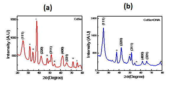
CdSe-DNA nanowire thin films were deposited onto Indium Tin Oxide (ITO) conducting glass substrates by a electro-technique cathodic deposition under galvanostatic condition from a solution containing CdSO4 (0.92 M), SeO2 (0.018 M), and ssDNA, Guanine (Poly-G- 60base pair) and oligonucleotides ssDNA, Cytosine (Poly-C- 60base pair) at room temperature [7]. Typically the electrolysis current density was 5mA/cm2 and the deposition time was kept as 2 minute to synthesize CdSe- DNA complex nanowires. Two samples were prepared for the investigation, namely, sample S1, CdSe nanocrystals analysis shows the XRD peaks at 2θ = 25 o, 42 o, and 49 o corresponds to the prominent planes (111) and (220) and (311), confirms the polycrystalline cubic phase in the deposit for both the samples. The peaks marked with a * corresponds to the X-Ray diffraction peaks of ITO substrate in both the sample. Note that for sample S1, the X-ray peak intensities are weak and broad compared to the CdSe-DNA complex suggesting the S1 crystallite sizes are small. Since the CdSe-DNA complex shows a wire like structure (discussed in the latter section), the observed XRD spectra shows intense X-ray peak intensities and narrow peak similar to bulk CdSe materials.
(NCs) without DNA and sample, S2, CdSe nanowire with DNA. The morphology and structural properties of samples were investigated by Atomic Force Microscopic (AFM), and X-ray diffraction XRD where X-ray diffractometer equipped with a monochromatic CuKα radiation source (1.54178Å ).
Results and Discussion
The XRD patterns of CdSe nanocrystalline (S1) and CdSe-DNA complex (S2) are shown in Figure1. Structural
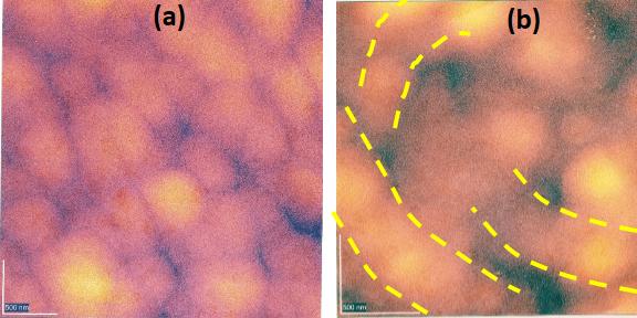
nanowire morphology as marked in dotted line in figure 2(b). The explanation for nanowire formation using ssDNA and its conjugate is that, initially the CdSe nanocrystals are tagged to ssDNA along its string then the interdiffucion of CdSe nanocrystals with its conjugate DNA make the whole system rigid to form a nanowire.
Conclusion
Biotemplated synthesis of CdSe nanowire is possible using single strand oligonucleotides (ssDNA) and its conjugate. The CdSe-DNA nanowire have been grown by an electro-deposition technique. XRD results identify the presence of cubic phase in both CdSe nanocrystal and biotemplated CdSe-DNA nanowire. The AFM results identify the shape of the CdSe in nanoparticle form whereas the shape of biotemplated CdSe-DNA complex is in nanowire form.
Acknowledgement
The author is grateful to the late Professor S. N. Sahu then at Institute of Physics for his support on this work.
References
-
Bruchez M, Moronne M, Gin P, Weiss S, Alivisatos AP (1998) Semiconductor nanocrystals as fluorescent biological labels. Science 281(5385): 2013-2016.
-
Mattoussi H, Mauro JM, Goldman ER, Anderson GP, Sundar VC, et al. (2000) Self-Assembly of CdSe−ZnS Quantum Dot Bioconjugates Using an Engineered Recombinant Protein. J Am Chem Soc. 122(49): 12142-12150.
-
Sarangi SN, Sahu SN, Nozaki S (2018) Physica E: Low- dimensional Systems and Nanostructures 97: 64-68.
-
Braun E, Eichen Y, Siran U, Ben-Yoseph G (1998) DNA-templated assembly and electrode attachment of a conducting silver wire. Nature 391(6669): 775- 779.
-
Coffer JL (1997) Approaches for Generating Mesoscale Patterns of Semiconductor Nanoclusters. J Cluster Science 8(9): 159-178.
-
Sirota M, Minkin E, Lifshitz E, Hensel V, Lahav M (2001) Spectroscopic Properties of Molecular- Wire/Semiconductor Nanocrystalline Superstructure s. J Phys Chem 105(29): 6792-6797.
-
Sarangi SN, Goswami K, Sahu SN (2007) Biomolecular recognition in DNA tagged CdSe nanowires. Biosens Bioelectron 22 (12): 3086-3091.
- Solution-Processed Chiral Perovskites for Biomedical Applications
- Nanotechnology in Health Chemistry and Medicine: Current Challenges and Future Directions
- Human Exposure to Micro- and Nanoplastics: Pathways, Toxicity, and Intervention Strategies
- Exosome Nanomedicine for Cancer Therapy
- Micro and Nanoplastics–Plastisphere, Biotoxicity, Impact on Human Health, and Mitigation Strategies
- Process Validation of Cefixime Powder for Suspension Dosage Form, 50 mL