Nanosilver Enabled Reactive Dyes for Antimicrobial Medical Textiles
The present study describes the preparation technique for silver nanoparticles incorporated fabrics for enhanced antimicrobial property for textile applications. The silver nanoparticles were prepared by the reduction of AgNO3 using non-toxic reducing agent under moderate conditions and the stable Ag colloidal solutions were obtained using poly vinyl alcohol. The particle size distribution of silver nanoparticles was found to be in the range from 20 to 50 nm with spherical morphology while observed from transmission electron microscopy and dynamic light scattering. The characterized colloidal silver nanoparticles were mixed with reactive dye namely Red brown ME RL, and made dyeing on cotton fabrics. The concentration of silver nanoparticles in the reactive dyes was observed to be varied in the mol percentage of 3, 5, 7, and 10 respectively. The characterization results of the prepared colloidal Ag nanoparticles with reactive dye, pre-treated and post-treated fabrics evidenced the presence of silver nanoparticles in the silver treated fabrics with enhanced fastness and light washing properties. The enhanced antimicrobial activity of the silver nanoparticles dyed cotton fabrics was evident against Gram-positive (Salmonella typhi and Bacillus subtilis) and Gram-negative (Escherichia coli and Klebisiella pneumoniae) bacteria. Thus, this study aids in the development of antimicrobial textile applications using silver nanoparticles.
Introduction
Nanotechnology is nowadays blooming in all fields of the most active research areas in modern science and technology. The properties of inorganic materials such as nobel metal nanoparticles differ from those of bulk particles even it has same atom [1]. Nanostructured materials which provide one of the greatest potentials for improving performance and extended capabilities of products in a number industries like textile [2], aerospace, tooling, automotive, recording, cosmetics, electric motor, biomedical [3], optical, magnetic and refrigeration [4, 5]. Nanoparticles do exhibit many interesting exotic properties such as improved physical, chemical, electrical and biological properties due to their nano scale size [6]. Hence, the unique and new properties of such nanomaterials have attracted not only scientists and researchers but also business due to their huge economic potential [7]. Currently, the exploitation of nanotechnology in textile industry has rapidly increased due to its unique and valuable properties [8, 9]. In the past, all textiles were made from natural fibers, including plant, animal, and mineral sources [10, 11]. Many antimicrobial agents are used in textile industry incorporated with textile substrates [7]. Cellulose cotton fabrics are used to make a number of textile products include for highly absorbent bath towels, jute and vest, jeans, cambric, popularly used in the manufacture of work shirts. The enhanced properties implemented to textile cotton fabrics includes water repellency, soil resistance, wrinkle resistance, antimicrobial activity, anti-static, UV-protection, flame resistance and improvement of dye ability [7, 10, 12]. Nanoparticles can provide high durability for fabrics, because it possesses high surface energy, better affinity to fabrics and leading to an increase in durability of the function, coating of nanoparticles without affecting their breath ability or hand feel [13]. The development of fabrics/yarns with antimicrobial agent coating and their applications are widespread in the area of textiles for the last few decades [14, 15]. To overcome the above problem most of the researchers are involved in the work regarding decolourisation, dye ability strength and removal of waste water using UV radiation but there is no such work carried out on the effect and antimicrobial on the dyeing behaviour of reactive dyes and vat dyes [7, 10, 16]. Reactive dyes (dichlorotriazine, monocholorotriazine) are generally used in the dyeing of cellulose, wool, nylon, silk, hair and leather. Antimicrobial effect of silver nanoparticles on textiles has already been shown by various researchers [8, 10]. But the influence of silver nanoparticles coated on the dyed fabrics on the physical and chemical properties of textiles is hardly found in the studies.
Nanosilver is of a great interest for their remarkable properties such as effective antimicrobial agent, good conductivity and catalytic properties against a broad range of Gram-negative (E.coli and Klebsiella pneumoniae) and Gram-positive bacteria (Staphylococcus aureus and Bacillius subtilis), yeast, filamentous fungi [7, 10, 11, 13]. The use of silver nanoparticles have attracted considerable interest for application in different fields such as clothing materials, semiconductor and nanocomposite materials preparation [16, 17]. Silver nanoparticles currently used to control bacteria growth in variety of applications including dental, catheters and burn wounds [15, 18, 19]. Silver nanoparticles are synthesised from using different methods such as chemical reduction, sol-gel, microwave dielectric, heating reduction, ultrasonic irradiation radiolysis, solvothermal, electrochemical and reverse micelles process [8, 16, 20]. Among the above methods, the chemical reduction is the best method [16] for long term stability of the colloidal nanoparticles solutions added with dye then coated on cotton fabrics and effect of good conditions. Reactive dye is a class of highly colored organic substances primarily used in tinting industries that attaches themselves to their cotton fabrics by chemical reactions that form a covalent bond between the dye (dichlorotriazine is a reactive linker between fabrics) molecule and the cotton fabric [2, 12]. In the present study, an attempt has been made to investigate the synthesis and characterisation of silver nanoparticles using chemical reduction method. In addition, dye doped cotton fabrics embedded with nanosilver is fabricated. The antimicrobial activity and UV protection potential of the fabrics are analysed using standard methods.
Materials and Methods
Silver nitrate (AgNO3, 99% purity, AR), poly vinyl alcohol (PVA, 89% purity, AR), sodium borohydride (NaBH4, 95% purity, AR), cotton fabric (100% purity), Bifunctional reactive dye stuff (Galofast’ME’ Dyes) C.I Reactive red brown ME RL (C3H4ClN5, 99% purity, AR), Glauber's salt sodium sulfate (Na2SO4, 99.5% % purity, AR) sodium carbonate (Na2CO3, 99.5% purity, AR), sodium hydroxide (NaOH, 98% purity, AR) and acetic acid (CH3COOH, 98% purity, AR) were obtained respectively from Loba, Merck, Fine, Lakshmi dyes and chemicals, India and employed without any additional purification.
Synthesis of Silver Nano particles
The colloidal silver nanoparticles were prepared by chemical reduction method [8, 16]. To synthesise the silver nanoparticles, an aqueous solution of the silver nitrate (AgNO3) was used as a metal salt precursor. In a typical experiment 15 mM of 0.254 g of AgNO3 was dissolved in 100 ml of double distilled water and stirred for 30 min. Then, 0.5 wt% of 0.25 g of PVA (stabilising agent) and 0.0056 g of NaBH4 (reducing agent) was dissolved separately in 100 ml of distilled water for homogeneous dispersion. The solution containing AgNO3 and PVA were mixed and stirred vigorously for 20 min. To these solutions, NaBH4 was added drop by drop to the solution and stirred for 10 min and continued until the yellow color colloidal solution was formed. The following is the schematic representation of the synthesis of silver nanoparticles: [8, 21]
AgNO3 + PVA (aq.) Ag+ + PVA Reduction [Agº/PVA] Ag° (i) AgNO3 + NaBH4 + PVA Ag (PVA) + 0.5 H2 + 0.5B2H6 + NaNO3 (ii)
Characterization of Silver Nanoparticles
Particle size distribution of the prepared colloidal silver nanoparticles was determined with a particle size analyser (Nanophox; Sympatec, Germany) according to the dynamic light-scattering (DLS) technique. The particle size of all the samples was measured in the range of 1 – 1000 nm at scattering angle of 90°. The zeta potential was calculated through Zetasizer (Malvern, Serial Number: MAL1037088, UK). The three dimensional characterisation of the synthesised colloidal silver nanoparticle was analysed using transmission electron microscopy (TEM, CM200; Philips, USA) operating at 200 kV. The surface morphology and elemental composition of the nanoparticles were analysed using scanning electron microscopy (SEM) coupled with energy dispersive X-ray analysis (EDX). Optical properties and transmittance of the silver nanoparticles were analysed using UV–Vis–NIR spectrometer (Lambda JASCO V-570, Perkin Elmer, USA). The absorbance maximum and the half width of the peak were determined between wavelengths from 700 nm to 350 nm. The concentration of metals in the colloidal silver nanoparticles was detected using inductively coupled plasma optical emission spectrophotometer (ICP-OES, Optima 2100 DV, Perkin Elmer, India).
Fabrication of Dyeing Cotton Fabric with Silver Nanoparticles
The cotton fabric material was weighed about 5g with a size 8.5 × 8.5 cm and then, the sample was soaked in wetting oil (textile yarn wetting agent) for few minutes. Then, the sample was again soaked in acetic acid (pH=4.0 - 4.5) until it reaches pH to 6. On the other hand, the bifunctional reactive dye stuff (C.I Reactive Red brown) of
45 ml was added in 40 ml of double distilled water and heated for 30 min at 35°C to form reactive dye. Silver nanoparticles at the different concentrations (x = 3, 5, 7 and 10 mol %) was added to reactive dye separately. Then, 80 g L-1 of Glauber’s salt (sodium sulfate) was mixed with the above solution and heated for 20 min at 60°C and the pH was allowed to increase to 6.7. The bleached cotton fabric was dipped in the prepared dye bath solution for dyeing at 60°C. 10 mL of dyeing soda (sodium carbonate 20 g L-1) and 4 mL of sodium hydroxide (2 g L-1) were added to the dye solution under stirring for 2 h at 60°C for fixation. The pH value of the dye solution was enhanced to 10.7-11.0. After 2 h, the treated cotton fabric sample was taken out for washing thrice with distilled water till it attains the pH 6. The sample was further neutralised with acetic acid (2 mL L-1) followed by usual cold washing and finally dried in air. The chemical reaction between the cotton fabrics and reactive dye molecules are schematically represented in the following equation:
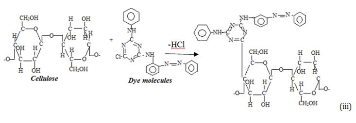
This procedure was followed for both bifunctional reactive dye stuff (galofast’me’ dyes) C.I Reactive Red brown ME RL as reported in previous studies [7, 8].
Characterization of Nano Silver Coated Dyed Cotton Fabrics
The functional groups of the coated fabric samples were revealed using Fourier transform infrared (FTIR)- attenuated total reflectance (ATR) sampler (Perkin Elmer spectrum 100, Waltham, USA) with the wave number range between 4000 to 500 cm-1. UV-blocking studies were analysed for un-coated, dyed and nanosilver coated dyed fabrics using UV-Visible Diffuse Reflectance spectroscopy (Lambda 35; PerkinElmer, USA) with the wavelength range between 270 and 800 nm. The ultraviolet protection factor (UPF) rating was calculated according to the ASTM D6603 standard. The surface morphology and elemental analysis of un-coated, dyed and nanosilver coated dyed cotton fabrics were analysed by SEM - EDX.
Bacterial Susceptibility Tests on Fabrics
Four selective bacterial cultures namely B.subtilis, E.coli, S.aureus and K.pneumoniae, were maintained in a specific agar media slants at 4°C. Antibacterial activity was screened using Kirby-Bauer disc diffusion method (1996) using Muller-Hinton Agar (HiMedia). About 15 ml of sterilised molten medium was poured into petri plates followed by swabbing of 0.1 ml inoculum (from 24 h old) uniformly over the agar. Silver nanoparticles at different concentrations were loaded individually on sterile discs (5 mm) followed by incubation of plates at 37°C for 24 h. After incubation, the incubation zone around the disc was measured with a transparent ruler and the experiment was performed in triplicates.
Results and Discussion
A colloidal solution of pale yellow indicates that the silver nanoparticles are formed which is clearly shown and confirmed from the earlier studies [22]. However, formed yellow color particles indicate the smaller size whereas bigger particles possess pale brown color [8, 16]. Particle size distribution of colloidal silver nanoparticles is shown in Figure 1a which reveals the particle size ranges from 20 (d10) to 85(d50) nm. However, a similar study shows particle size of colloidal solutions is in the range between 60 to 120 nm which is bigger particles than observed in our studies [8, 22, 23]. The zeta potential measurement of silver nanoparticles are graphically represented in Figure 1b. The zeta potential of the prepared colloidal solution is found to be -1.01 mV. However, a similar study on the silver nanoparticles are found to be -0.33 mV which is enhanced in our investigation [16, 22, 24]. The UV-Vis absorption spectra of silver nanoparticles formed in the reaction media has absorbance maxima at 407 nm as shown in Figure 1c. A remarkable broadening of peak around 350 nm to 480 nm indicates that the particles are polydispersed. It is observed that the peak is blue shifted in the absorption spectrum from 350 nm to 480 nm with an increase in reaction time. This results are correlated with an earlier investigation where colloidal silver nanoparticles solutions are prepared using NaBH4 as a reducing agent that increases the strength due to the surface plasmon absorption to 445 nm [23, 25, 26]. The chemical composition of the prepared silver nanoparticles solution analysed from ICP-OES is shown in Figure 1d. The silver content is found similar to the standard colloidal solutions and have element wavelength at 328 nm for the concentration of 468.9 mg L-1. TEM images of the colloidal silver nanoparticles obtained are observed to be uniform in size with spherical shape as shown in Figure 1e. The average particle size of the prepared silver nanoparticles is 20 nm. The selected area diffraction pattern (SAED) of the prepared nanoparticles (Figure 1e) reveals the crystalline nature.
![Figure 2: The peaks observed at 3342 and 2883 cm-1 correspond to the stretching modes of O-H and –C-H, respectively. The comparison of both the spectra shows the presence of an additional peak at 1710 cm-1 in treated fabrics. This band is attributed to -C=O stretching vibrations which indicates the presence of Ag coated on the dyed cotton fabric. The peak observed at 1642 cm-1 in all fabrics corresponds to the bending mode of water molecules (H-O-H) [15]. The above characteristic peaks with a slight shift of the peak 1325 to 1352 cm-1 corresponding to formed amide group. The band observed at 1107 cm-1 corresponds the asymmetric stretching of glucose ring, while the bands observed at 1026 and 1166 cm-1 correspond to the C-O stretching mode of cellulose [27]. In addition, the stretching vibration at 3414 corresponding to OH/NH2 groups is shifted to 3423 cm-1 which indicates the presence of functional groups of the bound silver particles. However, the peak observed at 795 cm-1 reveals the presence of Ag coated fabrics [28] as shown in Figure 2. The peaks seen at 654 and 795 cm-1 reveal the existence of Ag and dyed nanosilver coated fabrics.](/fulltextimages/825/fig_2.jpeg)
Performance of Nano Silver Coated on Dyed Cotton Fabric
The FTIR-ATR spectra of the un-coated, dyed and nano silver coated on dyed cotton fabrics are shown in Figure 2. The peaks observed at 3342 and 2883 cm-1 correspond to the stretching modes of O-H and –C-H, respectively. The comparison of both the spectra shows the presence of an additional peak at 1710 cm-1 in treated fabrics. This band is attributed to -C=O stretching vibrations which indicates the presence of Ag coated on the dyed cotton fabric. The peak observed at 1642 cm-1 in all fabrics corresponds to the bending mode of water molecules (H-O-H) [15]. The above characteristic peaks with a slight shift of the peak 1325 to 1352 cm-1 corresponding to formed amide group. The band observed at 1107 cm-1 corresponds the asymmetric stretching of glucose ring, while the bands observed at 1026 and 1166 cm-1 correspond to the C-O stretching mode of cellulose [27]. In addition, the stretching vibration at 3414 corresponding to OH/NH2 groups is shifted to 3423 cm-1 which indicates the presence of functional groups of the bound silver particles. However, the peak observed at 795 cm-1 reveals the presence of Ag coated fabrics [28] as shown in Figure 2. The peaks seen at 654 and 795 cm-1 reveal the existence of Ag and dyed nanosilver coated fabrics.
![Figure 3: The percentage of transmittance for UV-A and UV-B is almost constant in un-coated fabrics and the values are equivalent to 76%. The higher transmittance of UV indicates that the un-coated fabrics cannot be used to block both UV-A and UV-B radiations [29]. However, large reductions in transmittance is observed in cotton fabrics, which is due to the blocking of both UV-A and UV-B radiations. In both coated fabrics, the blocking rate of UV-B radiations is found higher than that of UV-A. The change of blocking rate of UV-A radiation in coated fabrics remain constant in the entire wavelength range from 315 to 400 nm whereas UV-A and UV-B radiations for nano silver dyed cotton fabrics is higher than pure dyed cotton fabrics. In addition, the blocking rate of UV radiation for nano coated fabrics after washing is almost constant, indicating that the subsequent washes lead to no change in the coating. Further, ultraviolet protecting factor (UPF) value is calculated using following equation [30] :](/fulltextimages/825/fig_3.png)
Performance of Nano Silver Coated UV- Protective Dyed Cotton Fabric
The wavelength dependent transmission spectra of un- coated, dyed and nano silver coated cotton fabrics is shown in Figure 3. The percentage of transmittance for UV-A and UV-B is almost constant in un-coated fabrics and the values are equivalent to 76%. The higher transmittance of UV indicates that the un-coated fabrics cannot be used to block both UV-A and UV-B radiations [29]. However, large reductions in transmittance is observed in cotton fabrics, which is due to the blocking of both UV-A and UV-B radiations. In both coated fabrics, the blocking rate of UV-B radiations is found higher than that of UV-A. The change of blocking rate of UV-A radiation in coated fabrics remain constant in the entire wavelength range from 315 to 400 nm whereas UV-A and UV-B radiations for nano silver dyed cotton fabrics is higher than pure dyed cotton fabrics. In addition, the blocking rate of UV radiation for nano coated fabrics after washing is almost constant, indicating that the subsequent washes lead to no change in the coating. Further, ultraviolet protecting factor (UPF) value is calculated using following equation [30] :
𝐸(𝜆)×𝑆(𝜆)𝑑(𝜆)
$$ \int_ {2 9 0} ^ {4 0 0} \frac {E (\lambda) \times S (\lambda) d (\lambda)}{E (\lambda) \times S (\lambda) \times \tau (\lambda)} \quad \mathrm {U P F} \quad (\mathrm {i v}) $$ = where (λ) represents the wavelength, E (λ) the relative erythermal effectiveness, s (λ) the solar UV spectral irradiance (W m-2nm-1), r (λ) the spectral transmittance of the specimen, and d (λ) the wavelength increment in nm [29, 31].
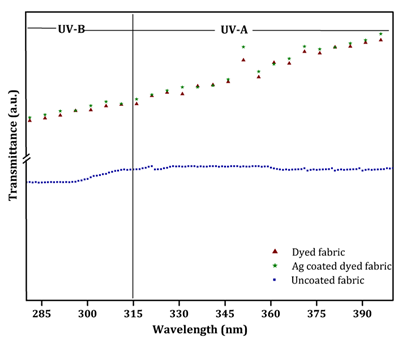
Figure 3: UV transmittance spectra of red color cotton fabric a) un-coated fabric b) Dyed cotton fabric and c) Nanosilver coated dyed fabric Fabrics assigned to UPF rating number and protection category are shown in Table 1. The test standard states that the highest UPF rating of 50. Garments made from fabrics with ratings higher than 50 are labeled as UPF 50+. The average UPF values of un-coated, dyed and nanosilver coated dyed cotton fabrics for red color is found to be 7.8, 30, and 35.2 respectively where as for yellow color fabric, the observed values are 7.8, 28.9, and 30.1 respectively. According to the Australia-New Zealand standard AS/NZS 4399:1996, the UV protection performance of cotton fabric is evaluated and expressed in terms of UPF. The UPF rating (Table 1) and transmittance results of the cotton fabric and nanosilver coated dyed cotton fabric before and after washing are shown in Figure 3 respectively. The results show that the dyed UV-protective nanosilver-coated cotton fabrics absorb UV-B better than UV-A. There is a slight decrease in the UPF rating of dyed cotton fabric and nanosilver- coated dyed cotton fabric but the fabric could still maintain very good protection against UV. The transmittance is slightly increased which shows the strong bonding existed between the dyed metal nanoparticles and cotton fabrics. The washing fastness of the nanosilver-coated dyed cotton fabric samples is observed to be satisfactory which demonstrates the high affinity of nanoparticles on the fabric.
| UPF Rating | Protection Category | UVR Blocked (%) |
|---|---|---|
| 25-24 | Good | 93.3-95.9 |
| 25-39 | Very Good | 96.0-97.4 |
| 40 and above | Excellent | 97.5 and more |
Table 1: Australia-New Zealand standard AS/NZS 4399:1996 UPF rating protection category and percentage of UV rays blocked.
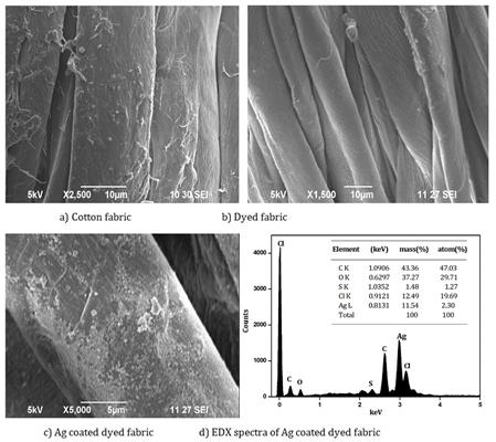
Table 1: Australia-New Zealand standard AS/NZS 4399:1996 UPF rating protection category and percentage of UV rays blocked. SEM image clearly shows the differences in the morphology patterns between the un-coated, dyed and nano silver coated dyed fabrics. The change surface morphology with slight agglomeration of particles coated on the cotton fabrics is confirmed from Figure 4. However, the particle size of silver nanoparticles used for coating is confirmed through by dynamic light scattering method. In addition, the presence of cellulose (carbon) and Ag are shown from the EDX (Figure 4d). These images show the durability of the silver nanoparticles coated on the dyed cotton fabrics that strongly adhered on the surface. The results support the presence of coating after subsequent washes.
Antibacterial Activity
The antibacterial activity of dyed and nano silver coated dyed fabric is shown in Table 2. The zone of inhibition at different concentrations of silver nanoparticles dyed fabric shows differences in zone of inhibition. Table 2 reveals that red dyed fabric coated with different concentrations (3, 5, 7 and 10 mol %) of silver nanoparticles show zone of inhibition (in mm) against Bacillus sp.(1.4±1.8), E. coli (0.9±1.2), Klebsiella sp. (1.0±1.3) and S.aureus (0.8±1.2). All the sample concentrations show better antimicrobial activity against Bacillus species and E. coli whereas; moderate activity is observed for strain such as Klebsiella sp. and S.aureus. It is clear that silver nanoparticles coated on dyed fabrics at different concentrations 0.7 mM and 1.9 mM inhibit growth of bacteria but the effect is more at less concentration. An increase in concentration of silver nanoparticles increases the bacterial inhibition.
| Bacteria | 3% Ag | 5% Ag | 7% Ag | 10% Ag | ||||||||||
| B. subtilis | 1.4 | 1.6 | 1.7 | 1.8 | ||||||||||
| E. coli | 0.9 | 1 | 1 | 1.2 | ||||||||||
| K. pneumoniae | 1 | 1.1 | 1.2 | 1.3 | ||||||||||
| S.aureus | 0.8 | 0.9 | 1.1 | 1.2 |
Table 2: Antibacterial activity of red color cotton fabrics in terms of zone of inhibition.
Conclusion
In the present study, silver nanoparticles are prepared from chemical reduction method and mixed with reactive dye (Red brown ME RL) on cotton fabrics. The Ag dye coated on cotton fabric is used to analyse its antimicrobial property and UV-protection ability. The nanoparticles binding capacity of dyed and Ag coated cotton fabric show better stability after washing, which is confirmed through SEM-EDX analysis. The antimicrobial activity results demonstrate that higher antibacterial activity and better UV-blocking properties are observed against Gram- positive (S.aureus and Bacillus) and Gram-negative (E.coli and Klebsiella) bacteria. Hence, such type of antimicrobial finish using nanoparticles can find wide application in the health and hygiene textile technology which can be further extended to polyester, silk and other fabrics.
Acknowledgements
One of the authors (Dr. R.S) is thankful to the University Grants Commission (UGC), New Delhi for granting Post-Doctoral Fellowship for Women (F.15-
1/2015-17/PDFWM-2015-17-TAM-36274 dt.12/10/2015).
Conflicts of Interest
Authors declare that we have no conflict of interest on submission of manuscript for publication.
Reference
1. Fujigaya T, and Nakashima N (2012) Soluble carbon nanotubes and nanotube-polymer composites. J Nanosci Nanotechnol 12(3): 1717-1738.
2. Anselmann R (2001) Nanoparticles and Nanolayers
In Commercial Applications. J Nanopart Res 3(4): 329-336.
3. Mohammedsadiq I, Dolly S, Chandrasekaran N,
Mukherjee A (2011) Ecotoxicity study of titania (TiO₂) NPs on two microalgae species: Scenedesmus sp. and Chlorella sp. Ecotoxicol Environ Safety 74(5): 1180-1187.
4. Parthasarathi V, Thilagavathi G (2011) Synthesis
and Characterization of Zinc oxide Nanoparticle and Its Application on Fabrics for Microbe Resistant Defence Clothing. International Journal of Pharmacy and Pharmaceutical Sciences 3(2).
5. Sharma VK, Yngard RA, Lin Y (2009) Silver
nanoparticles: green synthesis and their antimicrobial activities. Adv Colloid Interface Sci 145(1-2): 83-96.
6. Ali S, Ssleem S, Umbreen S (2009) ationizing efficiency and performance of antimicrobial agent on cotton fabric dyed with vinyl sulfone based reactive blue dye. Indian Journal of fiber & textile Research 34: 274.
7. Chatopadhyay DP, Patel BH (2009) D P Chatopadhyay and B H Patel, Indian Journal of fiber & textile Research. Indian Journal of fiber & textile Research 34: 368-373.
8. Ratyakshi, Chauhan Rp (2009) Colloidal synthesis of silver nanoparticles. Asian Journal of Chemistry 2: 113.
9. Chun DTW, Gamble GR (2007) Textile Technology
Using the Reactive Dye Method to Covalently Attach
Antibacterial Compounds to Cotton. The Journal of Cotton Science 11: 154-158.
10. Vesna LC, Saponjic Z, Vodnik V, Dimitrijevi SC, Jovan
PC, et al. (2012) The study of antibacterial activity and stability of dyed cotton fabrics modified with different forms of silver. Journal of Serbian chemical society 77(2): 225-234.
11. Malina D, Sobczak-Kupiec A, Wzorek Z, Kowalski Z
(2012) Silver Nanoparticles Synthesis with Different Concentrations of Polyvinylpyrrolidone. Digest Journal of Nanomaterials and Biostructures 7(4): 1527-1534.
12. Wang Q, Fan X, Gao W, Chen J (2006) Characterization of bioscoured cotton fabrics using FT-IR ATR spectroscopy and microscopy techniques. Carbohydrate Research 341(12): 2170- 2175.
13. Singha K, Maity S, Singha M (2012) The Salt-Free
Dyeing on Cotton: An Approach to Effluent Free Mechanism; Can Chitosan be a Potential Option? International Journal of Textile Science 1(6): 69-77.
14. Dastjerdi R, Montazer M (2010) A review on the application of inorganic nano-structured materials in the modification of textiles: focus on anti- microbial properties. Colloids Surf B Biointerfaces 79(1): 5-18.
15. Duran N, Priscyla DM, De Souza GIH, Oswaldo LA,
Esposito E (2007) Antibacterial Effect of Silver Nanoparticles Produced by Fungal Process on Textile Fabrics and Their Effluent Treatment. Journal of Biomedical Nanotechnology 3(2): 203- 208.
16. El-Shishtawy RM, Asiri MA, Nayera AMA, Maha M
AO (2011) In situ production of silver nanoparticle on cotton fabric and its antimicrobial evaluation. Cellulose 18: 82.
17. Benn TM, Westerhoff P (2008) Nanoparticle Silver
Released into Water from Commercially Available Sock Fabrics. Environmental science and Technology 42(11): 4133-4139.
18. Zhang L, Jiang Y, Ding Y, Daskalakis N, Jeuken L, et al. (2012) ZnO nanofluids–A potential antibacterial agent. Progress in Natural Science 18: 939-944.
19. Sangeetha G, Rajeshwari S, Rajendran V (2012)
Progress in Natural Science: Materials International 22(6): 693-700.
20. Chattopadhyay SN, Pan NC, Day A (2006) Bioresource Technology 97(1): 77-83.
21. Pinto VV, Ferreira MJ, Silva R, Santos HA, Silva F, et al. (2010) Long time effect on the stability of silver nanoparticles in aqueous medium: Effect of the synthesis and storage conditions. Colloids and Surfaces A: Physicochem. Eng Aspects 364:19-25.
22. Son WK, Youk J, Park WH (2012) Carbohydrate
polymers 87: 1419.
23. Xin JH, Daoud WA, Kong YY (2004) A new approach to UV-blocking treatment for cotton fabrics. Textile Research Journal 74(2): 97-100.
24. Cruickshank R (1968) Medical microbiology: a guide to diagnosis and control of infection. Edinburghand London: E&S. Livingston Ltd. pp: 888.
25. Popa M, Pradell T, Crespo D, Moreno JMC (2007)
Colloid surf A 30: 3184-3190.
26. Khan Z, Al Thabaiti SA, Yousif Obaid A, Al Youbi AO
(2011) Preparation and characterization of silver nanoparticles by chemical reduction method. Colloids and Surfaces B: Biointerfaces 82: 513-517.
27. Zhang Y, Yu L, Ke S, Shen B, Meng X, et al. (2011)
TiO2/SiO2 hybrid nanomaterials: synthesis and variable UV-blocking properties. J Sol-Gel Sci Technol 58: 326-329.
28. Bhatti IA, Adeel S, Siddique S, Abbas M (2012)
Journal of Saudi Chemical Society 12: 1319.
29. Guzmán MG, Dille J, Godet S (2009) International
Journal of Chemical and Biomolecular Engineering 2: 102.
30. Iqbal J, Bhatti IA, Adeel S (2008) Effect of UV
radiation on dyeing of cotton fabric with extracts of henna leaves. Indian Journal of fiber and Textile research 33: 157-162.
31. Abidi N, Hequet E, Tarimala S (2009) Journal of Applied Polymer Science 14: 3551-3556.
References
-
Fujigaya T, and Nakashima N (2012) Soluble carbon nanotubes and nanotube-polymer composites. J Nanosci Nanotechnol 12(3): 1717-1738.
-
Anselmann R (2001) Nanoparticles and Nanolayers In Commercial Applications. J Nanopart Res 3(4): 329-336.
-
Mohammedsadiq I, Dolly S, Chandrasekaran N, Mukherjee A (2011) Ecotoxicity study of titania (TiO₂) NPs on two microalgae species: Scenedesmus sp. and Chlorella sp. Ecotoxicol Environ Safety 74(5): 1180-1187.
-
Parthasarathi V, Thilagavathi G (2011) Synthesis and Characterization of Zinc oxide Nanoparticle and Its Application on Fabrics for Microbe Resistant Defence Clothing. International Journal of Pharmacy and Pharmaceutical Sciences 3(2).
-
Sharma VK, Yngard RA, Lin Y (2009) Silver nanoparticles: green synthesis and their antimicrobial activities. Adv Colloid Interface Sci 145(1-2): 83-96.
-
Ali S, Ssleem S, Umbreen S (2009) ationizing efficiency and performance of antimicrobial agent on cotton fabric dyed with vinyl sulfone based reactive blue dye. Indian Journal of fiber & textile Research 34: 274.
-
Chatopadhyay DP, Patel BH (2009) D P Chatopadhyay and B H Patel, Indian Journal of fiber & textile Research. Indian Journal of fiber & textile Research 34: 368-373.
-
Ratyakshi, Chauhan Rp (2009) Colloidal synthesis of silver nanoparticles. Asian Journal of Chemistry 2: 113.
-
Chun DTW, Gamble GR (2007) Textile Technology Using the Reactive Dye Method to Covalently Attach Antibacterial Compounds to Cotton. The Journal of Cotton Science 11: 154-158.
-
Vesna LC, Saponjic Z, Vodnik V, Dimitrijevi SC, Jovan PC, et al. (2012) The study of antibacterial activity and stability of dyed cotton fabrics modified with different forms of silver. Journal of Serbian chemical society 77(2): 225-234.
-
Malina D, Sobczak-Kupiec A, Wzorek Z, Kowalski Z (2012) Silver Nanoparticles Synthesis with Different Concentrations of Polyvinylpyrrolidone. Digest Journal of Nanomaterials and Biostructures 7(4): 1527-1534.
-
Wang Q, Fan X, Gao W, Chen J (2006) Characterization of bioscoured cotton fabrics using FT-IR ATR spectroscopy and microscopy techniques. Carbohydrate Research 341(12): 2170- 2175.
-
Singha K, Maity S, Singha M (2012) The Salt-Free Dyeing on Cotton: An Approach to Effluent Free Mechanism; Can Chitosan be a Potential Option? International Journal of Textile Science 1(6): 69-77.
-
Dastjerdi R, Montazer M (2010) A review on the application of inorganic nano-structured materials in the modification of textiles: focus on anti- microbial properties. Colloids Surf B Biointerfaces 79(1): 5-18.
-
Duran N, Priscyla DM, De Souza GIH, Oswaldo LA, Esposito E (2007) Antibacterial Effect of Silver Nanoparticles Produced by Fungal Process on Textile Fabrics and Their Effluent Treatment. Journal of Biomedical Nanotechnology 3(2): 203- 208.
-
El-Shishtawy RM, Asiri MA, Nayera AMA, Maha M AO (2011) In situ production of silver nanoparticle on cotton fabric and its antimicrobial evaluation. Cellulose 18: 82.
-
Benn TM, Westerhoff P (2008) Nanoparticle Silver Released into Water from Commercially Available Sock Fabrics. Environmental science and Technology 42(11): 4133-4139.
-
Zhang L, Jiang Y, Ding Y, Daskalakis N, Jeuken L, et al. (2012) ZnO nanofluids–A potential antibacterial agent. Progress in Natural Science 18: 939-944.
-
Sangeetha G, Rajeshwari S, Rajendran V (2012) Progress in Natural Science: Materials International 22(6): 693-700.
-
Chattopadhyay SN, Pan NC, Day A (2006) Bioresource Technology 97(1): 77-83.
-
Pinto VV, Ferreira MJ, Silva R, Santos HA, Silva F, et al. (2010) Long time effect on the stability of silver nanoparticles in aqueous medium: Effect of the synthesis and storage conditions. Colloids and Surfaces A: Physicochem. Eng Aspects 364:19-25.
-
Son WK, Youk J, Park WH (2012) Carbohydrate polymers 87: 1419.
-
Xin JH, Daoud WA, Kong YY (2004) A new approach to UV-blocking treatment for cotton fabrics. Textile Research Journal 74(2): 97-100.
-
Cruickshank R (1968) Medical microbiology: a guide to diagnosis and control of infection. Edinburghand London: E&S. Livingston Ltd. pp**:** 888.
-
Popa M, Pradell T, Crespo D, Moreno JMC (2007) Colloid surf A 30: 3184-3190.
-
Khan Z, Al Thabaiti SA, Yousif Obaid A, Al Youbi AO (2011) Preparation and characterization of silver nanoparticles by chemical reduction method. Colloids and Surfaces B: Biointerfaces 82: 513-517.
-
Zhang Y, Yu L, Ke S, Shen B, Meng X, et al. (2011) TiO2/SiO2 hybrid nanomaterials: synthesis and variable UV-blocking properties. J Sol-Gel Sci Technol 58: 326-329.
-
Bhatti IA, Adeel S, Siddique S, Abbas M (2012) Journal of Saudi Chemical Society 12: 1319.
-
Guzmán MG, Dille J, Godet S (2009) International Journal of Chemical and Biomolecular Engineering 2: 102.
-
Iqbal J, Bhatti IA, Adeel S (2008) Effect of UV radiation on dyeing of cotton fabric with extracts of henna leaves. Indian Journal of fiber and Textile research 33: 157-162.
-
Abidi N, Hequet E, Tarimala S (2009) Journal of Applied Polymer Science 14: 3551-3556.
- Solution-Processed Chiral Perovskites for Biomedical Applications
- Nanotechnology in Health Chemistry and Medicine: Current Challenges and Future Directions
- Human Exposure to Micro- and Nanoplastics: Pathways, Toxicity, and Intervention Strategies
- Exosome Nanomedicine for Cancer Therapy
- Micro and Nanoplastics–Plastisphere, Biotoxicity, Impact on Human Health, and Mitigation Strategies
- Process Validation of Cefixime Powder for Suspension Dosage Form, 50 mL