The Impact of the Notch1 on the Migratory Capacity and the Expression of E-Cadherin and CyclinD1 in Ameloblastoma Cells
Objective: This study aimed to explore the impact of the Notch signalling pathway on the migratory capacity of AM cells and the expression profiles of E-cadherin and cyclinD1 protein. Study Design: Experimental study. Place and Duration of the Study: Inner Mongolia Key Laboratory of Oral Craniomaxillofacial Diseases Research, Affiliated Hospital of Chifeng University, from June to October 2023. Methods: In vitro cultures of AM cells and dental follicle (DF) cells were established. Transwell assays were conducted to assess cell migration and invasion, while Western blot analysis was employed to evaluate the protein expression levels of Notch1, E-cadherin, and cyclinD1. The inhibition of the Notch signalling pathway was achieved using FLI-06, allowing for comparative analysis of the migration and invasion abilities of AM and DF cells, alongside the expression levels of E-cadherin and cyclinD1 proteins in AM cells. Results: AM cells exhibited significantly enhanced migration capacity compared to DF cells (P
Gabiyatu B¹, Tsagaankhuu S¹*, Li W², Tian Y², Li S², Baolidao², Wurihan³ and Boldbaatar D⁴
¹Department of Health Research, Graduate School of Mongolian National University of Medical Sciences, Mongolia ²Department of Oral Implantology, Affiliated Hospital of Chifeng University, China
³Department of surgery, Affiliated Hospital of Chifeng University, China ⁴Department of Physiology, Mongolian National University of Medical Sciences, Mongolia Keywords: Notch1; Ameloblastoma; Migration; E-cadherin; CyclinD1 Thesis
Abbreviations
AM: Ameloblastoma; DF: Dental Follicle; PMSF: PhenylMethylSulfonyl Fluoride; DMSO: DiMethyl SulfOxide; CDKs: Cyclin-Dependent Kinases.
Introduction
Ameloblastoma (AM), characterized by its benign nature yet aggressive behaviour, presents a formidable clinical challenge in the field of oncology [1, 2]. Despite advancements in surgical techniques, the high recurrence rates and associated morbidities underscore the urgency for novel treatment modalities. The conventional approach to treating AM, predominantly through complete osteotomy, while effective in tumor removal, often results in significant aesthetic and functional impairments [3, 4]. Facial deformities and jaw defects post-surgery not only impact the patient’s quality of life but also necessitate complex reconstructive procedures. Moreover, the propensity of AM for local recurrence and potential malignant transformation further complicates treatment strategies, necessitating a paradigm shift towards targeted therapies.
In recent years, the field of oncology has witnessed a surge in targeted therapies tailored to specific molecular pathways implicated in tumorigenesis. For AM, targeting key signaling pathways such as the Wnt/β-catenin pathway, which plays a crucial role in AM pathogenesis, holds immense promise [5, 6, 7, 8]. By inhibiting aberrant signaling cascades driving tumor growth, targeted therapies offer a more precise and effective alternative to traditional treatment modalities.
Studies have shown that various proteins in the body are involved in the metastasis of tumor cells. For example, reduced cell adhesion is associated with E-cadherin, while cell proliferation is linked to cyclinD [9, 10]. The Notch signaling pathway is a key pathway that regulates cell proliferation, differentiation, and apoptosis, playing a crucial role in development and tissue regeneration.
Recent research has indicated that the Notch signaling pathway is activated in AM cells and contributes to the development of the tumor [11, 12]. It has been suggested that Notch signaling may influence the migration and invasion of AM cells and impact the expression of E-cadherin and cyclinD1 [13, 14]. However, the precise mechanism remains unclear.
FLI-06 is a novel inhibitor that targets the Notch signaling pathway. Even at low doses, FLI-06 effectively inhibits Notch signaling and can serve as a blocker in relevant studies [15].
In this study, we treated cultured AM cells with the Notch signaling inhibitor FLI-06 to investigate its effects on cell migration, E-cadherin expression, and cyclinD1 expression. Understanding the role of the Notch signaling pathway in AM progression could provide new insights into targeted therapies for this condition. By inhibiting Notch1 with FLI- 06, we may be able to modulate cell migration and expression of key proteins involved in tumor growth. Further research in this area could lead to the development of more effective treatments for AM with fewer side effects.
Methodology
This study was approved by the Ethics Committee of the Mongolian National University of Medical Sciences (2023/3- 04). In vitro cultures of human-derived ameloblastoma (AM) cells and human-derived dental follicle (DF) cells were established. Transwell assays were conducted to assess cell migration and invasion, while Western blot analysis was employed to evaluate the protein expression levels of Notch1, E-cadherin, and cyclinD1. The inhibition of the Notch signalling pathway was achieved using FLI-06, allowing for comparative analysis of the migration and invasion abilities of AM and DF cells, alongside the expression levels of E-cadherin and cyclinD1 proteins in AM cells.
The FLI-06 protein quantitative reagent was procured from Sigma Company in the United States, Matrigel matrix adhesive was obtained from BD Company in the United States, DMEM medium and fetal bovine serum were acquired from GIBCO Company in the United States, and the Transwell lab was sourced from Costar Company in the United States. Western blot reagents were purchased from Millipore, Germany. The ECL chemiluminescence kit was bought from Millipore Company, trypsin from Invitrogen Company, Mouse anti-human Notch/E-cadherin from AMcam Company, Rabbit Anti-human cyclinD1 from Cell Signaling Corporation, the CO2 constant temperature incubator and enzyme labeling instrument from Thermo Corporation, Germany, the inverted microscope from Life Corporation, USA, and the gel imager from Bio-Rad Corporation, USA.
Ameloblastoma (AM) and dental follicle (DF) cells were cultured in DMEM medium supplemented with 10% fetal bovine serum in a CO2 incubator set at 37℃. Upon reaching the logarithmic growth phase, the cells were enzymatically dissociated using pancreatic enzymes and harvested for subsequent analysis (fourth generation).
For the assessment of cell migration ability, Transwell chambers were employed. Specifically, Matrigel was excluded for migration assays and included for invasion assays. Cells in logarithmic growth were enzymatically detached with pancreatic enzymes, suspended in serum-free medium, and enumerated under a high-power microscope. Subsequently, a 100 μL inoculum of cell suspension was added to the upper chamber of the Transwell apparatus, while 600 μL of DMEM medium (with 10% fetal bovine serum) was placed in the lower chamber. Following a 24-hour incubation period in a CO2 incubator, the media in both the Transwell chamber and the accompanying 24-well plate were aspirated and rinsed with PBS. The specimens were fixed with anhydrous ethanol at room temperature for 30 minutes, stained with 0.1% crystal violet, and rinsed with water after 10 minutes. Migration and invasion of AM and DF cells were observed under an inverted microscope (×200) in 8 randomly selected fields. Each experimental group was subjected to three independent replicates.
Cells in the logarithmic phase of growth were harvested from the pancreas following enzymatic treatment. A total of 150 μL of protein lysate containing phenylmethylsulfonyl fluoride (PMSF) was added, and the mixture was vortexed to ensure homogeneity before being subjected to low- temperature lysis for 30 minutes.
The cell lysate was then centrifuged at 12,000 rpm in a cold centrifuge for 10 minutes to obtain the supernatant. The total protein concentration was quantified using a BCA protein assay kit. Subsequently, the proteins in the cell supernatant were separated via 5% SDS-PAGE. After transferring to a polyvinylidene difluoride (PVDF) membrane, the membrane was washed with TBST buffer for 10 minutes, repeated three times.
A blocking solution containing 5% BSA was applied, and the membrane was incubated at room temperature for 1 hour. Following this, a diluted primary antibody (1:1000) was added and incubated overnight at 4°C. After washing three times with TBST for 10 minutes each, a secondary antibody diluted at 1:500 was added, and the membrane was incubated at room temperature for 1 hour. The membrane was then washed again with TBST for 10 minutes, repeated three times. The proteins were visualized using a hypersensitive ECL luminescence kit, and imaging was performed with a gel documentation system. Protein expression levels were quantified using ImageJ software. Each experiment was conducted independently in triplicate for each group.
FLI-06 was solubilized in dimethyl sulfoxide (DMSO) to form a 10 mM/L stock solution, which was then cryopreserved at -70℃. Prior to the commencement of the experiment, the stock solution of FLI-06 was further diluted to a concentration of 10 μM/L, while the control group remained untreated with FLI-06. The methodology for assessing cell migration and invasion followed the procedures outlined in the Cell Migration Test. The levels of Notch signaling pathway- associated proteins (Notch1, E-cadherin, and cyclinD1) were evaluated using Western blot analysis. Each experimental condition was independently replicated thrice.
Statistical analysis was conducted using SPSS software (version 22.0, SPSS Inc., Chicago, IL, USA) for data entry and statistical analysis in this study. Measurement data were presented as ¯x ±s, and the difference between the two groups was analyzed using LSD-t test, with α=0.05 as the significance level.
Results
Comparison of Migration and Invasion Ability of AM and DF Cells
The number of migration and invasion of the two kinds of cells was analyzed by LSD-t test, and the differences were statistically significant(P<0.05. The results are shown in Table 1.
| Cells | Migration | Invasion |
|---|---|---|
| AM | 147.40 ± 26.01 | 139.00 ± 26.11 |
| DF | 88.40 ± 10.70 | 80.40 ± 3.36 |
| T | 5.06 | 5.27 |
| P | 0.01 | 0.01 |
Table 1: Migration and invasion ability of AM and DF cells.
Expression of Notch Signaling Pathway in AM and DF Cells
Compared with DF cells, the expression levels of Notch1 protein and cyclinD1 protein in AM cells were significantly increased, while the expression level of E-cadherin was significantly decreased (P<0.05). The results are shown in Table 2.
| Cells | Notch1 | E-Cadherin | CyclinD1 |
|---|---|---|---|
| AM | 1.44 | 0.87 | 0.39 |
| DF | 0.41 | 1.23 | 0.19 |
| t | 24.35 | 11.43 | 14.24 |
| P | 0 | 0.01 | 0.01 |
Table 2: Expression of Notch signalling pathway related proteins in AM and DF cells.
Changes of Migration and Invasion Ability of AM Cells after Treatment with FLI-06
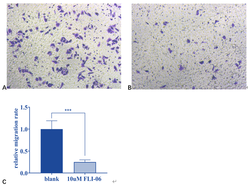
After Notch signaling pathway was blocked by FLI-06, the migration and invasion ability of AM cells was weaker than that of control group, and the differences were statistically significant (P < 0.05). The results are shown in Table 3 and Figure 1.
| AM | Control group | FLI-06 Treatment Group | T | P |
|---|---|---|---|---|
| Migration | 148.27 ± 28.16 | 40.40 ± 9.69 | 7.22 | 0 |
| Invasion | 137.20 ± 25.55 | 34.60 ± 6.95 | 8.21 | 0 |
Table 3: Migration and invasion ability of AM cells after FLI-06 treatment.
Expression Changes of Notch Signalling Pathway Related Proteins after FLI-06 Treatment
After AM was treated with FLI-06, the expression levels of Notch1 protein and cyclinD1 protein in AM cells were significantly decreased, while the expression level of E-cadherin was increased (P<0.05), as shown in Table 4 and Figure 2.
| AM | Notch1 | E-Cadherin | CyclinD1 |
|---|---|---|---|
| Control group | 1.44 | 0.87 | 0.39 |
| FLI-06 treatment group | 0.69 | 1.44 | 0.18 |
| T | 21.36 | 27.95 | 36.37 |
| P | 0 | 0 | 0 |
Table 4: Expression of Notch signaling pathway related proteins after FLI-06 treatment.
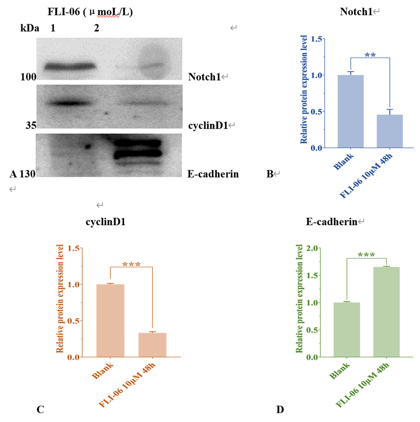
Figure 2: Expression changes of Notch1, cyclinD1 and E-cadherin proteins in AM cells treated with FLI-06 for 48h. (A) Western blot detection of the expressions of Nocth1, cyclinD1 and E-cadherin proteins (1, Blank. 2, FLI-06 10μmoL/L). (B) Gray ratio of Notch1 parameter before and after FLI-06 treatment. (C) The gray ratio of the parameters in cyclinD1 before and after FLI-06 treatment. (D) Gray ratio of E-cadherin parameter before and after FLI-06 treatment. Treatment at a concentration of 10μmol/L.
Discussion
Tumorigenesis is a complex process influenced by various factors, among which the Notch signaling pathway emerges as a key regulator in the differentiation and function of ameloblasts [11, 12, 16, 17]. Notch1, in particular, has been spotlighted for its indispensability in tooth morphogenesis and the determination of ameloblast fate [18, 19].
Previous studies have underscored the significance of Notch signaling in various developmental processes, including tooth development. Notch receptors, particularly Notch1, 3, and 4, alongside their cognate ligands, Jagged1 and Delta1, have been identified within the ameloblastic niche, implicating their involvement in regulating cellular behaviors [11, 12, 20]. Of note is the heightened expression of Notch4, hinting at its potential role in dictating ameloblast functionality.
In this study, the Transwell chamber assay was employed to assess the migratory and invasive properties of AM cells in comparison to DF cells, serving as a reference population. The experimental findings revealed a marked disparity in the migratory and invasive potentials between AM and DF
cells, with AM cells exhibiting significantly higher mobility and invasiveness. Upon treatment with FLI-06 to inhibit Notch signaling, a noteworthy reduction in the migration and invasion abilities of AM cells was observed, indicating the pivotal role of Notch signaling in orchestrating these cellular behaviors. These results underscore the functional relevance of the Notch pathway in regulating ameloblast migration and invasion, suggesting its potential as a therapeutic target for interventions aimed at modulating dental tissue remodelling and regeneration processes.
CyclinD1, a key player in the Notch signaling pathway, serves as a crucial regulator of the cell cycle by interacting with cyclin-dependent kinases (CDKs) and modulating the progression of various tumors [21, 22, 23, 24, 25]. This study employed Western blot analysis to scrutinize the expression profiles of Notch1, E-cadherin, and cyclinD1 in AM cells juxtaposed with dental follicle (DF) cells. The results delineated substantially elevated levels of Notch1 and cyclinD1 expression in AM cells in contrast to DF cells, while E-cadherin expression exhibited a marked diminution. Significantly, treatment with FLI-06, a potent inhibitor of the Notch signaling pathway, elicited a reduction in Notch1 and cyclinD1 expression levels in AM cells concomitant with a surge in E-cadherin expression. These findings underscore the pivotal involvement of Notch signaling pathway-associated proteins in the migration and invasion of AM cells, underscoring their potential as therapeutic targets.
The upregulation of Notch1 and cyclinD1 in AM cells signifies their pivotal roles in driving tumorigenesis and supports the notion that targeting these proteins could impede cancer cell proliferation and metastasis [26]. The inverse relationship between E-cadherin expression and tumor development further underscores the significance of cell adhesion molecules in tumor progression [27, 28, 29, 30]. The findings of this study elucidate the intricate interplay between Notch signaling components, cell cycle regulators, and cell adhesion molecules in orchestrating the migratory and invasive capacities of AM cells.
Furthermore, Studies have demonstrated that Notch1 may exhibit cross-regulatory interactions with the Wnt/β- catenin signaling pathway. Reduced expression of E-cadherin results in the dissociation of β-catenin from the cell membrane, enabling its translocation into the nucleus and subsequent activation of Cyclin D1 transcription. Notch1 may indirectly potentiate the β-catenin signaling by suppressing E-cadherin expression, thereby upregulating Cyclin D1 levels [12]. The positive correlation between Notch1 and cyclinD1 expression and the migratory and invasive capabilities of AM cells, coupled with the negative correlation between E-cadherin expression and these phenotypes, highlights the intricate regulatory network governing tumor progression in AM. These results not only deepen our understanding of the molecular mechanisms underpinning AM pathogenesis but also present a promising avenue for targeted therapy by modulating the Notch signaling pathway to impede tumor progression.
In conclusion, the findings of this study elucidate the critical role of the Notch signaling pathway in driving the migration and invasion of AM cells by modulating the expression of E-cadherin and cyclinD1. These insights not only shed light on the underlying molecular mechanisms of AM progression but also hold significant implications for the development of targeted therapies aimed at disrupting Notch signaling components to curb tumor growth and metastasis. By unraveling the intricate interplay between Notch1, E-cadherin, and cyclinD1, this study paves the way for novel therapeutic strategies in the management of AM and potentially other malignancies characterized by dysregulated Notch signaling. However, this study still has limitations. If Notch1 siRNA/Crispr is employed as the foundation, the knockout validation confirms that the observed effect of FLI-06 occurs via the Notch1 pathway. The experimental outcomes related to Notch1 are anticipated to improve. We plan to carry out further investigations in the future.
Acknowledgements
We appreciate Natural Science Foundation of Inner Mongolia Autonomous Region (grant no. 2021LHMS08026) , Program for Young people Talents of Science and Technology in Universities of Inner Mongolia Autonomous Region (grant no.NJYT 23064), and Natural Science Foundation International Cooperation and Exchange Project grant no.82361148724.
Competing Interests
The authors declare no competing interests.
References
-
Li WC, Ruan N, Tian Y, Zu W, Guan HJ, et al. (2019) Treatment for Unicystic Ameloblastoma by Adopting the Technique of Decompression by Fenestration with the Second Molar Preserved. SCIREA Journal of Clinical Medicine 4: 216-229.
-
Malphrus EL, Rivas E, Bryant JR, Oh AK, Rogers GF (2019) Hemimandibular Reconstruction of Pediatric Ameloblastoma with Templated Free Fibula Flap. Plastic and Reconstructive Surgery–Global Open 7(9): e2443.
-
Kreppel M, Zoller J (2018) Ameloblastoma-Clinical, Radiological, and Therapeutic Findings. Oral Dis 24: 63-6.
-
Effiom OA, Ogundana OM, Akinshipo AO, Akintoye SO (2018) Ameloblastoma: Current Etiopathological Concepts and Management. Oral Dis 24: 307-316.
-
Marquez C, Kirby J, Hunter KD (2024) Molecular Pathogenesis of Ameloblastoma. Journal of Oral Pathology & Medicine 53: 277-293.
-
Liu S, Liu D, Liu J, Liu J, Zhong M (2021) miR-29a-3p Promotes Migration and Invasion in Ameloblastoma via Wnt/beta-Catenin Signaling by Targeting Catenin Beta Interacting Protein 1. Head & Neck 43(12): 3911-3921.
-
Babichenko II, Tsimbalist NS, Rybalskaya VF, Sherstnev AA, Syomkin VA (2018) The role of Wnt/beta-catenin signaling pathway in ameloblastoma formation. Stomatologiia (Mosk) 97: 22-24.
-
Dutra SN, Pires FR, Armada L, Azevedo RS (2017) Immunoexpression of Wnt/beta-catenin signaling pathway proteins in ameloblastoma and calcifying cystic odontogenic tumor. J Clin Exp Dent 9(1): e136-E140.
-
Rubtsova SN, Zhitnyak IY, Gloushankova NA (2022) Dual role of E-cadherin in cancer cells. Tissue Barriers 10(4): 2005420.
-
Dai W, Dai YG, Ren DF, Zhu DW (2023) Dieckol, a natural polyphenolic drug, inhibits the proliferation and migration of colon cancer cells by inhibiting PI3K, AKT, and mTOR phosphorylation. Journal of Biochemical and Molecular Toxicology 37(5): e23313.
-
Pazhani J, Veeraraghavan VP, Jayaraman S (2023) Molecular docking analysis of cetuximAM with NOTCH signalling pathway targets for oral cancer. Bioinformation 19(4): 471-473.
-
Hashemi M, Hasani S, Hajimazdarany S, Mirmazloomi SR, Makvandy S, et al. (2022) Non-coding RNAs targeting notch signaling pathway in cancer: From proliferation to cancer therapy resistance. International Journal of Biological Macromolecules 222(Part A): 1151-1167.
-
Siar CH, Nakano K, Ng KH, Tomida M, Nagatsuka H, et al. (2010) Squamous odontogenic tumor of the mandible: a case report demonstrating immunoexpression of Notch1, 3, 4, Jagged1 and Delta1. European Journal of Medical Research 15: 180-184.
-
Siar CH, Nakano K, Han PP, Nagatsuka H, Ng KH, et al. (2010) Differential expression of Notch receptors and their ligands in desmoplastic ameloblastoma. Journal of Oral Pathology & Medicine 39(7): 552-558.
-
Gan RH, Lin LS, Xie J, Huang L, Ding LC, et al. (2019) FLI-06 Intercepts Notch Signaling and Suppresses The Proliferation and Self-renewal of Tongue Cancer Cells. OncoTargets and Therapy 12: 7663-7674.
-
Guo MZ, Niu Y, Xie M, Liu XS, Li XC (2023) Notch signaling, hypoxia, and cancer. Frontiers in Oncology 13: 1078768.
-
Li XX, Yan XC, Wang YF, Kaur B, Han H, et al. (2023) The Notch signaling pathway: a potential target for cancer immunotherapy. Journal of hematology & oncology 16: 45.
-
Yoshizaki K, Hu LZ, Nguyen T, Sakai K, Ishikawa M, et al. (2017) Mediator 1 contributes to enamel mineralization as a coactivator for Notch1 signaling and stimulates transcription of the alkaline phosphatase gene. Journal of Biological Chemistry 292(33): 13531-13540.
-
Costa NMMD, Fialho ADV, Proietti CC, Kataoka MSDS, Jaeger RG, et al. (2016) Role of hypoxia-related proteins in invasion of ameloblastoma cells: crosstalk between NOTCH1, hypoxia-inducible factor 1α, a disintegrin and metalloproteinase 12, and heparin-binding epidermal growth factor. Histopathology 69(1): 99-106.
-
Gabiyatu BB, Li WC, Wu RH, Tian Y, Li SH, et al. (2023) Expression of Notch1 in Ameloblastoma and Correlation with CBCT Imaging Subtypes of Ameloblastoma. Central Asian Journal of Medical Sciences 9(4): 182-188.
-
Li Y, Cheng XY, Chen CG, Wu HJ, Zhao H, et al. (2020) a flavonoid constituent derived from P. villosa, inhibits hepatocellular carcinoma cell growth by CyclinD1/CDK4 regulation via p38 MAPK-p21 signaling. Pathology - Research and Practice 216(1): 152701.
-
Febres CA, Chang JC, Ptashkin R, Wang YH, Gedvilaite E, et al. (2022) Rb Tumor Suppressor in Small Cell Lung Cancer: Combined Genomic and IHC Analysis with a Description of a Distinct Rb-Proficient Subset. Clinical Cancer Research 28(21): 4702-4713.
-
Sun HB, Han XH, Zhong M, Yu DJ (2020) Linc00703 suppresses non-small cell lung cancer progression by modulating CyclinD1/CDK4 expression. European Review for Medical & Pharmacological Sciences 24: 6131-6138.
-
Wang J, Zhang JX, Ma QL, Zhang SS, Ma FD, et al. (2023) Influence of cyclin D1 splicing variants expression on breast cancer chemoresistance via CDK4/CyclinD1- pRB-E2F1 pathway. Journal of Cellular and Molecular Medicine 27(7): 991-1005.
-
Ding C, Wei RQ, Rodriguez RA, Mullo MDMR (2019) LncRNA PCAT-1 plays an oncogenic role in epithelial ovarian cancer by modulating cyclinD1/CDK4 expression. Int J Clin Exp Pathol 12(6): 2148-2156.
-
Xu K, Zhang LF (2020) Inhibition of TUG1/miRNA- 299-3p Axis Represses Pancreatic Cancer Malignant Progression via Suppression of the Notch1 Pathway. Digestive Diseases and Sciences 65: 1748-1760.
-
Aborisade A, Akinyele A, Aregbesola B, Adesina O, Ladeji A (2022) Immunohistochemical expression of E-cadherin, N-cadherin and Snail/slug in ameloblastoma. Journal of Stomatology, Oral and Maxillofacial Surgery 123(6): e801-e807.
-
Mendonsa AM, Na TY, Gumbiner BM (2018) E-cadherin in contact inhibition and cancer. Oncogene; 37: 4769- 4780.
-
Rubtsova SN, Zhitnyak IY, Gloushankova NA (2022) Dual role of E-cadherin in cancer cells. Tissue Barriers 10(4): 2005420.
-
Kielbik M, Kielbik I, Klink M (2022) E-Cadherin Expression in Relation to Clinicopathological Parameters and Survival of Patients with Epithelial Ovarian Cancer. International Journal of Molecular Sciences 23(22): 14383.
- Diagnosis and Management of Mental Nerve Paresthesia Secondary to Apical Periodontitis of Mandibular Second Premolar: A CBCT Based Case Report
- A Randomized, Double Blinded Clinical Trial to Compare the Effect of Oral Premedication (Diclofenac Potassium or Dexamethasone) on Post-Operative Pain Following Pulpectomy
- Modified Lip Repositioning Technique for the Management of Excessive Gingival Display
- Integral Role of Non-Dental Providers and Fluoride Dissemination
- Root Canal Treatment Rate in Deciduous Teeth Among 6-Year- Olds in the Era of Discontinuing Water Fluoridation - Historical Cohort Study
- Treatment of Temporomandibular Disorder and Orofacial Pain in Unified Health System in Brazil