Acute Myocardial Infarction in Labor: A Case Report
Risk of AMI increased 3-4 fold just by pregnancy. Pregnancy related AMI mostly occurs due to spontaneous coronary artery dissection (SCAD). 37 years old, multiparous women, at 39 weeks of gestation was diagnosed with AMI due to thrombosis. Management of AMI is not clear. Vaginal delivery is may be safe and should be postponed for 2 or 3 weeks because of the increased risk of reccurrent myocardial ischemia or acute heart failure, if possible. If not, cesarean delivery might be an option. Adeaquate analgesia must be provided to avoid increased myocardial oxygen requirement.
Introduction
Acute myocardial infarction (AMI) incidence reported as 2,8- 6,2/100.000 deliveries [1]. Risk of AMI increased 3-4 fold just by pregnancy [1]. Factors such as hypertension, trombophilia, diabetes mellitus, tobacco use, maternal obesity, blood transfusion, postpartum infection, family history, hyperlipidemia, increased maternal age are associated with increased risk of pregnancy related AMI [2]. Less than 1 in 10 000 pregnancies are complicated with AMI. Unlike AMI that happens mostly because of plaque rupture, pregnancy related AMI occurs due to spontaneous coronary artery dissection (SCAD) [3]. Thanks to diagnostic and therapeutic developments, maternal mortality rates after AMI decreased to 5.1-7.3% [1]. Although pregnancy related AMI is reported throughout all trimesters and during postpartum period, it most commonly occurs during third trimester and postpartum period [4].
Management of AMI during labor is quite rare in literature. This case report is about management and considerations of AMI during labor and postpartum period.
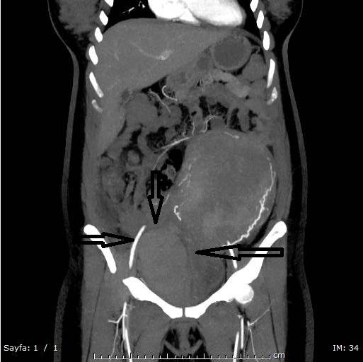
Gravida 2 para1, 37 years old lady, at 39 weeks’ gestation was presented with retrosternal chest pain to emergency department. She did not have any complications during prior normal vaginal delivery 14 years ago. Comprehensive physical examination including cardiac system osculation was performed and provided no additional finding. Electrocardiogram (ECG) records revealed ST segment elevation in leads II, III and aVF. Blood pressure (BP) was 130/85 mmHg and pulse 70 beats per minute and respiration 22 breaths per minute. She did not have any documented known risk factors for AMI. The patient’s history revealed that she had mitral valve replacement surgery four years ago due to embolism. Cardiology consultation notes showed that at early pregnancy the patient was recommended to replace warfarin sodium (Coumadin 5mg tb/ Zentiva /Czech Republic) with enoxoparin sodium (Clexane® 6000 anti- Xa / 0.6 ml/Sanofi Aventis/France). At 13 week’ gestation warfarin sodium oral tablet was added to existing treatment. Blood investigations showed elevated Troponin-T levels. She was admitted to cardiology inpatient unit and was prepared to coronary angiography. Coronary catheterization was performed through right radial artery access after shielding her back and abdomen with lead aprons to minimize fetal radiation exposure. Coronary angiogram showed total occlusion of posterolateral branch of right coronary artery. Medical follow-up decision was made and heparin infusion was started. During angiography procedure spontaneous rupture of amniotic membrane was suspected by the cardiologist and thus she was consultated to us. The cervix was three cm dilated 70% effaced and spontaneous flow of amniotic fluid was detected. Obstetric ultrasound reflected fetus at 38 weeks’ gestation. Heparine infusion was stopped after six hours of angiography procedure and vaginal delivery decision was made without any further medication. Patient was given labetolol tab. and maternal and fetal monitorization was performed. Six hours after the initial event, the cervix was fully dilated to 10 cm. After extensive consultation with the cardiologist, the decision to perform vaginal delivery was made. During an uncomplicated labor, a girl of 3000gr was born with an Apgar score of 9/10. Vacuum or forceps were not used. There was no observed tachycardia and hypertension during the delivery. After the delivery there wasn’t any laseration and heavy bleeding from the vagina. Abdominal ultrasonography was performed and there was no sign of bleeding; coagulation and placental retention. A total of five IU of oxytocin (Synpitan forte enj./Deva Inc./Turkey) was given in 500 cc Ringer lactate to run in two hours. Patient was admitted to coronary intensive care unit after two hours when there was no sign of active vaginal bleeding. At the second day, transesophageal echocardiography revealed thrombosis of the mechanical mitral valve prosthesis especially in the ventricular side. Mild to moderate paravalvular mitral valve insufficiency was detected. Patient suffered abdominal pain at third day of admission. Abdominal and pelvic examination was performed. Bimanuel examination showed solid lesion at the sides and in front of the vagina. Organized hematoma, about nine cm in diameter, at the right parametrial space was demonstrated by transvaginal ultrasonography. Antibiotherapy and analgesic treatment was given. Initial evaluation with CT showed that a hematoma was located at the right hemipelvis which was approximately 11 cm in diameter. Extravasation of radiographic contrast material to the abdomen was not seen on CT angiography and that was consistent with the CT which also was showed no active bleeding (Figure 1). Patient follow-up was done to check any progression or regression of the present hematoma with transvaginal and abdominal ultrasonography daily. There was no progression of the hematoma. Drainage was not seen appropriate because of localization of heamatoma. When the systemic hemodynamic state of the patient was stable and she felt good at eighth day, patient was discharged with warfarin sodium and clopidogrel (Plavix 75 mg tb/Sanofi Aventis/France). After one week, pelvic examination showed heamatoma was regressed from ten cm to five cm in diameter and there was no sign of ischemia on ECG. Pelvic examination or transvaginal ultrasonography showed no hematoma at the one month follow up.
Discussion
Not only atherosclerotik coronary artery disease is the most common reason of AMI in general population but it also is responsible one third of acute MI cases during pregnancy. SCAD is the most common cause of AMI in pregnancy. AMI due to coronary emboli has been described in only few case reports. Prostetic valves, mitral stenosis, vegetations in infective endocarditis, intracavitary thrombus in peripartum cardiomyopathy or paradoxial embolism might be the source of embolism. In our case, the origin of emboli was thought as prosthetic mitral valve.
ECG is a safe and noninvasive test to determine cardiac problems. Cardiac troponins (troponin I and T) are the most sensitive and specific markers of myocardial necrosis. Although CK-MB concentrations tend to elevate during pregnancy, troponins are still sensitive for pregnancy related AMI [5]. CT coronary angiography is an alternative and safe method to obtain anatomical information. But it’s use is limited during pregnancy because of fetal radiation risks and the need for excessive doses of B blockers for heart rate control. It is of the importance to minimize the fetal radiation while performing coronary angiography. Therefore appropiate abdominal protection, brachial or radial approach and short-term flouroscopy must be used. Estimated values of fetal radiation exposure are between 0.02 to 0.1 mSv during coronary intervention [6]. In cases of pregnancy related AMI, angiographic intervention is recommended above thrombolysis and surgery [3]. A safe method for maternal and fetal survival is thought to be percutaneous coronary intervention (PCI). It is unknown which drug regimen is the safest and most efficient one. Heparin use is safe during pregnancy. Cessation of heparin is required at least 6 hours prior delivery. Protamine sulphate’s antagonistic effect might be required to reduce the risk of bleeding which can be occur during delivery, operation or spinal/epidural anesthesia [7]. Ergot derivatives support uterine contractions and control hemorrhage after labor and abortion, however they might cause prolonged spasm and iatrogenic AMI in otherwise normal coronary arteries. There are several types of beta blocker but in this case labetolol could be a better choice. Except in patients who have stenotic or advanced cardiac disease, vaginal delivery is preferable. Delivery should be postponed for two-three weeks after initial event. If patient is unstable or there is an obstetrical indication, either an instrumental vaginal delivery or cesarean delivery could be performed, with measures taken to avoid increased cardiac workload such as epidural anesthesia, supplemental oxygen, left lateral position, treatment of hypertension and tachycardia. Cesarean section should be preferred in patients on clopidogrel and patients with stenotic or advanced cardiac disease. Vaginal delivery is tolerable for most of the patients with coronary artery diseases [3]. Pain could increase maternal cardiac rate and myocardial oxygen demand that’s why early continuous epidural anesthesia should be performed to minimize pain.
Obstetricians should be in cooperation with cardiologists, anesthesiologist and patients should be treated in an intensive care unit. Pregnancy related AMI is a rare and serious condition that special attention should be paid and needs multidisciplinary approach. Fortunately, the outcomes of maternal and fetal mortality rates decreased dramatically from %30 to %5-10 and %17 to %9 respectively [3]. Maternal mortality in patients with AMI in the peri-partum period is twice as higher than ante-postpartum periods [5]. The most critical time period in patients with pregnancy related AMI is the first 48 hours and these patients should be monitorized in coronary intensive care unit. The most approapiate approach is unclear and conclusions from a nationwide population based study would surely prove more reliable in clarifying the features of pregnancy related AMI.
References
-
James AH, Jamison MG, Biswas MS, Brancazio LR, Swamy GK, et al. (2006) Acute myocardial infarction in pregnancy a United States population-based study. Circulation 113(12): 1564-1571.
-
Kealey AJ (2010) Coronary artery disease and myocardial infarction in pregnancy: a review of epidemiology, diagnosis, and medical and surgical management. Canadian Journal of Cardiology 26(6): 185-189.
-
Roth A, Elkayam U (2008) Acute myocardial infarction associated with pregnancy. Journal of the American College of Cardiology 52(3): 171-180.
-
Roth A, Elkayam U (1996) Acute myocardial infarction associated with pregnancy. Annals of Internal Medicine 125(9): 751-762.
-
Ray P, Murphy GJ, Shutt LE (2004) Recognition and management of maternal cardiac disease in pregnancy. British Journal of Anaesthesia 93(3): 428- 439.
-
Conti, C Richard (2009) Cardiovascular studies and the radiation dose. Clinical cardiology 32(2): 56-57.
-
Jaiswal A, Rashid M, Balek M, Park C (2013) Acute myocardial infarction during pregnancy: a clinical checkmate. Indian heart journal 65(4): 464-468.
- Postpartum Maternal Mental Health - A Narrative Review
- Beta HCG in Cervico-Vaginal Secretion as a Predictor of Preterm Delivery
- Successful Management of Mid Trimester Foetal Death with Major Placenta Previa by Expectant Management Followed by Induction of Labour
- To Evaluate the Expression of Egr2 Gene in Term Low Birth Weight Newborns
- Impact of Maternal Obesity on Maternal and Foetal Outcomes: A Prospective Cohort Study from Northern India
- ‘’Benefit of Pulsatile GnRH Therapy in Treatment of Functional Hypothalamic Amenorrhea (FHA) and Congenital Hypogonadotropic Hypogonadism(CHH) in Infertile Patients Over Canonical Gonadotropins with IVF –A Short Communication’’