Peritonitis Leading to Sepsis Resulting from Postcoital Vaginal Laceration
Vaginal lacerations following consensual intercourse, while well described, are uncommon. Presenting symptoms are more commonly vaginal bleeding resulting in significant blood loss or hemorrhagic shock. Perforation into the abdominal cavity resulting in significant infectious etiology has been less described. We present a case of vaginal laceration following consensual intercourse resulting in peritonitis sepsis requiring prolonged hospitalization.
Introduction
Genital tract injuries following consensual intercourse are very uncommon, and when they occur are usually seen in postmenopausal women or those with weakened tissue [1]. Vaginal injuries often result from penetrating trauma, including non-consensual or forceful consensual coitus. Risk factors include first coitus, hypoestrogenic states, intoxication, pelvic radiation and anatomic abnormalities [2]. The most common sites for coital vaginal lacerations are the posterior fornix and lateral vaginal walls [2]. Treatment of the lacerations frequently requires surgical repair. Delay in diagnosis and treatment may result from the sensitive nature of the injury [3]. However, with delay in treatment vaginal bleeding may be profuse and lead to transfusion of blood products or hemorrhagic shock [3]. While most described complications are a result of blood loss, or damage to surrounding organs, significant infectious sequelae are less often described. In fact, for routine vaginal lacerations repairs antibiotics are not recommended [2]. We report a case of vaginal laceration following consensual intercourse resulting in purulent peritonitis leading to sepsis.
A 21-year-old Gravida 0 female, without significant past medical or surgical histories, presented to the emergency department with a chief complaint of abdominal pain and constipation. The patient stated that she had progressively worsening diffuse abdominal pain, flank pain and dysuria over 4 days. She also endorsed associated fever, malaise, nausea and vomiting. On physical exam the patient had bilateral flank pain and diffuse abdominal tenderness. The
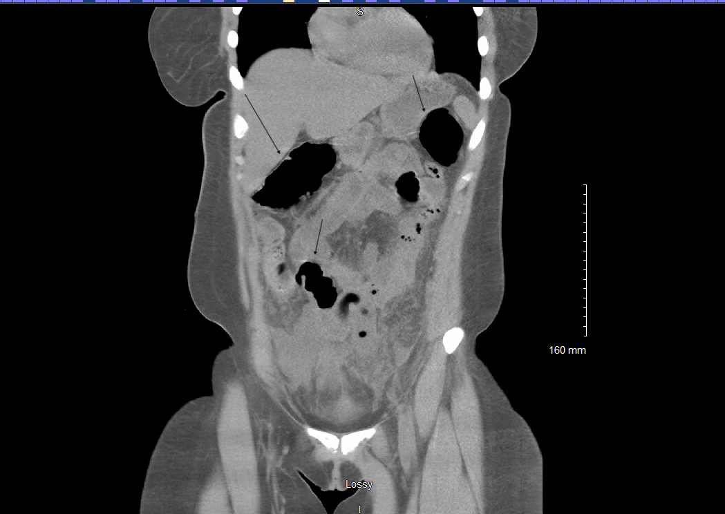
patient was febrile at 38.1 degrees Celsius (°C), tachycardic with heart rate (HR) in 110s beats per minute (bpm) and blood pressure (BP) was 90s/60s mmHg which responded to intravascular (IV) fluids rising to 130s/60s mmHg. Laboratory work up results showed a leukocytosis: white blood cell counts (WBC) 18.9 x10^3/ul with neutrophils 89.0 percent (%), Hemoglobin 11.5g/dL, international normalized ratio (INR) 1.5, Creatinine 1.76mg/dL, alanine aminotransferase (ALT) 21 unit/L, aspartate aminotransferase (AST) 24 unit/L, total bilirubin 2.7mg/dL, lactic acid 2.1mmol/L, human chorionic gonadotropin (HCG) negative, urinalysis from voided urine showed positive nitrites, WBC 283/high power fields (HPF), 287/HPF red blood cells, negative bacteria, and 5/HPF squamous epithelial cells. Urine culture was negative. The patient was admitted to the general medicine service with primary diagnosis of sepsis suspected to be secondary to pyelonephritis. Computerized tomography (CT) abdomen/pelvic was then performed and findings were read as pneumoperitoneum with a constellation of findings most compatible with enteritis, probable peritonitis with suspected bowel perforation, site uncertain. Appendix not visualized. Reproductive organs were within normal limits and kidneys showed no sign of hydronephrosis (Figure 1). Patient was started on IV antibiotics (Cefepime and Metronidazole). General surgery was consulted with a plan to proceed with diagnostic laparoscopy due to concern for intra-abdominal process. Risks, benefits and alternatives for the procedures were discussed with the patient and the patient signed the consent with a witness present.
CT showing pneumoperitoneum with a constellation of findings most compatible with enteritis (arrows), probable peritonitis with suspected bowel perforation, site uncertain. Appendix not visualized. Reproductiveorgans were within normal limits and kidneys showed no sign of hydronephrosis.
The patient was taken to the operating room where diagnostic laparoscopy was performed. Upon entry, the patient was found to have purulent fluid in the peritoneal cavity which was suctioned and sent for culture. Adhesions were noted in the pelvis between the omentum and the abdominal wall. After clearing the adhesions, more purulent fluid/pus was found in the pelvis. The tip of the appendix was noted to be inflamed and appendectomy was performed by general surgery. While the small bowel appeared to be inflamed, no bowel perforation was noted. After further exploration, an area of defect was found in the posterior fornix of the vagina. Gynecology (GYN) was consulted intraoperatively. The vagina was prepped with betadine and pelvic exam was performed revealing a 5 centimeter (cm) x 0.5 cm midline defect in the posterior fornix (intraoperative photos not available). The laceration was closed in a running locking fashion with absorbable suture. The abdominal cavity was irrigated with 80 milligrams of Gentamicin in sterile water, a Jackson Pratt (JP) drain was placed in the pelvis and the procedure was concluded.
After the procedure, a further history was taken from the patient. Patient admitted to having intercourse (consensual) for the first time 4 days prior. She denies any history of trauma or assault. She reported mild to moderate vaginal bleeding since intercourse she believed was normal.
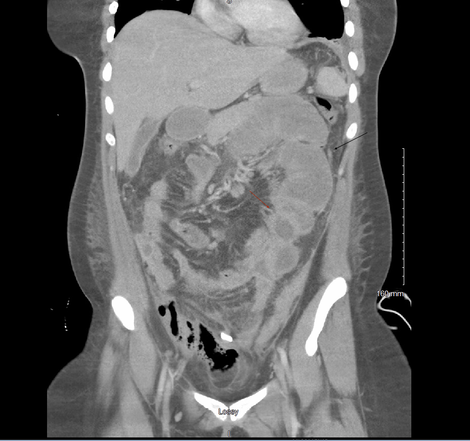
Postoperatively the patient continued to have intermittent fevers and leukocytosis ranging from WBC of 19-29 x10^3/ul and neutrophils of 78.0-90.6%. Patient also experienced an increase in her liver function tests (LFT) reaching a max of ALT/AST 233/356 unit/L. From the gynecology standpoint, the patient remained stable with minimal vaginal bleeding. Infectious disease was consulted, and the antibiotics were broadened with the addition of IV Vancomycin. Intraoperative intraabdominal cultures showed no growth, despite a positive gram stain. The patient continued to have fevers to a maximum temperature of 39.0°C, leukocytosis and elevated LFTs. Repeat CT scan was performed and found innumerable small abscesses throughout the abdomen and pelvis, the majority of which were too small for drainage or inaccessible (Figure 2).
The innumerable small abscesses throughout the abdomen and pelvis, the majority of which were too small for drainage or inaccessible. Dominant collection inseparable from bowel (red arrow). Generalized ileus also noted (black arrow).
CT was suggestive of a generalized ileus. Patient’s antibiotics were changed to IV Ertapenem, Daptomycin and Fluconazole. JP drain cultures were performed, with no growth noted. Blood cultures were repeated three times throughout the patient’s admission and showed no growth. Interventional Radiology (IR) was consulted, and conservative measures were recommended. The antibiotics were again changed to IV Meropenem, Micafungin and Daptomycin. Despite the need for continued broad spectrum antibiotics, the patient having no clinical complaints but, she continued to have persistent leukocytosis and fevers. A multi-disciplinary meeting was held with the patient’s care teams and the decision was made to transfer the patient to a higher level of care center.
The patient was transferred to a tertiary care center for drainage of abscesses by IR. Patient did not have any further procedures done after transfer of her care and IR drainage was not completed at the tertiary care center due to the proximity of the abscesses to the small bowel and the small size. She continued IV antibiotics and a peripherally inserted central catheter (PICC) line was placed. Vaginal examination was performed which revealed well healing vaginal tissue. She was discharged home from the tertiary care center on one month of IV Ceftriaxone and Metronidazole. The patient was seen in the office for a postoperative visit. At the postoperative visit her vitals were: BP 130/90mmHg and temperature 36.7°C. She reported no residual abdominal pain, no dyspareunia and had returned to regular menses. Pelvic examination was completed, revealing well healed vaginal laceration without defect.
Discussion
Vaginal lacerations following intercourse are not uncommon; however, usually result from non-consensual intercourse, or those with risk factors such as hypoestrogenic states, anatomic abnormalities, or pelvic radiation [2]. During coitus, the posterior fornix is weakened and subject to direct trauma as the upper vagina lengthens and the lower vagina contracts, increasing the risk of injury to the area [4]. Posterior vaginal fornix injuries have been reported to occur with consensual intercourse, but the incidence is higher during first intercourse, at age extremes, those with atrophic vaginal tissue or with genital size discrepancy [3, 5]. However, posterior fornix perforation is still rare and thought to occur in less than 1% of non-obstetric genital tract injuries [6]. Other risk factors for posterior fornix laceration include retroverted uterus and failure of normal vaginal lubrication or dilation, the latter of which is thought to be associated with more severe lacerations [3]. Recognition of these injuries can often be delayed due to the youth of the patient combined with the sensitive nature of the injury [3].
A common presentation of the injury is vaginal bleeding which may be severe enough to result in anemia, hypovolemic shock or even hemoperitoneum [3, 4, 7]. However, vaginal bleeding in reproductive aged women can be normal, and therefore in patients who are hemodynamically stable, with moderate vaginal bleeding, a high index of clinical suspicion is needed for the diagnosis of vaginal lacerations. Especially in the absence of any other gynecologic complaints. Other cases have been presented where patients present with peritoneal signs and lack of gynecologic complaints [6]. As in our case, this can lead to a delay in diagnosis of vaginal injury. Patients
who are hemodynamically stable, with minimal bleeding and low risk for infection may be treated conservatively [4]. However, those with ongoing inflammation, bleeding or infection often require surgical intervention. Repair may be completed vaginally, laparoscopically or via laparotomy depending on the patient’ symptoms and clinical stability [4]. In the case presented above the laceration was repaired vaginally; however, laparoscopic evaluation was required given the patient’s presentation.
In our case, the patient was found to have intraperitoneal perforation into the abdominal cavity. Though rare, intraperitoneal perforation can lead to pneumoperitoneum causing the patient to present primarily with peritoneal signs [5, 6, 8]. However, fewer reports exist of patients who develop significant infection or sepsis from perforation. Literature review found two similar cases where patients presented with suspected bowel perforation and sepsis [8, 9]. In both cases patients were taken to the operating room where purulent fluid was found in the abdomen and the cause was noted to be a perforating laceration from the posterior fornix. As in our case, patients required inpatient care and treatment with IV antibiotics following the procedure. However, our case differs in that the patient required prolonged hospitalization with formation of multiple abdominal abscesses and treatment with multiple IV antibiotic regimens and antifungal agents.
The diagnosis of vaginal lacerations can be difficult, especially when the patient is in the reproductive age and presents with few gynecologic complaints. The sensitive nature of the injury can lead to a delay in diagnosis: therefore, it is important to take a thorough history and have a high index of suspicion. It is important to remember that vaginal injuries with perforation into the abdominal cavity can have complications beyond acute blood loss and hemoperitoneum. Patients can develop peritonitis and sepsis leading to prolonged hospitalization and can even lead to more serious complications such as intra-abdominal abscesses.
Conclusion
While less described, vaginal lacerations with perforation into the abdominal cavity can result in significant infectious sequelae, including peritonitis and sepsis. Our case outlines rare complications that can arise from a seemingly straightforward condition. Therefore, it is important to monitor patients for continued complications following this diagnosis.
Declaration of Patient Consent
The author certifies that she had obtained all appropriate patient consent forms. In the form the patient has given her consent for her images and other clinical information to be reported in the journal. The patient understands that her name and initials will not be published, and due efforts will be made to conceal her identity, but anonymity cannot be guaranteed.
References
-
McLean I, Roberts SA, White C, Paul S (2011) Female genital injuries resulting from consensual and non- consensual vaginal intercourse. Forensic Sci Int 204: 27- 33.
-
Laufer MR, Makai G (2024) Evaluation and management of female lower genital tract trauma. UpToDate.
-
Frioux SM, Blinman T, Christian CW (2011) Vaginal lacerations from consensual intercourse in adolescents. Child Abuse and Neglect 35(1): 69-73.
-
Lal P, Mohan P, Sharma R, Sehgal A, Aggarwal A (2001) Postcoital vaginal laceration in a patient presenting with signs of small bowel perforation: report of a case. Surg Today. 31(5): 466-467.
-
Cohen A, Ulrich A, Semenyuk N (2020) A Laparoscopic Approach to Postcoital Vaginal Perforation in an Adolescent with peritonitis and Hypovolemic Shock. Journal of Pediatric and Adolescent Gynecology 33(5): 594-598.
-
Fletcher H, Bambury I, Williams M (2013) Post-Coital posterior fornix perforation with peritonitis and haemoperitoneum. International Journal of Surgery Case Reports 4(2): 153-155.
-
Astrup BS, Lykkebo AW (2015) Post-Coital Genital Injury in Healthy Women: A Rewiew. Clinical Anatomy. 28(3): 331-338.
-
Khosla AH, Singhal S (1997) Coital Tear: A Rare Cause of Secondary Peritonitis. Australia and New Zealand Journal of Obstetrics and Gynecology 37(2): 243-244.
-
Cheng AY, Cooley AS, Sulton CD (2019) Vaginal laceration in an Adolescent Girl Presenting with Abdominal Pain. Clinical Pediatrics 58(14): 1547-1549.
- Postpartum Maternal Mental Health - A Narrative Review
- Beta HCG in Cervico-Vaginal Secretion as a Predictor of Preterm Delivery
- Successful Management of Mid Trimester Foetal Death with Major Placenta Previa by Expectant Management Followed by Induction of Labour
- To Evaluate the Expression of Egr2 Gene in Term Low Birth Weight Newborns
- Impact of Maternal Obesity on Maternal and Foetal Outcomes: A Prospective Cohort Study from Northern India
- ‘’Benefit of Pulsatile GnRH Therapy in Treatment of Functional Hypothalamic Amenorrhea (FHA) and Congenital Hypogonadotropic Hypogonadism(CHH) in Infertile Patients Over Canonical Gonadotropins with IVF –A Short Communication’’