Rare Presentation of Ureterocele in Adult Male Patient
Background: Ureterocele presented with urine retention in a 38-year-old male patient is so rare to be seen. Case presentation: Male 38 Years old presented with severe lower abdominal pain and urine retention, Urine for c/s & urine routine analysis were normal. Abdomen & pelvis u/s revealed retention and cystic lesion in bladder mostly ureterocele with mild left hydroureteronephrosis. Foley’s catheter was inserted and evacuated more than 900cc urine and patient underwent urgent ureterocele resection and DJ stent insertion under GA. Conclusions: Ureterocele can present in such adult age in rare presentation like urine retention.
Introduction
A ureterocele is a cystic out-pouching of the distal ureter into the urinary bladder. Ureteroceles are swellings at the bottom of one of the ureters and they may cause a diagnostic and therapeutic dilemma, with perplexing clinical symptoms resulting from a spectrum of abnormal embryogenesis associated with abnormal development of the intravesical ureter, the kidney, or the collecting system.
Ureteroceles may be classified according to their relationship with the renal unit or on distal ureteral configuration and location.
Epidemiology
The incidence of ureteroceles has been reported as 1 in 500 cases [1]. Ureteroceles occur five times more frequently in females than in males, and more commonly in White individuals than in other races [2]. Unilateral ureteroceles occur with similar incidence on the right and left, and in 10 percent of patients there is bilateral involvement.
Classification
The classification system of the Section on Urology of the American Academy of Pediatrics divides ureteroceles based on their location to intravesical or ectopic [3]. Sometimes, to differentiate between intravesical and ectopic ureteroceles may be difficult.
Presentation
Ureterocele presented with variable spectrum of symptoms, however urine retention in adult male patients was so rare to be seen by many experienced urologists and it is necessary to be documented.
Male 38 Years old visited our clinic before one year with complaint of burning urination and left loin pain for 1 week, negative medical and surgical history apart from hyperlipidemia on Crestor 10 mg.
Urine for c/s & urine routine analysis were normal and Abdomen & pelvis ultrasound was normal, KUB and CT scan was inconclusive apart from mild left hydroureteronephrosis without presence of stones Figure 1.
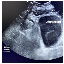
Pre-operative ultrasound revealed retention and cystic lesion in bladder mostly ureterocele with mild left hydroureteronephrosis. Pre-operative labs (CBC was normal, Creatinine = 1.2, Uric acid = 6.5, Urinalysis normal) (Figure 2).
Management and Outcome
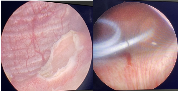
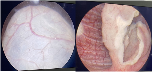
Urine retention due to obstructed ureterocele was relieved by foley’s catheter inserted and evacuated more than 900cc urine and patient underwent urgent ureterocele resection and DJ stent insertion under general anesthesia (Figure 3).
Discussion
Many ureteroceles are detected incidentally on antenatal ultrasonography [4]. Hydroureteronephrosis and bladder outlet obstruction are very common presentations of ureteroceles. This may lead to significant deterioration in kidney function [5]. The most common presentation after birth is during investigation for urinary tract infection (UTI) in the first year of life [6].
Obstruction of the bladder outlet is commonly caused by prolapse of the ureterocele into the urethra. A prolapsing ureterocele in a female patient may cause physical obstruction of the bladder neck. Anatomic obstruction of the bladder neck by the cystic ureterocele may cause obstructive voiding symptoms or may lead to acute urinary retention in both pediatric and adult patients [7].
A vulval mass caused by prolapse of the ureterocele is highly rare presentation. During per vaginal examination, a prolapsing cystic mass may be seen prolapsing from the meatus in both young and adult females. Bladder neck obstruction can even be caused by a large simple nonprolapsing intravesical ureterocele; it has been reported in an adult male [8].
Conclusions
Ureterocele can present in such adult age in rare presentation like urine retention.
Consent
No written consent has been obtained from the patient as there is no patient identifiable date included in this case report.
Conflicts of Interest
All authors have no conflict of interest related to the current manuscript.
References
-
Uson AC, Lattimer JK, Melicow MM (1961) Ureteroceles in infants and children: a report based on 44 cases. Pediatrics 27(6): 971-983.
-
Shokeir AA, Nijman RJ (2002) Ureterocele: an ongoing challenge in infancy and childhood. BJU Int 90(8): 777- 778.
-
Glassberg KI, Braren V, Duckett JW, Jacobs EC, Kinget LR, et al. (1984) Suggested terminology for duplex systems, ectopic ureters and ureteroceles. J Urol 132(6): 1153- 1154.
-
Bascietto F, Khalil A, Rizzo G, Makatsariya A, Buca D, et al. (2020) Prenatal imaging features and postnatal outcomes of isolated fetal duplex renal collecting system: A systematic review and meta-analysis. Prenat Diagn 40(4): 424-431.
-
Dada SA, Rafiu MO, Olanrewaju TO (2015) Chronic renal failure in a patient with bilateral ureterocele. Saudi Med J 36(7): 862-864.
-
Coplen DE, Duckett JW (1995) The modern approach to ureteroceles. J Urol 153(1): 166-171.
-
Zerin JM, Baker DR, Casale JA (2000) Single-system ureteroceles in infants and children: imaging features. Pediatr Radiol 30(3): 139-146.
-
Pike SC, Cain MP, Rink RC (2001) Ureterocele prolapse- rare presentation in an adolescent girl. Urology 57(3): 554.
- Results of 6-Month Follow-Up of Patients After B-Turp and Thulep
- The Effect of Drinking Water with a High Content of Antimony and Arsenic on the Dynamics of their Distribution in the Kidneys and the Renal Excretory Function in Rats
- Effectiveness and Safety of Tansurethral Thulium Laser Enucleation of the Prostate in the Treatment of BPH: Review
- A Systematic Review on Molecular Pathophysiology Involved in Chronic Kidney Disease and the Role of Animal Models in Drug Discovery to Manage in Chronic Kidney Disease - An Update
- Functional Development of Kidneys in Human Ontogenesis
- Testicular Metastasis: Uncommon Prostate Cancer Case Report