Changes in Thiols Indicators in the Lens of the Eye of White Rats under the Exposure of Radiation during Operation of a Cellular Mobile Phone
The results of previous work carried out at the Laboratory of Biophysics of Cellular Metabolism of the Institute of Physiology named after A.I. Karaev, which concerned the study of the effect of EMR on LPO processes and the antioxidant system in visual structures, pointed to the oxidative nature of the implementation of EMR exposure. Since thiol compounds in living tissues primarily interact with peroxide products, it was assumed that the effect of EMR should be reflected in the thiol content of the lens. For this purpose, a group of white outbred rats (40 species, half of which were identified as the control group) were studied, which were daily irradiated with EMR from a working cell phone. On the 7th, 14th, 21st and 28th day of the experiment, 5 animals from the experimental group and 5 animals from the control group were slaughtered. The lens of the eye was removed from the animals, in which the level of thiols was determined according to the modified Lindsey-Sedlak method based on the Ellman method. The results of the study showed that common and readily available (cytoplasmic) thiols in the nucleus of the lens of the eyes of rats irradiated with mobile phone EMR, in general, have similar dynamics of quantitative changes depending on the duration of exposure. Hidden thiols behave similarly. In the lens cortex, the nature of changes in thiols is not so pronounced, but generally similar to the results in the nucleus. The authors believe that these results may indicate the antioxidant properties of thiols in LPO reactions.
Introduction
Studies on various animal species show that microwave radiation of varying intensity causes significant physiological changes in their visual systems [1, 2]. In our previous studies conducted at the Laboratory of Biophysics of Cellular Metabolism of the Institute of Physiology named after A.I.
Karaeva, data were obtained indicating that exposure of white rats to high-intensity electromagnetic radiation (EMR) slows down the processes of lipid peroxidation (LPO) in the lens [3, 4]. In the format of expanding research on this topic, we conducted a series of experiments to study the effect of mobile phone radiation on the content of thiols in the substructures of the lens of the eye in albino rats.
The Aim of the Study
The purpose of this work was to study the indicators of thiols in the cortex and the nucleus of the lens of albino rats, depending on the duration and power of exposure of animals to EMR, which is formed during the operation of a mobile phone.
Materials and Research Methods
We studied outbred white rats 3 months old (males). The study generation (40 individuals) was divided into a control group (20 individuals) and 4 experimental groups (5 individuals in each group). Animals on each experiments which were carried out were irradiated using a standard mobile phone daily for 20 minutes. To do this, a call was made to a mobile phone located directly next to the animals from another phone and a connection was established for 20 minutes (i.e., the phone was in active mode for 20 minutes). Then after 20 minutes the telephone connection was interrupted. With the indicated daily exposure, the experimental groups were irradiated for 7, 14, 21 and 28 days.
The control group was kept in similar conditions except for the phone, which was not turned on. After the radiation exposure necessary for the experiments, the selected (irradiated) group of animals and 5 individuals from the control group were slaughtered on the seventh day of the experiments. The remaining part of the animals continued to be irradiated in accordance with the intended number of days (they were killed in the above order on the 14th, 21st and 28th day from the start of the experiments).
In slaughtered rats, the lens of the eye was removed by preparation for research. The content of sulfhydryl groups (thiols) was determined by the modified Lindsey-Sedlak method based on the Ellman method [5]. The principle of this method is to reduce 5,5’-dithiobis-(2-nitrobenzoic) acid with sulfhydryl groups to form a yellow colored compound having an absorption maximum at 412 nm. Calculations of the content of sulfhydryl groups were carried out according to the difference in optical densities at the first and 15th minutes (for easily accessible ones), at the first and 30th minutes (for masked ones).
In both cases, when measuring SH-groups in the lens, 20 µl of Ellman’s reagent was added due to the saturation of the lens with SH-groups. The content of hidden (masked) SH-groups was determined from the difference between the amount of total and easily accessible sulfhydryl groups. The content of SH groups in the studied lens substructures was expressed in nmol/mg protein. The numerical indicators obtained during the experiments were analyzed using simple methods of variation statistics [6].
Results and Discussion
The results of the study showed that in animals slaughtered after 7 days of irradiation, the content of readily available and total thiols, both in the nucleus and in the lens of the eye, changes slightly. The difference between the numerical indicators is not statistically significant, and in this case we can only speak of a slight trend towards an increase in the content of SH-groups in the studied lens substructures.
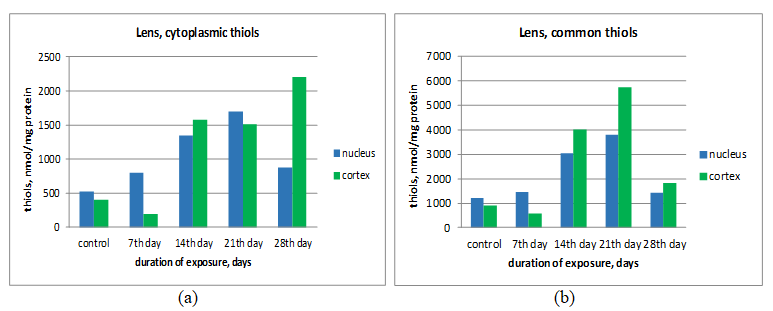
The amount of total and cytoplasmic thiols significantly increases in both substructures of the lens (indicators are statistically significant at p<0.05). Interesting in the results for this group of animals is a more pronounced change in the thiols of the cortex compared to the SH-groups of the nucleus, while in rats irradiated for 7 days, sulfhydryl groups, as well as in the experimental series, contain more in the nucleus.
The content of sulfhydryl groups in the nucleus and cortex of the lens of animals slaughtered after 21 days of irradiation also significantly exceeds the values of the control group. At the same time, the amount of cytoplasmic thiols exceeds the control values in both substructures by more than 200%, and total thiols in the nucleus increase by more than 350%, and in the cortex by almost 500%. However, the qualitative ratio of the amount of cytoplasmic thiols in the nucleus and lens cortex of the experimental animals again becomes the same as in the control ones (there are more SH- groups in the nucleus).
For common thiols, however, the tendency for a higher content of SH groups in the core than in the core is retained. Note that this trend for total thiols does not change in animals slaughtered after 28 days of exposure. In addition, this duration of irradiation is characterized by a drop in the content of total thiols in the nucleus to figures close to the control ones (the content of total thiols is almost the same as after 7 days of irradiation), while in the cortex the amount
Conclusions
Electromagnetic radiation from mobile phones affect the content of thiols in the substructures of the lens of the eye in albino rats. The noted phenomenon is characterized by an increase in the amount of reduced thiols in this tissue. The authors believe that this observation may reflect the antioxidant properties of thiols in LPO reactions.
References
-
Ismailova LF, Gadzhiev AM (2002) Influence of decimeter microwaves on the state of lipid peroxidation in the tissues of the visual system in young and adult rats. physiology and biochemistry 20: 117-125.
-
Bayramova SD, İbragimova JM (2019) Alteration in redox homeostasis of the rabbits eye lens. IV International Congress of the Georgian Society of Physiologists, IS Beritashvili, Biomedical series, 45(3-4): 417.
-
Ibragimova ZhM (2009) Study of the influence of decimeter electromagnetic radiation of non-thermal intensity on the state of oxidative stress in the lens of the eye and the possibility of its correction. Diss Candidate of Medical Sciences, Baku, pp: 144.
-
Ibragimova JM, Muxtarov MM, Bayramova SD (2021) Study of sulfhydryl groups in the eye lens of white rats subjected to electromagnetic radiation. XVII International Interdisciplinary Congress, Neuroscience for medicine and psychology, Sudak, pp: 159.
-
Sedlak J, Lindsey R (1968) Estimation of Total, Protein- Bound and Nonprotein Sulfhydryl Groups in Tissue with Ellman’s Reagent. Anal Biochem 25(1): 192-205.
-
Glantz ST (2011) Primer of Biostatistics. 7th (Edn.), McGraw Hill Professional, pp: 320.
-
Grigoriev YuG (2000) Long-term consequences of the biological action of electromagnetic fields. Radiation biology. Radioecology 40(2): 217-225.
-
Zubkova SM (2002) Biophysical and physiological mechanisms of the therapeutic effect of electromagnetic radiation. Physiotherapy, balneology and rehabilitation 2: 3-9.
-
Spector A (1995) Oxidative stress-induced cataract: mechanism of action. FASEB J 9(12): 1173-1182.
-
Hossmann KA, Hermann DM (2003) Effects of electromagnetic radiation of mobile phones on the central nervous system. Bioelectromagnetics 24(1): 49- 62.
- Sense, Gravity, Parity & Chirality in Mathematical Physics
- Quantum Lattice Simulations PHYSICS: Microcircuit Particle Formation and Observable Macroscopic Irreversible Time - A Discrete Lagrangian with Cellular Automata Framework
- Quantum Biology from Biomacromolecule to Cell, and Central Dogma Described by Quantum Theory
- Focus, Agility, Speed and Technology (FAST) for Sustainability and Growth
- Square Root Metric Geometry and Pati-Salam Model in Curved Space-Time
- A Simple System Demonstrating the Mpemba Effect in Classical Mechanics