Technical Parameters Selection on CT Scans Doses Used for Adult Diagnosis in Some Governmental Hospitals in Addis Ababa, Ethiopia
This study had assessed a technical parameters selection used on CT scan for adult diagnosis doses in Tikur Anbesa and Yekatit 12 specialized hospitals in Addis Ababa, Ethiopia. Data on Head, Chest, Abdomen-Pelvic, Abdomen, Brain, HRCT-chest, L-Sacral, and PNS scan regions were collected from a sample of 132. It was analyzed through descriptive statistics on Microsoft Excel 2016. The results were presented in the table, pie chart, line graph, and histogram. The parameter, Eff.mAs, pitch, table feed, and rotation time have strong correlations with CTDIvol, r(132) =0.749, α = 0.00, r (132) = -0.794, α = 0.00, r (132) = -0.708 α = 0.00, and r (132)=0.723, α = 0.00 respectively. Eff. mAs and DLP deviate by far from mean whereas KV, Rotation time, pitch and ED were almost nearly equal. The effects of technical parameters on doses descriptors agree on the theoretical facts, but for different scan regions 120KV was used. The dose descriptors of this study were greater than the studies in other countries. The medical technologist should have selected the appropriate KV based on scan regions. Relevant authorities should monitor the radiation management implementation in CT scan medical imaging procedure in hospitals where CT scan machines are employed.
Introduction
In medicine, CT uses special x-ray equipment and some mathematical algorithm to produce across-sectional imaging representing a slice of the person being image [1]. It exams at very high resolution that completed within a few seconds are routinely available today and offer excellent diagnostic capabilities combined with high patient comfort [2]. In CT; CTDI which is a measure of the dose deposited in a single axial slice of the patient. In order to calculate the total absorbed dose for a complete CT examination, the size of the body being scanned must be taken into account and influenced by a number of factors scanning mode, technical parameters, protocols, and others [3, 4, 5]. During a CT procedure; per- scan conditions checked, types of examination identified, the patient laid on the table, the table pass slowly through the center of large x-ray machine and, area of interest identified. With some types of CT scanner, the table stays still and the machine moves around the person. The person might hear whirring sounds during the procedure. During scanning procedure by selecting technical parameters according to the examination types and patients, the patient may be asked to hold the client breath to prevent blurring the images. The radiation exposure health risks are issue of concern and managing the radiation dose irrespective of its diagnoses benefit is found to be important.
This concerned issue resolved by adjusting the dose contributing control factors specifically the CT scan technical parameters, KV, mA, pitch, table feed and rotation time by the medical technologist. These factors are controlled based on patients’ age, sex, and weight. The technologists should have knowledge on how the technical factors affect the patient dose without affecting image quality and the radiation health risk of the patient. Therefore, knowing the effects of CT scan technical parameters factors on diagnosing dose facilitates the health diagnosis progress through imaging modalities in our working environment. In this way, now this study has tried to examine the relationship of the effects of the technical parameter on adult CT scan diagnosis dose under helical scanning mode. Nowadays, most referral hospitals in Ethiopia implementing the CT scan diagnosis services. Thus, identify whether the technical parameters of the CT scan parameters are scientifically implemented. So, the intention of this study was to assess the effects of CT scan technical parameters selection used on CT scan for adult diagnosis in Tikur Anbesa and Yekatit 12 specialized hospitals.
Materials and Methods
The study sought to assess the effects of technical parameters selection used on CT scan for adult diagnosis doses under helical scanning mode. More specifically, the study aimed at answering the following basic questions: How to describe the extent of the effects of CT scan technical parameters selection used on CT scan for adult diagnosis doses in Black lion and Yekatit 12 Special Referral Hospitals And how could the output diagnostic doses of CT scan parameters be interpreted in a perspective of other research results and estimated effective dose levels? Simple statistics and correlational analysis were used to reveal the status of the technical parameters selection and their outputs doses descriptors. To assess the issue under study, mixed research (explanatory design) approaches have been employed. Thus, of 132 sampled clients those underwent the CT scan for different examinations were taken. In order to address the significance of this study, a multistage sampling technique has been used. Firstly, purposive sampling technique has applied to identify the specialized referral hospitals. Next to this draw sampling technique was applied to scanning regions and sex types for adults. The primary data were obtained from the inputs of CT machine and the result of the inputs that displayed as output. The measurements were scan region of Head, Neck, Abdomen, Pelvic and Chest each using GE- 64 slice CT scan machine. Specifically, adult patients underwent the CT scan for Abdomen- Pelvic, Abdomen, Brain and C-spine, Brain, Chest, HRCT-chest, Head and Neck, L-Sacral, Neck, Pelvic, and PNS examinations. The average age of adults was 42- years-old with maximum age of 93 -years -old. The technical parameters used were KV, mA, Eff. mAs, pitch, rotation time, and table feed whereas the doses descriptors were the output radiation CTDIvol, DLP and calculated ED. The study used mixed data analyses techniques which have been fit to the qualitative and quantitative data. In this case, quantitative data analysis techniques namely simple statics and Microsoft excel of 2016 were implemented to analyses the data. Tables, pie chart, line graph, bar graphs and charts were employed to describe the results of analyzed data. In imaging CT scan diagnosis, the radiation doses descriptors are influenced by; KV, mA, Eff.mAs, pitch, rotation time and table feed.
Results
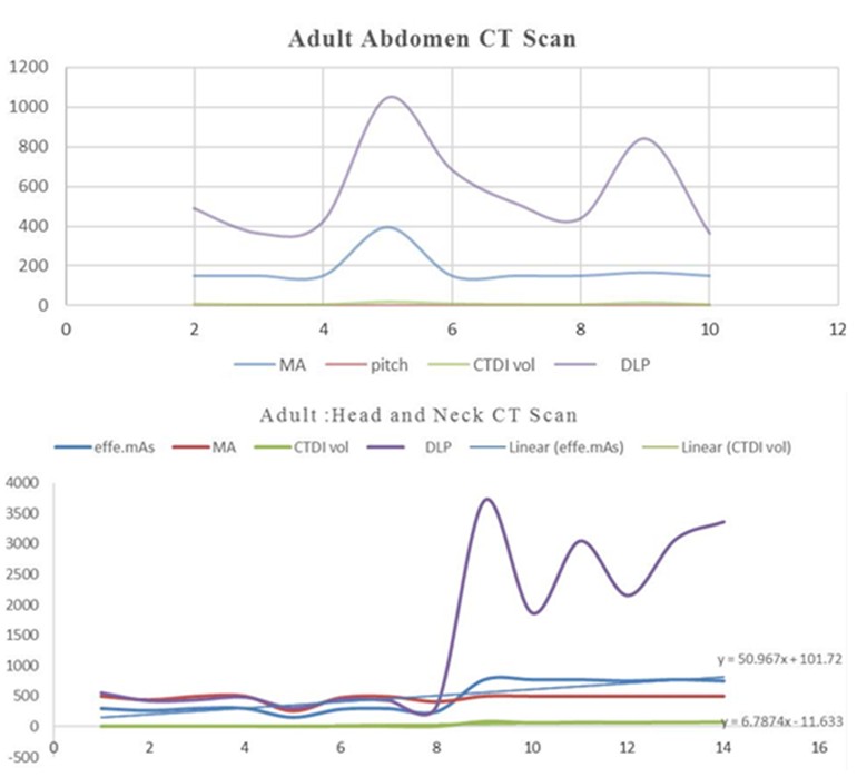
The results the data of 132 adults who underwent the CT scan for Abdomen-Pelvic, Abdomen, Brain and C-spine, Brain, Chest, HRCT-chest, Head and Neck, L-Sacral, Neck, Pelvic, and PNS CT examinations for different diagnosis to Tikur Anbesa and Yekatit 12 specialized hospitals in 2017 were presented as follows. The extent of CT scan technical parameters used effect adult CT scans diagnosis doses in Tikur Anbesa and Yekatit 12 special referral hospitals. In Adult abdomen CT scan diagnosis; constant 120KV, 396,150,167 mA, rotation time 0.8 sec, table feed with 39.4mm/rot and 55mm/rot, 0.984 and 1.375 pitch were used for acquisition. Thus, results an average estimated ED 8.6 mSv with max ED 15.7 mSv and min ED 5.47 mSv. As illustrated in figure 1:2 DLP increase as mA increase and decrease with other (i.e., 1049.9 mGy,843.11 mGy,469.23 mGy and 396mA,167mA,150mA respectively). In the same way, this relation was true for CTDIvol but pitch decrease as mA, CTDIvol, and DLP increase keeping other parameters constant.
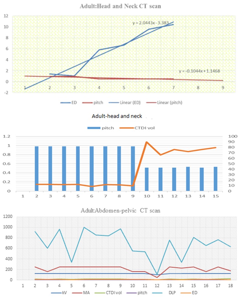
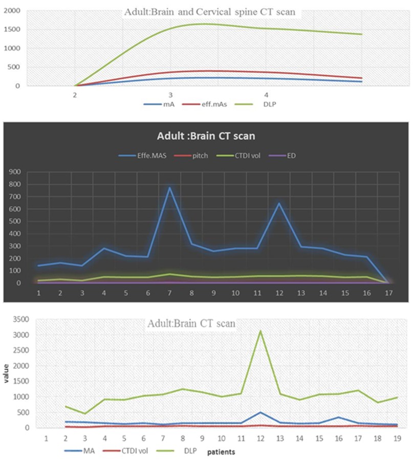
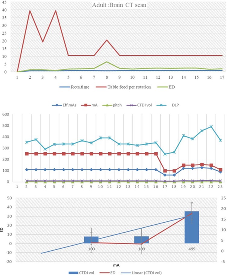
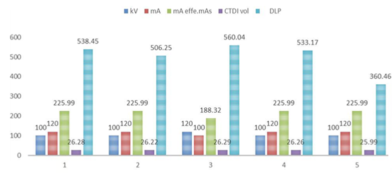
As observed from Figure 1:1 Eff.mAs and mA were increasing and decreasing simultaneously. The DLP also sharply increase and decrease simultaneously with them (i.e., Eff.Mas:751.79, 289.63, 267.68,250,157 and mA: 499,475,439,410,259 and DLP: 3363.94, 451.47, 424.45, and 309 respectively). Similarly, this fact was true for the Eff.mAs and CTDIvol. Thus, the linear Eff.mAs and CTDIvol equations were increasing and decreasing with each other for the same given value. As illustrated in Figure 1:2, ED increase significantly from point 3 to 4,5 to 6 and pitch decreases significantly at these points. Similarly, the linear lines equations were inversely proportional to each other. As seen in Figure 1:3, it shows that the pitch decreases sequentially as CTDIvol increases which was proportionally to adult head and neck CT scan. An estimated average ED was 4.3 mSv with max ED 11.5mSv and min ED 0. 96mSv. From the above Figure 1:4; there was significant variation in DLP and mA on the graph. Hence, as mA increase DLP also increase and vice- versa. From point 4 to 9 at constant KV and mA; pitch decrease as ED increase. At point 12, DLP and mA sharply decreased as KV decrease from 120 to 100 KV. And the average estimated ED 10.1 mSv obtained. The adult brain and cervical CT scan underwent at constant 120KV, pitch 0.969, table feed per rotation in mm 19.4 and rotation time 0.8 sec. The mA for this parameter was 200 and 115, and obtained the average estimated ED was 2.45mSv. As illustrated in Figure 1:5, the technical acquisition parameters mA and Eff.mAs decrease as DLP decreases. The technical parameters used were able to deliver the average estimated ED of 2.098975 with max ED 6.5816 mSv and 0.9698 mSv. Constant 120KV, mA varies from 499 to 113, pitch varies from 0.984 to 0.516, table feed per rotation from 39.4 mm to 10.6mm, rotation time from 1 to 0.8 sec. As seen on Figure 1:6 at constant 120 KV, as DLP and CTDIvol increases mA increase and vice versa but with different degree. This shows that direct relationship of DLP and mA was observed. As illustrated on Figure 1:7 table feed per rotation and ED significantly changes with each other linearly. In the adult chest, CT scan acquisition, 11male and 11 female patients whose age ranges between 20-years- old and 70 -years-old underwent with GE -64 slice CT scan in helical scanning mode. Constant 120 KV and varies mA from 100 to 250 used. The average doses descriptors yield CTDIvol, DLP and ED were 9.39 mGy, 355.7 mGy-cm and 4.9 mSv respectively. Further, the technical parameter used yields max ED 6.9 mSv and min-ED 3.5mSv.
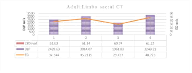
As Figure 1:8 illustrates, Eff.mAs, mA and CTDIvol increase and decrease with together (i.e.Eff.mAs 82.56,89.99,411.97 and mA: 100,109,499 and CTDIvol:7.79,7.89,35.56 respectively). In the adult, high-resolution chest CT scan acquisition 3 female patients whose age ranges between 50-year-old and 70 -years -old underwent with GE 64 slice machine in helical scanning mode. This CT scan diagnosis under went under constant technical parameters of, 120 KV, rotation time 0.8 sec and pitch 0.969 and the variations of mA from 100 to 499. The average doses descriptors yield CTDIvol, DLP and ED were 17.08 mGy, 598.94 mGy-cm and 8.38 mSv respectively. As illustrated on Figure 1:9 CTDIvol increase with mA and ED 109 mA to 499 mA ED also increases linearly. In this study, two female and two male patients underwent CT scan to limbo sacral CT examination under constant technical parameters of, table speed 20.6mm/rot, 120KV, rotation time 1sec, 12.5 mm slice thickness, 360mA, and 0.516 pitch. Hence, these parameters yield the average estimated ED of 40.17638 mSv with max ED 48.723mSv and min-ED 29.427 mSv. As Figure 1:10 shows, the output doses have positive relations with each other. Thus, with minimal changes in CTDIvol; ED has linearly changed with DLP. In this study, two male and three female patients, who underwent to Para nasal sinus CT examination in the helical mode were considered. In adult Para nasal sinus CT scan the technical parameters used were constant rotation time one sec, pitch 0.531, table feed 10.6 mm/rot and head size. The average estimated ED 1.05 mSv yielded as results of the selected parameters.
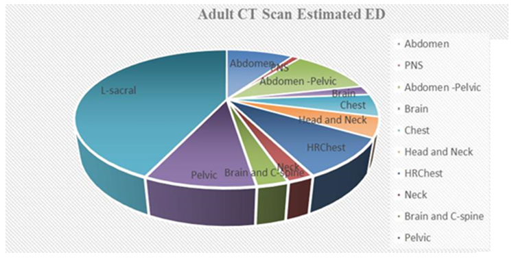
As showed on Figure 1:11, both mA and KV affect CTDIvol nearly by equal value if their value interchanges. Thus, both technical parameters affect the doses descriptors straight forwards. As shown in Figure 1:12 L-sacral, pelvic, abdomen- pelvic, abdomen and HRCT- chest of adults exposed to high estimated ED than others region. The Table 1:1 shown, Eff. mAs, pitch, Table feed, and rotation time have strong correlations with CTDIvol, r (132) =_0.749, α=0.00, r (132) = -0.794, α=0.00, r (132) = -0.708 α=0.00, and r (132) = 0.723, α=0.00 respectively. Similarly, Eff.mAs and pitch have strong correction with DLP, r (132) = 0.868, α=0.00 and r (132) = -0.543, α=0.00. This indicate that as Eff.mAs and Rotation time increase CTDIvol increase but it decreases as pitch and Table feed increase. And as Eff.mAs increase DLP increase but decrease as pitch increase. Whereas mA and CTDIvol have weak correlation with r (132) =0.184, α=0.022, KV and CTDIvol have insignificantly correlated with r (132) = 0.063, α=0.439. KV and CTDIvol have insignificantly correlated with r (132) =0.130, α= 0.107. Eff.mAs and DLP have strong correlation with DLP, r (132) =0.868, α=0. 00. And mA, Pitch, table feed and rotation have moderate correlation with DLP, r (132) = 0.467, α= 00, r (132) = -0.543, α= 0.00 and r (132) = -0.413, α= 0.00 and r (132) = 0.436, α=0.00 respectively. Eff.mAs and mA have moderate correlation with ED, r (132) =0.404, α=0.00 and with r (132) =0.317, α= 0.00 respectively. Whereas KV, pitch, table feed, and rotation time were insignificantly correlated with ED with r (132) =0.145, α=0.073, r (132) =0.031, α =0.701, r (132) =0.114, α=0.159, and r (132) =0.029, α=_0.719 respectively. The existence of significant correlation within the independent variables was checked using assumption test and reveal that it wouldn’t affect the results. As depicted by above Table 1:2. Eff.mAs and DLP deviate by far from mean whereas KV, Rotation time, pitch and ED were almost nearly equal. The outputs diagnosis doses of CT scan parameters interpretation in a perspective of other research results and estimated dose Levels. As can be seen from Table 1:3 the chest CT scan CTDIvol of this study was greater than Luxembourg and Ireland and less than Turkey and Italy. The HRCT-chest CT scan CTDIvol of this study was larger than Turkey and Luxembourg. The CTDIvol of Turkey and Italy were greater than this study but the CTDIvol value obtained from this study was greater than Ireland. As can be seen from Table 1:4 the DLP of adult chest of this study was larger than DLP of Turkey and Ireland but smaller than Italy in the literature. As shown in Table
1:5, ED of adult chest CT scan of this study was larger than in Turkey, in Syria and Ireland but smaller than in Italy in literature. The ED of HRCT-chest of adults in this study was twice of the ED adults in Turkey in literature. Similarly, the Abdominal CT scan of adult this study was larger ED adults in Turkey, Italy, Syria, and Ireland. Similarly, ED of pelvic in this study was larger than ED in Turkey, Syria, and Ireland smaller than Italy in the literature. As seen on Table 1:6, in dose value; max CTDIvol of this study was larger than the other dose values in Al-Ribat University Hospital Khartoum, Sudan. Similarly, the max and min DLP values were larger than the value obtained from the brain CT scan of the Sudan and the ED of this study were larger than the ED of the brain CT scan obtained in Al-Ribat University Hospital Khartoum. For acquisition parameters, table feed of this study was larger than that of the Al-Ribat University Hospital but the time of rotation was smaller than it in the adult case. In the pediatric brain CT scan, the value of rotation time and table feed in Al-Ribat University Hospital was larger than the value of this study. Similarly, the values of CTDIvol and DLP in Al- Ribat University Hospital were larger than the value obtains in this study. But ED of this study was larger than the value obtained in Al-Ribat University Hospital Khartoum, Sudan this show other acquisition parameters highly dominate the parameter illustrated in this table. Dose Level; minimal dose: less than 0.1 mSv, low Dose: 0.1 to 1 mSv, medium dose: 1 to 10mSv, High dose: 10 mSv and over [4]. As depicted in the above Table 1:7, most ED values of pediatric fall under medium dose and few under high dose. Similarly, the ED values of adult falls under medium dose and some under high dose.
| Independent variables | Dependent Variables | |||
|---|---|---|---|---|
| CTDIvol | DLP | ED | ||
| KV | Pearson Correlation | .063 | .13 | .145 |
| Sig. (2-tailed) | .439 | .107 | .073 | |
| mA | Pearson Correlation | .184* | .467** | .317** |
| Sig. (2-tailed) | .022 | .000 | .000 | |
| Eff.mAs | Pearson Correlation | .749** | .868** | .404** |
| Sig. (2-tailed) | .000 | .000 | .000 | |
| Pitch | Pearson Correlation | -.794** | -.543** | .031 |
| Sig. (2-tailed) | 0 | .000 | .701 | |
| Table feed | Pearson Correlation | -.708** | -.413** | .114 |
| Sig. (2-tailed) | 0 | .000 | .159 | |
| Rotation time | Pearson Correlation | .723** | .436** | .029 |
| Sig. (2-tailed) | .000 | .000 | .719 | |
| N | 154 | 154 | 154 |
| Input Variable | Age | N | Mean | Std. Deviation | |
|---|---|---|---|---|---|
| KV | 16-93 | 132 | 118.9916 | 4.39471 | |
| mA | 16-93 | 132 | 214.7311 | 112.66161 | |
| Eff.mAs | 16-93 | 132 | 231.3966 | 159.8836 | |
| Rotation time | 16-93 | 132 | 0.8303 | 0.14875 | |
| pitch | 16-93 | 132 | 0.8662 | 0.32869 | |
| Table feed | 16-93 | 132 | 30.2874 | 17.49779 | |
| Adult Examination | This Study | Turkey | Italy | Luxembourg | Ireland |
| 2014 | 2014 | 2014 | 2012 | ||
| Chest | 11.25958 | 11.6 | 15 | 6.8 | 9.3 |
| HRCT-chest | 12.34703 | 11.3 | - | - | 6.6 |
| Abdominal | 11.23444 | 13.3 | 18 | 9.6 | 12.3 |
| Pelvic | 13.23 | 19.4 | 18 | - | 12.3 |
- Adult Examination this study
- Turkey
- Italy
- EC
- Ireland
- 2014
- 2014
- 2011
- 2012
- Chest
- 454.3588
- 289
- 569
- 394
- 393
- HRCT-chest
- 598.94
- 283
- -
- -
- 276
- Abdominal
- 575.3489
- 204
- 555
- 464
- 598
- Pelvic
- 568.05
- 421
- 360
- 434
- 598
Table 3: 4: DLP for adults in this study and other countries [5].
| Adult Examination | In this study | In Turkey | In Italy | In Syria | In Ireland |
|---|---|---|---|---|---|
| 2014 | 2014 | 2009 | 2012 | ||
| Chest | 6.361052 | 4.1 | 8 | 5.4 | 6.2 |
| HRCT-chest | 8.382407 | 4 | - | - | - |
| Abdominal | 8.628667 | 3.1 | 7.8 | 7.7 | 8.3 |
| Pelvic | 8.5208 | 6.3 | 8.9 | 6.8 | 8.2 |
| Value | Adult Doses and Acquisition parameters | |||||||||
|---|---|---|---|---|---|---|---|---|---|---|
| CTDIvol | DLP | ED | Rotation time | Table feed | ||||||
| In Khartoum | this study | In Khartoum | this study | In Khartoum | this study | In Khartoum | this study | In Khartoum | this study | |
| Ave. | 67.43 | 46.204 | 1000.25 | 997.98 | 0.48 | 2.099 | 2.4 | 0.942 | 17.84 | 18.16 |
| Max | 69.12 | 84.25 | 1419 | 3134.1 | 0.49 | 6.58 | 2.5 | 1 | 19.5 | 39.4 |
| Min | 63 | 22.75 | 323 | 461.82 | 0.44 | 0.97 | 1 | 0.8 | 1 | 10.6 |
| Examination | Adult ED | Rank |
|---|---|---|
| Abdomen | 8.62 | medium |
| PNS | 1.05 | medium |
| Abdomen -Pelvic | 10.31 | High |
| Brain | 2.09 | medium |
| Chest | 4.98 | medium |
| Head and Neck | 4.31 | medium |
| HRCT- Chest | 8.38 | medium |
| Neck | 2.08 | Medium |
| Brain and C-spine | 2.45 | Medium |
| Pelvic | 8.52 | Medium |
| L-sacral | 40.17 | High |
Discussion
- The extent of CT scan technical parameters used affect an adult CT scans diagnosis doses in Tikur Anbesa and
- Yekatit 12 special referral hospitals. As depicted with Figure
- 1:2, CTDIvol and DLP increases with mA and agree with facts that CTDIvol was directly proportional to mA [7] and pitch decreases with increasing of CTDIvol which support the concept if pitch increases, CTDIvol decrease [8-10]. Figure
- 1:3 Eff.mAs and CTDIvol have a linear relationship as their lines equation reveal. Hence, Eff.mAs increase with CTDIvol and similarly mA has a linear relationship with CTDIvol.
- This finding agrees CTDIvol was directly proportional to Eff. mAs [9,10]. With Figure 1:7 as mA and Eff.mAs decreasing
- DLP was decreasing. Figure 1:9 also shows that as mA increases, DLP and CTDIvol increase with different degree.
- That is DLP significantly changing with mA. CTDIvol is approximately proportional to the square of the percentage change in KV [11]. As of Figure 1:8, as Eff.mAs increasing then CTDIvol increasing and the inverse also true. Similarly,
- CTDIvol increase as Eff.mAs increase and decrease as Eff. mAs decrease as in Figure 1:11.Figure 1:12 CTDIvol increase as mA increasing. Figure 1:4 ED and pitch has opposite line equation that as one increases the other would become decreasing. Figure 1:5 as pitch was decreasing CTDIvol would become increasing. This finding agrees with facts in literature if pitch increase, CTDIvol decrease and patient dose [8]. Figure 1:6 ED increase as KV increased, decrease as pitch increase and ED was increased and decreased with increasing and decreasing of DLP respectively. Figure 1:10 ED increase as rotation time increase and decrease as rotation time decrease. With Figure 1:13, it is indicated that CTDIvol,
- DLP and ED increase and decrease with each other. With
Table 7: 1 it is indicated that Eff. mAs, pitch, Table feed, and
rotation time have strong correlations with CTDIvol, r (132) =0.749, α=0.00, r (132) = -0.794, α=0.00, r (132) = - 0.708 α=0.00, and r (132) =0.723, α=0.00 respectively. Similarly, Table 1:2 Eff.mAs and DLP deviate by far from mean whereas KV, Rotation time, pitch and ED were almost nearly equal. Interpretation of output diagnosis dose descriptors of used technical parameters in respective of other research results and estimated effective dose levels. As indicated in Table 1:3 the chest CT scan CTDIvol of this study was greater than Luxembourg and Ireland and less than Turkey and Italy in the literature. The HRCT-chest CT scan CTDIvol of this study was larger than Turkey and Luxembourg. The CTDIvol of Turkey and Italy were greater than that of this study but CTDIvol value obtained from this study was greater than that of Ireland. As indicated in Table 1:3, Table1:4 showed that the DLP of the adult chest of this study was larger than DLP of Turkish and Ireland’s but smaller than that of Italy in the literature. Table 1:5 signifies that the ED of adult chest CT scan of this study was larger than Turkish, Syrian, and Ireland`s but smaller than Italy in the literature [12, 13, 14, 15]. The ED of HRCT-chest of adults in this study was twice of the ED of adults of Turkish study in literature. Similarly, the Abdominal CT scan of adult this study was with larger ED that of Turkish, Italy’s, Syrian and Ireland’s studies. Similarly, ED of pelvic in this study was larger than ED in Turkey, Syria, and Ireland smaller than Italy in the literature. In dose value, Table 1:6 shows that max CTDIvol of this study was larger than that of dose values in Al-Ribat University Hospital Khartoum’s study. Similarly, the max and min DLP values were larger than the value obtained from the brain CT scan of the Sudan and the ED of this study were larger than the ED of the brain CT scan obtained in Al-Ribat University.
For acquisition parameters, table feed of this study was larger than that of the Al-Ribat University Hospital’s but the time of rotation was smaller than it was in adult case. In the pediatric brain CT scan, the value of rotation time and table feed per rotation in Al-Ribat University Hospital was larger than the value of this study. Similarly, the values of CTDIvol and DLP in Al-Ribat University Hospital was larger than value obtain from this study. But, ED of this study was larger than the value obtained from the study of Al-Ribat University Hospital of Khartoum. The study of estimate effective radiation doses from CT examination of both adult and pediatric in Japan and Impacts of various scan parameters on ED results show that the mean EDs for chest and abdominal examinations using 80-110 KV were significantly lower than those using 120 KV but there was no statistically significant difference for head scans in employing 80-110 KV and 120 KV. Hence, the mean EDs for the adult head, chest and abdominal CT examinations were 2.9, 7.7 and 10.0 mSv, respectively. As depicted in Table 1:7, most ED for pediatric fall under medium dose and few under high dose. Similarly, the ED for adults falls under medium dose and some under high dose. This contradicts with the principle of radiation usage thus use low-dose limit Figure 1:14.
Summary
This study was purposefully assessed the CT scan technical parameters selection used on CT scan for adult diagnosis doses in Tikur Anbesa and Yekatit 12 specialized hospitals in Addis Ababa. The variables were KV, mA, Eff.mAs, rotation time, Table feed, pitch, and output doses. This study was the descriptive mixed research. The sample size of this study was 132 adult patients. The main sources of the data were inputs of CT machine and the result of the inputs that displayed as outputs and data that obtained from DLP and k-factors results. The doses descriptors of the adult in Chest, HRCT-chest, and abdominal pelvic, were larger than that of others countries studies. The technical parameters selection on CT scan used for adult CT scan diagnosis doses agree with the theoretical facts of doses descriptors but 120 KV used for different scan regions which violated the radiation safety principle .The exposed dose was high in Limbo sacral, pelvic and abdomen-pelvic CT scan for adults. The estimated average effective dose levels were also medium. Generally, the CTDIvol, DLP and ED values in this study were larger than corresponding values in other Asia and Eastern Europe countries. This would form a basis for future studies on dose management in CT scan. Thus, some recommendations are forwarded. The radiation quality assurance monitoring organization should re-check the implementation of the CT scan technical parameters to set the needed protocols for the parameters; Formal professional experience sharing on radiation protection should be scheduled and run by the technologist with the radiology departments.
References
-
Malecki A, Julia Herzen (October 28, 2015) X-Ray Computed Tomography.
-
Kalender WA (2014) Dose in x-ray computed tomography. Phys Med Biol 59(3): R129-R150.
-
The surprising dangers of CT scans and X-rays -consumer reports.
-
Atac GK, Parmaksiz A, Inal T, Bulur E, Bulgurlu F, et al. (2015) Patient doses from CT examinations in Turkey. Diagn Interv Radiol 21(5): 428-434.
-
Tamboul JY, Yousef M, Suleiman A, Suliman S (2014) Measurement of adult and pediatric patient doses during head CT scan. J AM Sci 10(2): 19-23.
-
http://www.aapm.org/pubs/CTProtocols/documents/ EducationSlides.pptxviewed in January 2017.
-
Michael McNitt-Gray [2012] Key CT Parameters - What Are They Called and What Do They Mean? Department of Radiology, Director, Biomedical Physics Graduate Program David Geffen School of Medicine at UCLA.
-
INF-GEO4310: Lecture notes on Medical imaging, spring 2011.
-
AAPM (2010) Comprehensive Methodology for the Evaluation of radiation Dose in X-ray computed tomography.
-
Stabin MG (2016) Doses from Medical Radiation sources.
-
Muhogora WE, Ahmed NA, AlSuwaidi JS, Beganovic A, Ciraj-Bjelac O, et al. (2010) Pediatric CT Examinations in 19 Developing Countries: Frequency and Radiation Dose. Radiat Prot Dosimetry 140(1): 49-58.
-
Fursevich DM, LiMarzi GM, O’Dell MC, Hernandez MA, Sensakovic WF (2016) CT scan imaging: Challenges and Solutions. RSNA 36(4).
-
Storrs C (2013) How Much DO Ct Scans Increase the Risk of Cancer? Researchers reevaluate the safety of radiation used in medical imaging. Scientific American.
-
Zewdneh D, Dellie ST, Ayele T (2012) A study of knowledge and awareness of medical doctors towards radiation exposure risk at Tikur Anbesa specialized referral and teaching hospital, Addis Ababa, Ethiopia. IOSRJPBS 2(4): 1-5.
-
Christner JA, Kofler JM, McCollough CH (2010) Estimating ED for CT using dose–length product compared with using organ doses: consequences of adopting international commission on radiological protection publication 103 or dual-energy scanning. AJR Am J Roentgenol 194(4): 881-889.
- Sense, Gravity, Parity & Chirality in Mathematical Physics
- Quantum Lattice Simulations PHYSICS: Microcircuit Particle Formation and Observable Macroscopic Irreversible Time - A Discrete Lagrangian with Cellular Automata Framework
- Quantum Biology from Biomacromolecule to Cell, and Central Dogma Described by Quantum Theory
- Focus, Agility, Speed and Technology (FAST) for Sustainability and Growth
- Square Root Metric Geometry and Pati-Salam Model in Curved Space-Time
- A Simple System Demonstrating the Mpemba Effect in Classical Mechanics