Aqueous Suspension of Ocimum sanctum on Alloxan Induced Diabetic Rats through Lipid Peroxidation and Biochemical Parameters in Blood
Diabetes is a disease associated with glucose metabolism resulting from defect in insulin secretion, action or both.Hyperglycemia was induced oxidative stress and causes of complication in diabetes. The present study was aimed to investigate the effect of Ocimum sanctum on alloxan induced diabetic rats, insulin secretion, metabolic disorder and oxidative stress in diabetic rats. The present study includes blood glucose, total cholesterol, triglyceride, uric acid, creatinine, insulin, c-peptide, and lipid peroxidation level and tissue histology in all groups. Diabetic rats increase the level of blood glucose, total cholesterol, triglyceride, uric acid, creatinine, MDA and 4HNE and decrease the level of insulin, c-peptide and change tissue architecture of pancreas as compared to respective control group. Diabetic rats treated with aqueous suspension of Ocimum sanctum improve the secretion of insulin, repaired metabolic disorder, reduce oxidative stress and prevented all these observed abnormalities. Aqueous suspension of Ocimum sanctum showed antidiabetic properties
Introduction
Diabetes mellitus is defined as a group of metabolic disorder caused by altered metabolism of carbohydrate, lipid and lipoprotein resulting from the defect in insulin secretion, action and both. It is characterized by symptoms like hyperglycemia, glycosuria, polyphagia, polyurea, polydipsia, gradual loss of weight, fatigue, cramps, blurred vision, constipation and candidiasis are prominent. It is the most common chronic disease in the world affecting almost 300 million persons of the population where 10-15% having type 1 while 85-90% of them suffers from type 2 diabetes mellitus. Diabetes leads to many health complications such as hyperlipidemia, hypertension and atherosclerosis [1, 2]. The complications of diabetes are linked to oxidative stress induced by hyperglycemia and it is decrease the body natural antioxidant system [3, 4]. Alloxan, a chemical used to induce experimental diabetes mellitus, causes the beta cells of the islets of langerhans to swell and finally degenerate. Alloxan diabetic rats have been reported to have increased vascular permeability, nerve fibber loss. The mechanism of action of this chemical is via free radical induced tissue damage, causing oxidative stress in the tissue involved, specifically the pancreas. Pancreatic beta cells are especially sensitive to ROS and RNS, because their natural enzymatic antioxidant defenses are lower compared to other tissues such as liver and brain. Moreover, they lack the ability to adapt their low enzyme activity levels in response to stress such as high glucose or high oxygen [5]. Glucose enters to the beta cell in an insulin independent fashion, because besides providing energy, glucose sensing in the beta cell is crucial for insulin secretion. It has been suggested that hyperglycemia can generate chronic oxidative stress by the glucose oxidation pathway [6], leading to an excess in mitochondrial superoxide production, which further activates uncoupling protein-2 (UCP-2). This protein lowers ATP/ADP relationship through proton leak in the beta-cell, which reduces insulin secretion [7]. ROS also increase the stress signaling pathways in the beta cells, such as NF-κB activity, which potentially leading to beta cell apoptosis [8]. Rats in the experimental group showed a significantly high blood glucose level following alloxan induction revealing the toxicity of the chemicals. Treated group, showed the protective effect of the extract on diabetic rats, plant extract has been characterized to contain antioxidants including, flavonoids, phenolics, alkaloids and some glycosides [9]. There is a positive correlation between flavonoids component of a plant extract and its ability to restore free radicals damaged cells. There are about 800 plants which have been reported to show antidiabetic potential [10]. A wide collection of plant derived active principles representing numerous bioactive compounds has established their role for possible use in the treatment of diabetes [10]. The most common and effective antidiabetic medicinal plants of Indian origin are Babul (Acacia arabica), bael (Aegle marmelose), church steeples (Agrimonia eupatoria), onion (Allium cepa), ghritakumarai (Aloe vera), neem (Azadirachta indica) etc. All these plants are a rich source of phytochemicals having medicinal value. It is commonly known as Tulsi. Since ancient times, this plant is known for its medicinal properties. The aqueous extract of leaves of Ocimum sanctum showed the significant reduction in blood sugar level in both normal and Alloxan induced diabetic rats [11]. Significant reduction in fasting blood glucose, uronic acid, total amino acid, total cholesterol, triglyceride and total lipid indicated the hypoglycemic and hypolipidemic effects of tulsi in diabetic rats [12]. The aim of the present study was to investigate the potential of an aqueous extract of Ocimum sanctum and determine the antidiabetic and lipidemic propertes of Ocimum sanctum in diabetic rats, compared to respective control and untreated diabetic rats.
Materials and Methods
Experimental Animal
Male albino rats of Wistar strain (weight 120 ± 20g) was used in the proposed study. Animals were obtained from the animal facilities of Defence Research and Development Establishment, Gwalior, India, and was maintained under controlled conditions of temperature (25 ± 2°C), relative humidity of (50 ± 15%), and normal photoperiod (light-dark cycle of 12 hrs) in the animal room of our department on standard pellet diet and tap water ad libitum. Animals was housed throughout the experiment in polypropylene cages containing paddy husk as bedding and allowed to acclimatize to the environment of animal room for 7 days before the start of experiment. Animals were handled, ethically treated and humanly killed as per the rules and instructions of Ethical Committee of Animal Care of Jiwaji University, Gwalior, India, in accordance with the Indian National law on animal care and use.
Experimental Design
Twenty four rats were randomly divided into four groups of six rats each. Animals were divided into four groups and were given following treatments: Group 1: Control (normal blood glucose level). Group 2: Treated control group (treated with Ocimum sanctum extracts 2.5 mg/kg body weight). Group 3: Diabetic (I.V. injection of alloxan 75 mg/kg body weight). Group 4: Treated diabetic group (treated with Ocimum sanctum extracts 2.5 mg/kg body weight).
Induction of Experimental Diabetes and Plant Extract Treatment
Type1 diabetes was induced by giving single intravenous injection of alloxan monohydrate 75 mg/kg body weight, dissolved in 0.9% solution of sodium chloride [13]. The animals were checked for blood glucose level 48 h after alloxan injection, and blood sugar level above 200 mg/dl was used for the experiment. Ocimum sanctum (Tulsi) leaves were found from the botanical garden in school of studies in Botany, Jiwaji
University Campus, cleaned and aqueous suspension of Ocimum sanctum was prepared and 2.5 mg/kg body weight was given orally to the rats of group 2 and 4 with the help of cannula, daily for two weeks. Rats were humanly killed 24 h after the last treatment by cervical dislocation; different tissues were excised off, washed with 0.9% NaCl and used for different estimations. Animals were handled, ethically treated and humanly killed as per the rules and instructions of Ethical Committee of Animal Care (Ref No. IAEC/JU/2012/01) of Jiwaji University, Gwalior, India, in accordance with the Indian National law on animal care and use.
Blood Sample Collection
Blood was collected at after 14 days from eye orbital of rats with the help of capillary glass tube and centrifuged 1000 rpm for 10 min. at 40C and serum sample collected. Serum sample all groups were analysed for various biochemical parameters at same time after 14 days of feeding.
Biochemical Parameters
Fasting blood glucose levels were estimated by glucose oxidase peroxidase reactive strips [14] (Accu-Chek, Roche Diagnostics, USA). The biochemical parameters evaluated were serum lipid profiles [15, 16] (Triglyceride and Total cholesterol), kidney biomarkers such as creatinine [17] using diagnostics kits. The c-peptide and insulin level was estimated by ELISA using Rat Insulin Kit.
Estimation of Glucose, Total Cholesterol, Triglyceride Uric Acid and Creatinine
The lipid profile parameters such as total cholesterol (Cholesterol oxidase- peroxidase method) and serum triglyceride (GPO-POD Method) were calculated from Freidewald’s Formula all the estimation were carried out fasting serum samples using commercial kits manufactured by Crest Biosystems India Pvt. Ltd. The kidney function test such as serum Uric acid, creatinine (Mod. Jaffe’s Kinetic method) were estimated by using kits manufactured by Crest Biosystems, India, Pvt. Ltd.
Estimation of Lipid Peroxidation
Malondialdehyde _(_MDA) and 4-hydroxynonanal (4HNE), the two major end products of lipid peroxidation, was estimated by the method of Jacobson, et al. [18] with minor modification. Blood sample collect and centrifuged at 1000 rpm at 4°C for 10 min and clear serum sample was used for the assay. Briefly 200 µL of supernatant was transferred to 650 µL of 10.3 mM 1-methyl-2- phenylindole in acetonitrite and vortex mixed. To assay MDA + 4HNE, 150 µL of 15.4 M methane sulfonic acid (MSA) was added, vortexed and incubated at 45°C for 40 min. To assay MDA alone, 150 µL of 37% HCl was added instead of MSA, vortexed, incubated at 45°C for 60 min. After incubation sample were kept on ice, centrifuged at 9500 g for 5 min and absorbance was measured at 586 nm. The levels of MDA and 4HNE are expressed as nmol g-
1 tissue using extinction coefficient 1.1×10⁵ M-1 cm-1.
Estimation of Insulin and C-peptide
Quantitative estimation of serum insulin was done by rat insulin ELISA kits. The sensitivity of the kit is 0.025 μg/l. It is a solid phase two-site enzyme Immunoassay. It is based on the direct sandwich technique in which two monoclonal antibodies are directed against separate antigenic determinants on the insulin molecule. During incubation, insulin in the sampler acts with peroxidase- conjugated anti-insulin antibodies and anti-insulin antibodies bound to microtitration well. A simple washing step removes unbound enzyme loaded antibody. The bound conjugate was detected by reaction with 3, 3’, 5, 5’- tetramethylbenzidine.The reaction was stopped by adding acid and read using an Elisa reader.
Histopathology
For histopathological analyses, tissues were collected at the time of sacrifice, freed from fat bodies, washed with normal saline and fixed in Bouin’s fluid for 12–24 h. After fixation, tissues were washed overnight under running tap water to remove excess fixative, and embedded in paraffin blocks. Paraffin blocks were cut at 4 µm for liver and pancreas and at 12 µm for brain with the help of semi automated microtome (Leica EG 1106 Microtome). Six slides per tissue were prepared and stained with hematoxylin and eosin [19]. Stained tissue sections were mounted in DPX, covered with cover slip and viewed under light microscope at 10x magnification (Leica Optiphase microscope).
Results and Discussion
The induction of experimental diabetes in the rats using chemicals which selectively destroy pancreatic β cells is very convenient and simple to use. Type 1 diabetes mellitus (T1DM) is an autoimmune disease caused by absolute insulin deficiency due to destruction of the pancreatic β cells. Type I is characterized by progressive β cell failure and recent treatments are focusing on enhancing endogenous β cell function and regeneration. Because apoptosis is probably the main form of β cell death, apoptosis-caused β cell mass decreasing and function impairing has provided a hopeful target in diabetes treatment. It is suggested that cytokines, lipotoxity and glucotoxity are three main stimuli for β cell apoptosis [20]. ROS cause lipid peroxidation and damage protein by chemical modifications through cross-linking and fragmentation. Therefore oxidative stress has been considered to contribute to the pathological processes of diabetic complications. The present results revealed significant elevation in the level of blood MDA. Treatment of diabetic rats with Ocimum sanctum extract reduced blood glucose, MDA, 4HNE and increase antioxidant enzyme potential as compared to control rats. This elevation is more notable in long duration of diabetic and is due to increased peroxidation of lipid in plasma membranes [21]. Herbal extract of various plants examined were probably interfere with either food intake or gastrointestinal glucose absorption [22]. The herbal treatment could also lead to decrease the tendency for the formation of carcinogenesis. Various drugs show a dose dependent reduction in the blood glucose level and also compared well with the effect of insulin [23]. Blood glucose level, cholesterol, triglycerides and blood pressure reduction had also been noted well when oyster mushrooms administrated in the diet of diabetic patient [24]. Various extract of medicinal plants shows anti- hyperlipidemic and anti-peroxidative effect in patients. The blood glucose level of all the rats was tested by taking the blood from the tail vein and using electronic glucometer. The anti diabetic effects of the extracts on the fasting blood sugar levels of diabetes (Table 1) and administration of alloxan (70 mg/kg, i.v) led to 4 fold elevation of fasting blood glucose levels, which was maintained for period of 14 days. It was observed that oral administration of Ocimum sanctum extracts significantly decreased the blood glucose levels in diabetic rats. The present study results showed that oral administration of Ocimum sanctum extracts daily for 14 days, to the diabetic rats caused 18.2% decrease on 7th day and 45.5% decrease in the blood glucose level on 14 days of the start of treatmet (Table 1).
| Groups | 0 day | 7th day | 14th day | |
|---|---|---|---|---|
| 1 | Control | 97.67±2.4 | 100.67±2.19 | 104.33±2.4 |
| 2 | Control + Ocimum sanctum | 98.67±0.88# | 95.33±0.88# | 90.67±0.58* |
| 3 | Diabetic | 410.00±1.15*** | 415.33±1.45*** | 421.67±2.4*** |
| 4 | Diabetic + Ocimum sanctum | 411.67±1.76*** | 339.67±3.28*** | 230.00±2.89*** |
Table 1: Effect of oral treatment of Ocimum sanctum extracts on glucose concentration in alloxan induced diabetic rats.
Table 1: Effect of oral treatment of Ocimum sanctum extracts on glucose concentration in alloxan induced diabetic rats. Few drop of blood was taken from the tail vein and glucose level was measured using electronic glucose meter. Blood glucose levels are expressed as mg/ dl. Results are mean ± S.E. of four set of observation. p>0.05 #, p<0.05 *, p<0.01 , p<0.001 * when compared with respective control, comparison between diabetic and diabetic + treatment group; Control and diabetic rats were given aqueous suspension of Ocimum sanctum extracts orally, 2.5 mg/kg body weight with the help of gastric tube or gavage tube daily for 14 days. The results clearly showed the hypoglycemic potential of Ocimum sanctum extracts.Diabetic rats treated with oral administration of Ocimum sanctum extracts for 14 days showed that significantly increased the level of insulin and c-peptide 61.2% and 51.1% in diabetic rats as compared with diabetic control rat (Table 2).
| S No. | Groups | Insulin in ng/ml | C-peptide in pmol/l |
| 1 | Control | 1.87±0.04 | 244.33±2.33 |
| 2 | Control + Ocimum sanctum | 2.12±0.04# | 257.00±3.61# |
| 3 | Diabetic | 0.85±0.07*** | 147.00±4.36*** |
| 4 | Diabetic + Ocimum sanctum | 1.37±0.09** | 220.67±3.28** |
Table 2: Effect of Ocimum sanctum extracts for 14 days in experimental rats on the levels of insulin and C-peptide in Table 2: Ef
Table 2: Effect of Ocimum sanctum extracts for 14 days in experimental rats on the levels of insulin and C-peptide in Table 2: Effect of Ocimum sanctum extracts for 14 days in experimental rats on the levels of insulin and C-peptide in normal and diabetic rats. Insulin concentration is expressed as ng/ml and C- peptide concentration is expressed as pmol/L. Results are mean ± S.E. of four set of observation. * P < 0.05, ** P <
0.001, *** P < 0.0001 and # P > 0.05 when compared with respective control, comparison between diabetic and diabetic + treatment group; Control and diabetic rats were given aqueous suspension of Ocimum sanctum extracts orally, 2.5 mg/kg body weight with the help of gastric tube or gavage tube daily for 14 days. The results showed total Cholesterol, triglyceride Uric acid and creatinine levels were increased in alloxan induced diabetic rats. Total cholesterol, triglyceride, uric acid and creatinine levels were increase 76%, 101%, 94.2% and 101% in alloxan induced diabetic rats, compared with control rats (Table 3).
| S No. | Experiment | Control | Control+ Ocimum sanctum | Diabetic | Diabetic+ Ocimum sanctum |
| 1 | Total cholestrol | 141.67±2.03 | 132±2.31* | 249.33±6.6*** | 165±1.73*** |
| 2 | Triglyceride | 91.67±2.03 | 87.67±1.45# | 184.33±2.33*** | 111.00±2.08*** |
| 3 | Uric acid | 1.21±0.02 | 1.11±0.02* | 2.35±0.03*** | 1.43±0.03*** |
| 4 | Creatinine | 0.92±0.02 | 0.88±0.03# | 1.85±0.03*** | 1.45±0.03** |
Table 3: Effect of Ocimum sanctum extracts for 14 days in experimental rats on the levels of Total Cholesterol, Table 3: Effect o
Table 3: Effect of Ocimum sanctum extracts for 14 days in experimental rats on the levels of Total Cholesterol, Table 3: Effect of Ocimum sanctum extracts for 14 days in experimental rats on the levels of Total Cholesterol, Triglyceride, uric acid and Creatinine in normal and diabetic rats. Total cholesterol, Triglyceride, Uric acid and creatinine concentration is expressed as mg/dl. Results are mean ± S.E. of four set of observation. * P < 0.05, P < 0.001, * P < 0.0001 and # P > 0.05 when compared with respective control, comparison between diabetic and diabetic + treatment group; Control and diabetic rats were given aqueous suspension of Ocimum sanctum extracts orally, 2.5 mg/kg body weight with the help of gastric tube or gavage tube daily for 14 days. Diabetic rats treated with oral administration of Ocimum sanctum extracts for 14 days, caused 33.8%, 39.8%, 39.1% and 21.6% decrease total cholesterol, triglyceride, uric acid and creatinine levels as compared with diabetic rats.
Lipid peroxidation
The results showed increased lipid peroxidation (LPO) of alloxan induced diabetic rats. The results of the presnt study clearly showed that alloxan adminstration in rats caused accumulation of malondialdehyde (MDA) and 4- hydroxynonanal (4HNE), the two major end products of lipid peroxidation, in the rats when comapared with control. MDA and 4HNE levels were increased 63.4% and 59% in the diabetic rats when compared with control (Table 2). When the diabetic rats were given Ocimum sanctum treatment for two weeks, 24% and 24.2% decrease in MDA and 4HNE levels were observed when compared with diabetic group (Table 4).
| S No. | Groups | MDA | 4HNE |
| 1 | Control | 24.67±0.88 | 13.00±1.15 |
| 2 | Control + Ocimum sanctum | 18.67±1.2* | 10.67±0.88# |
| 3 | Diabetic | 40.33±1.2** | 20.67±1.45* |
| 4 | Diabetic + Ocimum sanctum | 30.67±1.45* | 15.67±0.88* |
Table 4: Effect of Ocimum Sanctum extract in 14 days experimental animals on Malondialdehyde and 4-hydronanonal Table 4: Effect o
Table 4: Effect of Ocimum Sanctum extract in 14 days experimental animals on Malondialdehyde and 4-hydronanonal Table 4: Effect of Ocimum Sanctum extract in 14 days experimental animals on Malondialdehyde and 4-hydronanonal level in alloxan induced diabetic rats. Malondialdehyde and 4-hydronanonal concentration is expressed as n mole/gm. Results are mean ± S.E. four set of observation. * P < 0.05, P < 0.001, * P < 0.0001 and # P > 0.05 when compared with respective control, comparison between diabetic and diabetic + treatment group; Control and diabetic rats were given aqueous suspension of Ocimum sanctum extracts orally, 2.5 mg/kg body weight with the help of gastric tube or gavage tube daily for 14 days. The results clearly showed antioxidative potential of Ocimum sanctum.
Histological Analysis
Result from the pancreatic control and control treatments (Ocimum sanctum) tissue rats showed normal acini, architecture of pancreas (Figure a,b) and no histopathological alterations were observed in these animals. Pancreatic section from diabetic rats (Figure c) showed extensive damage to the islets of langerhans and reduced dimensions of islets. When the diabetic rats were given_Ocimum sanctum_ oral treatment for two weeks and its is show regenerating tiny islets (Figure d) which could be comparable to that of non-diabetic control rats. These histological observations showed the protective role of polyphenolic extract on pancreas in alloxan induced diabetic rats (Figure1).
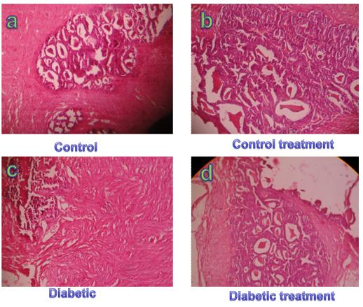
Figure 1: Histology of pancreas tissue of control (Figure c), control treatment (Figure c), Diabetic rat’s tissue (Figure c) showed extensive damage to the pancreatic beta islets of Langerhans and reduced the dimensions of pancreatic beta islets cells. When the diabetic rats were received aqueous suspension of Ocimum sanctum for two weeks and its showed regenerating tiny islets (Figure d) as compared with diabetic group.
Conclusion
Alloxan induced diabetic rats showed negative effects on the activities of insulin secretion, increase lipid peroxidtion and oxidative stress in diabetic rats. Oral administration of raw extracts of Ocimum sanctum increased insulin secretion, decrease lipid peroxidtion and decrease metabolic complications along with oxidative stress in diabetic rats. The results clearly showed the hypoglycemic potential ofaquous suspension of Ocimum sanctum and regenration of panceatic beta cells.Further studies are necessary to found the active component of Ocimum sanctum, role of these herbal drugs in controlling type I diabetes and its complications and action.
Acknowledgments
Mr. SKJ has received stipend from the Department of Higher Education, Government of M.P., Bhopal, India. The financial support from the Department of Science and Technology, N. Delhi, India, in the form of FIST grant to the school is thankfully acknowledged.
Conflict of Interest
The authors declare no conflict of interest.
References
-
American diabetes association (2007) Clinical practice recommendation. Standard of medical care in diabetes. Diabetes care 30 (1): S4-41.
-
Harris MI, Flegal KM, Cowie CC, Eberhardt MS, Goldstein DE, et al. (1998) Prevalence of diabetes, impaired fasting glucose, and impaired glucose tolerance in U.S. adults: the Third National Health and Nutrition Examination Survey, 1988-1994. Diabetes Care 21(4): 518-524.
-
Kikkawa R, Koya D, Haneda M (2003) Progress of diabetic neuropathy. Am. J Kidney Dis 41: 19-21.
-
Udoh AE, Ntu I, Essien O, Ndon M (2007) Red cell catalase activity in diabetics. Pak J Nutr 6(5): 511- 515.
-
Tiedge M, Lortz S, Drinkgern J, Lenzen S (1997) Relation between antioxidant enzyme gene expression and antioxidative defense status of insulin-producing cells. Diabetes 46(11): 1733-1742.
-
Robertson RP, Harmon J, Tran PO, Tanaka Y, Takahashi H (2003) Glucose toxicity in beta-cells: type 2 diabetes, good radicals gone bad, and the glutathione connection. Diabetes 52(3): 581-587.
-
Brownlee M (2003) A radical explanation for glucose- induced beta cell dysfunction. J Clin Invest 112(12): 1788-1790.
-
Rhodes CJ (2005) Type 2 diabetes-a matter of beta- cell life and death? Science 307(5708): 380-384.
-
Youdim K, McDonald J, Kalt W, Joseph J (2007) Potential role of dietary flavonoids in reducing micro- vascular endothelium volubility to oxidative and inflammatory results. J Nutr Biochem 13(5): 282-288.
-
Patil R, Patil R, Ahirwar B, Ahirwar D (2011) Current status of Indian medicinal plants with antidiabetic potential: a review. Asian Pacific Journal of Tropical Biomedicine 1(2): S291-S298.
-
Vats V, Grover JK, Rathi SS (2002) Evaluation of antihyperglycemic and hypoglycemic effect of _Trigonella foenumgraecum_ Linn, _Ocimum sanctum_ Linn and _Pterocarpus Marsupium_ Linn in normal and alloxanized diabetic rats. J Ethnopharmacol 79(1): 95- 100.
-
Rai V, Iyer U, Mani UV (1997) Effect of Tulsi (_Ocimum_ _sanctum_) leaf powder supplementation on blood sugar levels, serum lipids and tissue lipid in diabetic rats_._ Plant Foods Hum Nutr 50(1): 9-16.
-
Rahimi RS, Larijani B, Abdollahi M (2005) A review on the role of antioxidants in the management of diabetes and its complications. Biomed Pharmacother 59(7): 365-373.
-
Trinder P (1969) Determination of glucose in blood using glucoseoxidase with an alternative oxygen acceptor. Ann Clin Biochem 6: 24-27.
-
Van Handel E, Zilversmit DB (1957) Micro method for direct 12. Determination of serum triglycerides. J Lab Clin Med 50(1): 152-157.
-
Allain CC, Poon LS, Chan CSG (1974) Enzymatic determination of total serum cholesterol. Clin Chem 20(4): 470-475.
-
Bowers LD (1980) Kinetic serum creatinine assays I. The role of various factors in determining specificity. Clin Chem 26(5): 551-554.
-
Jacobson SOP, Cassel GE, Person SA (1990) Increased levels of nitrogen oxides and lipid peroxidation in the rat brain after soman induced Seizures. Arch Toxicol 73(4-5): 269-273.
-
McManus JFA, Mowry RW (1960) Staining methods: histologic and histochemical, paul B Hoeber, Inc, USA. _20._ Cerasi E, Kaiser N, Leibowitz G (2000) Type 2 diabetes and beta-cells apoptosis. Diabetes Metab 26(3): 13-16_._
-
Kesavulu MM, Rao BK, Giri R, Vijaya J, Subramanyam G (2001) Lipid peroxidation and antioxidant enzyme status in Type 2 diabetics with coronary heart disease. Diabetes Res Clin Pract 53(1): 33-39.
-
Musabayane CT, Cooper RG, Rao PV, Balment RJ (2000) Effects of ethanol on the changes in renal fluid and electrolyte handling and kidney morphology induced by long-term chloroquine administration to rats. Alcohol 22(3): 129-138.
-
Gundidza M, Masuku S, Humphrey G, Magwa M (2005) Anti-diabetic activity of Aloe excelsa. Cent Afr J Med 51(11-12): 115-120.
-
Khatun K, Mahtab H, Khanam PA, Sayeed MA, Khan KA (2007) Oyster mushroom reduced blood glucose and cholesterol in diabetic subjects. Mymensingh Med J 16(1): 94-99.
- Gallic and Citric Acid Present in the Peels of Tropical Fruits as an Alternative in the Fight against Cancer
- Treating the Forehead Lines with Combination of Forehead and Glabellar Botulinum Toxin Among Japanese Patients
- Clinical Evaluation of Patients Suffering from Breast Cancer & Determination of Treatment Therapies and Better Strategies Related to Breast Cancer
- Medieval Recipes by Al-Zahrāwī for Heart Palpitations Treatment
- Etiology and Prescription Errors of Myocardial Infarction in Different Health Care Systems of Azad Kashmir
- Early Diagnosis and Multidisciplinary Management of Turner Syndrome: A Paediatric Case Study