Molecular Identification of Lactic Acid Bacteria Isolated From Regional Yoghurt Samples of Bangladesh Using 16S Rdna Sequencing
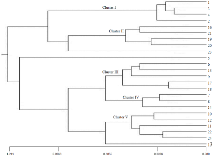
Lactic acid bacteria (LAB) are a group of probiotic organisms that play significant roles in the fermentation of sugars into lactic acid as end product and they are extensively employed in dairy industrial applications since they were categorized as ‘generally recognized as safe’ (GRAS) micro-organisms. The aim of this study was to identify and characterize probiotic LAB strains from traditional dairy product like yoghurt through molecular approaches. A total of 24 isolates (previously isolated from five regional yoghurt samples of Bangladesh) were analyzed using RAPD - PCR, where 5 primers were used to produce reproducible polymorphic (100%) banding patterns in discriminating LAB isolates followed by 16S rDNA sequencing. On the basis of UPGMA dendrogram, it was observed that the isolates originated from the samples of the same geographical location grouped in the same cluster. 10 representative isolates from each group were analyzed and identified as Pediococcus acidilactici (8 out of 10 isolates) and Enterococcus faecium (2 out of 10 isolates). These identified bacteria can be introduced as valuable for further probiotic food development.
Introduction
Lactic acid bacteria (LAB) play comprehensively an important role in food fermentation techniques in industrial perspective [1, 2]. According to international dairy federation and international organization for standardization, LAB are described as a group of Gram- positive, cocci or rods, non-spore forming and anaerobic micro-organisms that produce lactic acid as the end Adv Pharmacol Clin Trials
product mostly throughout the course of fermentation of carbohydrates. Members of this LAB perform a major role in the human and animal gastrointestinal tract as well as in the production of many foods, feeds and beverages. As the probiotic capacities are strain-dependent, methods for credible identification of LAB at the strain level are very important, especially for the description of new strains [3].
The LAB are categorized as probiotic organisms which induce biosynthesis in dairy products [4, 5]. Probiotics can be defined as ‘live microorganisms which when administered in adequate amounts confer a health benefit on the host’ [6]. Probiotics keep broadly a significant role in our health by inhibiting growth of harmful bacteria, promoting immune status and progression of resistance to infection [7, 8, 9]. The traditional dairy products such as yoghurt, cheese worldwide are the treasure house of natural probiotic bacterial resources [10]. Yoghurt is prominent source of lactic acid bacteria which has worthy fermentation properties and probiotics beneficial to the health [11, 12].
In Bangladesh, yoghurt is likely the oldest traditional fermented milk product which is widely consumed by a large scale of population in their daily diet. In most of the areas of Bangladesh, different types of conventional yogurts are noticed but their probiotic role and identification of LAB have not much studied. Isolation, identification and characterization of probiotic microorganism (LAB) from indigenous yoghurt are the demand of time. Then identified LAB strain can be incorporated into suitable food products and certainly be augmented for the health status of mass Bangladeshi people.
Conventional morphological, physiological and biochemical approaches are often conducted but are ambiguous and time-consuming identification of the LAB. Therefore, the prime focus for the identification has changed from phenotypic to genotypic methods because of more sensitive and accurate results, as reported for lactic acid bacteria [13]. RAPD analysis is effective to identify genetic diversity in a wide variety of plants, fungi and bacteria [14, 15, 16, 17, 18]. RAPD markers have been successfully employed in classifying and determining genetic variation and divergence of many bacteria to the strain level [19]. 16S rDNA sequencing is well-established, authentic and universal gold standard method for the identification and phylogenetic grouping of prokaryotic species, genera and families [20, 21].
Therefore, the aim of this study was to conduct molecular approaches for the identification of microorganisms found in fermented regional yoghurt of Bangladesh expecting to give better understanding of microbial community specially probiotics.
Materials and Methods
Sample Preparation
Twenty four isolates of lactic acid bacteria isolated previously using morphological and biochemical assay from regional yoghurt samples (Bogura, Sirajgonj, Jashore, Khulna and Sathkhira) of Bangladesh was used in this study. All isolates were grown in selective media MRS agar plate through streaking method (de Man Rogosa and Sharpe; Merck KGaA, Darmstadt, Germany) and incubated under anaerobic environment at 37°C for 48h. Then overnight cultures ware performed at 37°C in MRS liquid medium.
Extraction of Genomic DNA
The total genomic DNA of each isolate was extracted from 1ml MRS liquid culture medium grown overnight at 37°C following bacterial DNA preparation kit manufactured by Jena Bioscience, Germany. The kit (100 preps) comprised of cell suspension solution, lysozyme, cell lysis solution, RNase A, protein precipitation solution, washing buffer and DNA hydration solution. Briefly, for each of the isolates, the culture was centrifuged at 15000 g for 1 min and discarded the supernatant. The cell pellet was resuspended in 300 µl of cell resuspension solution and added 2 µl of lysozyme solution with mixing by inverting and then centrifuged at 15000 g for 1 min for getting pellet which was resuspended again in 300 µl of cell lysis solution. Next, 1.5 µl of RNase A solution was added and mixed by inverting and incubated at 37°C for 30 min and then cooled on ice for 1 min. Subsequently, 100 µl of protein precipitation solution was added and vortexed for 30 sec. and centrifuged at 15000 g for 5 min. Then, the supernatant was transferred to a clean 1.5 ml micro-tube containing 300 µl isopropanol (99%). The sample was mixed by inverting gently for 1 min and centrifuged at 15000 g for 1 min. Washing buffer about 500 µl was added to the tube and inverted several times to wash the DNA pellet after discarding the supernatant. Afterwards, the tube was centrifuged at 15000 g for 1 min and discarded the ethanol carefully and dried at room temperature for 15 min. Finally, 50 µl of DNA hydration solution was added to the dried DNA pellet and incubated at 65°C for 60 min for each sample. The extracted DNA samples were stored at -20°C.
RAPD - PCR
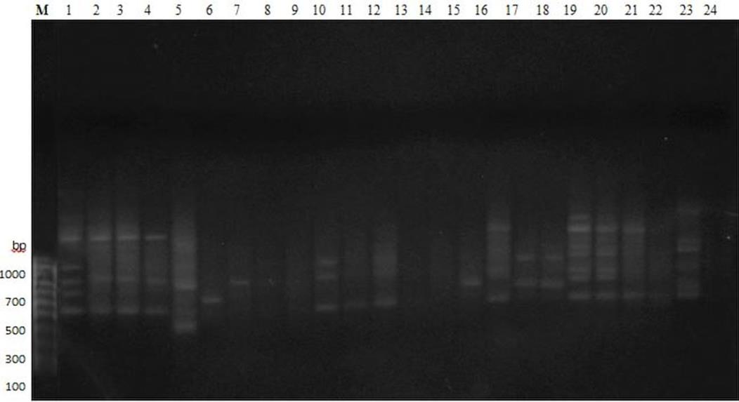
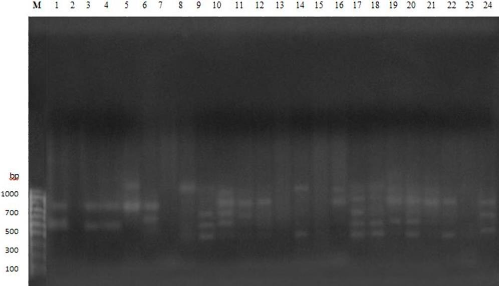
The primers used for RAPD banding pattern were OPA 1 (5’-CAGGCCCTTC-3’), OPA 2 (5’-TGCCGAGCTG-3’), OPA 3 (5’- AGT CAG CCA C-3’), OPA 4 (5’- AAT CGG GCT G -3’), OPA 5 (5’- AGG GGT CTT G -3’). RAPD was conducted on the basis of Chao et al. (2008). The PCR reactions were executed in 20 µl of a mixture containing 0.20 µl of 250 µM dNTPs, 0.30 µl of 1.5 U Tag DNA, 2.40 µl of 3 mM MgCl2, 2.00 µl of 1X PCR buffer, 1.00 µl of 10 Pmol primer, 1.00 µl of 5 ng/µl DNA template and 13.10 µl PCR grade water. The PCR condition for DNA amplification were 30 cycles of denaturation for 2 min at 94°C, annealing for 1 min at 37oC and extension for 2 min at 72°C. The cycles were initially denatured at 94°C for 3 min followed by final extension for 5 min at 72°C. The PCR amplified product was separated by electrophoresis on 1.5% (w/v) agarose gel using 1X TAE buffer and the gel was stained with ethidium bromide solution. Finally, results were visualized under UV transilluminator [22], (Table 1).
Banding patterns of DNA fragments were scored manually (1 for presence and 0 for absence). The genetic similarity and distance among the isolates, Nei’s genetic diversity Nei [23] and Shannon information index Shannon & Weaver [24] were calculated using POPGENE ver. 1.32 (1997). Dendrogram was depicted on the basis of unweighted pair group method of arithmetic mean (UPGMA).
| Primer code | Sequences (5'-3') | Total number of bands scored | Number of polymorphic bands | Proportion of polymorphic loci (%) |
| OPA01 | 5'-CAGGCCCTTC-3' | 16 | 16 | 100 |
| OPA02 | 5'-TGCCGAGCTG-3' | 8 | 8 | 100 |
| OPA03 | 5'- AGT CAG CCA C-3' | 10 | 10 | 100 |
| OPA04 | 5'- AAT CGG GCT G -3' | 6 | 6 | 100 |
| OPA05 | 5'- AGG GGT CTT G -3' | 7 | 7 | 100 |
| Average | 9.4 | 9.4 | 100 | |
| Total | 47 | 47 |
Table 1: Banding patterns of RAPD-PCR for 24 isolates using universal primers.
16S Rdna Amplification
Fragments of the 16S rDNA genes of lactic acid bacterial isolates were amplified using the Universal primer 27F (5’-AGAGTTTGATCCTGGCTCAG-3’) and 1492R (5’GGTTACCTTGTTACGACTT-3’). PCR reaction for amplification of 16S rDNA genes were in 25 µl of mixture volume comprised of Hot Start Green Master Mix, (Promega, USA) 12.5 µl, template 1 µl (25 ng/µl), primer 27F 1 µl (10 pmol) and 1492R 1 µl (10 pmol) and PCR grade water 9.5 µl (Table 2).
| Sequence ID | Isolate no. | Species | Identity | Accession no. |
| 2888787_1_27_F | 1 | Pediococcus Acidilactici | 99% | MK640923 |
| 2888789_2_27_F | 2 | Pediococcus Acidilactici | 99% | MK640924 |
| 2888791_3_27_F | 3 | Pediococcus Acidilactici | 99% | MK640925 |
| 2888793_8_27_F | 8 | Enterococcus faecium | 99% | MK640926 |
| 2888795_10_27_F | 10 | Pediococcus Acidilactici | 99% | MK640927 |
| 2888797_13_27_F | 13 | Pediococcus Acidilactici | 99% | MK640928 |
| 2888799_17_27_F | 17 | Enterococcus faecium | 99% | MK640929 |
| 2888801_19_27_F | 19 | Pediococcus Acidilactici | 99% | MK640930 |
| 2888803_20_27_F | 20 | Pediococcus Acidilactici | 99% | MK640931 |
| 2888805_21_27_F | 21 | Pediococcus Acidilactici | 99% | MK640932 |
Table 2: Identification of 10 lactic acid bacteria by 16S rDNA sequencing.
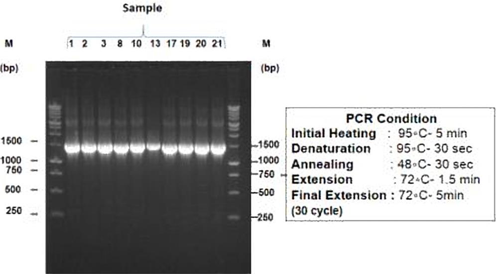
The PCR conditions were initial heating at 95°C for 5 min followed by 30 cycles of denaturation at 95°C for 30 sec, annealing at 48°C for 30 sec, extension at 72°C for 1.5 min and final extension at 72°C for 5 min. The PCR product was electrophoresed in 1.5% (w/v) agarose gel using 1X TAE buffer and the gel was stained with ethidium bromide solution. Marker 1 kb Ladder was used as standard DNA size. Finally, results were visualized under UV trans-illuminator [22].
Sequencing of PCR Product and Phylogenetic Evaluation
PCR products of ten isolates were sent to 1st base laboratories, Malaysia for sequencing. The sequencing results were analyzed applying Basic Local Alignment Search Tool (BLAST) in NCBI website and phylogenetic tree was constructed using MEGA 6 software. Tamura, et al. [25], Sunil & Vora [26] and sequencing results were compared with available sequences in GeneBank database using BLAST program at NCBI.
Results
The five primers (OPA-1, OPA-2, OPA-3, OPA-4 and OPA-5) were used in this study produced reproducible bands (Figures 1-3). Total 47 bands were observed and all (100%) were polymorphism. Among the primers OPA-1 produced highest number of bands (16) and lowest number of bands was produced by the primers OPA-4 (6).
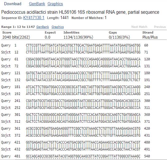
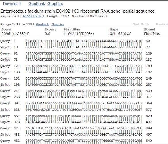
Ten isolates of each cluster were used for amplification of 16S rDNA region using 27F and 1492R primers. The isolates of lactic acid bacteria collected from yoghurt sample of different region of Bangladesh were successfully amplified in the above PCR amplification procedures. All of isolates showed single 16S rDNA band and the lengths of amplified DNA fragments were approximately 1500 bp which were preliminary identified as 16S rDNA (Figure 4).
which were collected from Sathkhira region.
The phylogenetic tree was made from sequenced 16s rDNA region of two representative isolates [isolate no. 2 (288789_2_27_F) and 8 (2888793_8_27_F)] with related reference strains of NCBI GeneBank (Figures 5 & 6).
Among the 10 lactic acid bacterial strain isolated from yoghurt samples of different region of Bangladesh, isolate no. 8 and 17 belonged to Enterococcus faecium strain and rest of the 8 isolates were included as Pediococcus acidilactici strain. All isolates of complete 16S rDNA gene sequence showed high level of similarity (99%) with the reference strains of NCBI GeneBank database.
Discussion
Among the dairy products yoghurt is widely acceptable product as probiotic for their health benefit [27, 28]. Lactic Acid Bacteria (LAB) is the most useful starters in fermentation of dairy products like yoghurt, cheese etc [29].
In this study, isolates from traditional dairy product like yoghurt samples were analyzed for the identification of the potential probiotic bacteria by molecular techniques in order to gain knowledge about specific characteristics of LAB and utilize them more progressively in food products of Bangladesh.
24 isolates of yoghurt samples originated from different region of Bangladesh were extracted DNA genome and to determine DNA concentrations, spectrophotometric analysis was performed applying UV- VIS spectrophotometer that measured absorbance at 230 nm, 260 nm and 280 nm. The results of A260/280 ratio were within 1.7 - 2.1. An A260/280 ratio of 1.8-2.0 is the expected DNA concentration mentioned by Sambrook, et al. [22]. Similarly, the ratio of A260/230 was within 1.6 - 2.2. The values of A260/230 more than 1.8 are considered compatible for analysis according to Mustopa, et al. [30]. So identification and characterization of probiotic bacteria from conventional dairy products using molecular techniques for future utilization in food industries are the present demand [31].
RAPD fingerprinting is sensitive and fruitful method in comparison with biochemical method to measure genetic diversity among LAB. Therefore, molecular characterization of LAB was performed in this research using its powerful discriminative capability in typing of bacteria in strain level following Danova, et al. [32]. This technique is readily admissible for initial screening and isolates selection originated from different source Abdollahniya, et al. [33]. A total number of 5 primers were used in the present study producing reproducible polymorphic (100%) banding patterns in discriminating LAB isolates. Abdollahniya, et al. [33] stated that among 42 primers, all except one primer showed 100% polymorphism in distinguishing Lactobacillus isolates and also reported that highest genetic similarity and same grouping was investigated among the isolates collected from geographically less distant location and sources. Amirbozorgi, et al. [29] reported that the genetic diversity of LAB species in dairy products was variable and area specific. On the basis of UPGMA dendrogram in this study, it was observed that isolates originated from the samples of same geographical location grouped in the same cluster.
16S rDNA is well established and authentic-universal- gold standard method for the identification and phylogenetic grouping of prokaryotic species, genera and families [20]. In present study, 10 representative isolates from each group were analyzed by 16S rDNA gene sequencing and identified predominant strain as Pediococcus acidilactici (8 out of 10 isolates) and Enterococcus faecium (2 out of 10 isolates). Ekinci, et al. [34] found that traditional dairy products such as yoghurt, cheese in Iraq contain Pediococcus spp. and Leuconosto_c spp. identified by applying molecular techniques and 34 isolates of them were _Pediococcus spp. whilst 4 of them were designated as Leuconostoc spp. Alnakip, et al. [35, 36] reported that Enterococcus is the predominant Lab genus followed by Lactococcus, Pediococcus, Lactobacillus, Leuconostic in cow’s raw milk in Egypt. Lu Ren & Huayi [10] investigated that Enterococcus was the major lactic acid bacteria in yak yoghurt in western Sichuan region. Mustopa, et al. [30] mentioned that strains of LAB and other microorganisms differed depending on the place from where product was originated and found that identified three isolates were Lactobacillus fermentum, Pediococcus acidilactici and Pediococcus pentosaceus identified the isolated strains as leuconostoc, lactobacillus, Enterococcus and Pediococcus genus in Camel milk.
Conclusion
A total of 24 LAB isolates (previously isolated from five regional yoghurt samples) were analyzed using RAPD - PCR and 16S rDNA gene sequencing. A total number of 5 primers used in the present study produced reproducible polymorphic (100%) banding patterns in discriminating LAB isolates. On the basis of UPGMA dendrogram, it was observed that isolates originated from the samples of same geographical location grouped in the same cluster. 10 representative isolates from each group were analyzed by 16S rDNA gene sequencing and identified predominant strain as Pediococcus acidilactici (8 out of 10 isolates) and Enterococcus faecium (2 out of 10 isolates). These identified bacteria can be introduced as valuable sources for further probiotic food development.
Consent for Publication
Not applicable.
Availability of Data and Materials
The data that support the findings of this study are available from the corresponding author upon reasonable request.
Competing Interest
The authors participating in this research article have no competing interest.
References
-
Sudhamani M, Ismaiel E, Geis A, Batish V, Heller KJ (2007) Characterisation of pSMA23, a 3.5 kbp plasmid of Lactobacillus casei, and application for heterologous expression in Lactobacillus. Plasmid 59: 11-19.
-
Korhonen J (2010) Antibiotic resistance of lactic acid bacteria. [Dissertations] Finland: University of Eastern Finland, pp: 71.
-
Weiss A, Lettner HP, Kramer W, Mayer HK, Kneifel W (2005) Molecular methods used for the identification of potentially probiotic Lactobacillus reuteri strains. Food Technology and Biotechnology 43(3): 295-300.
-
Chen X, Du X, Wang W (2010) Isolation and identification of cultivable lactic acid bacteria in traditional fermented milk of Tibet in China. Int J Dairy Technol 63(3): 437-44.
-
Liu SQ, Holland R, Crow V (2004) Esters and their biosynthesis in fermented dairy products: a review. Int Dairy J 14: 923-945.
-
FAO, WHO (2002) Joint FAO/WHO Working Group Report on Drafting Guidelines for the Evaluation of Probiotics in Food. London, Ontario, Canada.
-
Amara A, Shibl A (2015) Role of Probiotics in health improvement, infection control and disease treatment and management. Saudi Pharm J 23(2): 107-14.
-
Parvez S, Malik K, Ah Kang S (2006) Probiotics and their fermented food products are beneficial for health. J Appl Microbiol 100: 1171-1185.
-
Perdigon G, Fuller R, Raya R (2001) Lactic acid bacteria and their effect on the immune system. Curr Issues Intest Microbiol 2: 27-42.
-
Lu Ren, Huayi SUO (2017) Molecular Identification of Lactic Acid Bacteria Isolated from theTraditional Fermented Yak Yogurt in Western Sichuan Region. Advances in Computer Science Research (ACSR) 76.
-
Zhang H, Xu J, Wang J (2008) A survey on chemical and microbiological composition of kurut, naturally fermented yak milk from Qinghai in China. J Food Control 19(6): 578-586.
-
Chen ZL, Cheng C, Ma K (2008) Isolation and identification of lactic acid bacteria from fermented yak milk products in tibet area. J Food Science 29(12): 408-412.
-
Callon C, Millet L, Montel MC (2004) Diversity of lactic acid bacteria isolated from AOC Salers cheese. Journal of Dairy Research 71(2): 231-244.
-
Assigbetse, KB, Fernandez D, Dubois MP, Geiger JP (1994) Differentiation of Fusarium oxysporum f. sp. vasinfectum races on cotton by random amplified polymorphic DNA (RAPD) analysis. Phytopathology 84(6): 622-626.
-
Balardin RS, Jarosz AM, Kelly JD (1997) Virulence and molecular diversity in Colletotrichum lindemuthianum from South, Central, and North America. Phytopathology 87(12): 1184-1191.
-
Kelly A, Alcalá-Jiménez AR, Bainbridge BW, Heale JB, Perez-Artes E, et al. (1994) Use of genetic fingerprinting and random amplified polymorphic DNA to characterize pathotypes of _Fusarium_ _oxysporum_ f. sp. ciceris infecting chickpea. Phytopathology 84(11): 1293-1298.
-
Babaei A, Nematzadeh GA, Hashemi H (2011) Molecular RAPD markers analysis of Sange-tarom and Taromhashemi cultivars (_Oryza sativa_ L.) in M2 population. Annals of Biological Research 2(4): 24-30.
-
Gupta S, Preet S (2012) Protocol optimization for genomic DNA extraction and RAPD-PCR in mosquito larvae (Diptera: Culicidae). Ann Biol Res 3(3): 1553- 1561.
-
Kwon OS (2000) Characterization of isolated Lactobacillus spp. and classification by RAPD-PCR analysis. The Journal of Microbiology 38(3): 137-144.
-
Patel JB (2001) 16S rRNA gene sequencing for bacterial pathogen identification in the clinical laboratory. Mol. Diag 6(4): 313-321.
-
Janda JM, Abbott SL (2007) 16S rRNA gene sequencing for bacterial identification in the diagnostic laboratory: pluses, perils, and pitfalls. Journal of Clinical Microbiology 45(9): 2761-2764.
-
Sambrook J, Fritsch EF, Maniatis T (1989) Molecular Cloning a Laboratory Manual, 2nd (Edn.), Cold Spring Harbour Laboratory Press, New York.
-
Nei M (1978) Molecular Evolutionary Genetics, Columbia University Press- New York.
-
Shannon CE, Weaver W (1949) The Mathematical Theory of Communication, University of Illinois Press- Urbana.
-
Tamura K, Stecher G, Peterson D, Filipski A, Kumar S (2013) MEGA6: Molecular Evolutionary Genetics Analysis version 6.0,” Molecular Biology and Evolution 30(12): 2725-2729.
-
Sunil N, Vora JD (2013) Licensed Under Creative Commons Attribution CC BY Analysis of 16S rRNA Gene of Lactic Acid Bacteria Isolated from Curd 5: 2319-7064.
-
Behnsen J, Deriu E, Sassone-Corsi M, Raffatellu M (2013) Probiotics: properties, examples, and specific applications. Cold Spring Harb Perspect Med 3(3): a010074.
-
Ozen M, Dinleyici EC (2015) The history of probiotics: the untold story. Benef Microbes 6(2): 159-165.
-
Amirbozorgi G, Samadlouie H, Shahidi SA (2016) Identification and Characterization of Lactic Acid Bacteria Isolated from Iranian Traditional Dairy Products. Int Biol.Biomed J 2(1): 47-52.
-
Mustopa AZ, Puspitasari IFP, Fatimah Triratna L, Kartina G (2018) Genetic diversity of mastitis cow’s milk bacteria based on RAPD-PCR. Biodiversitas 19(5): 1714-1721.
-
Coeuret V, Dubernet S, Bernardeau M, Gueguen M, Vernoux JP (2003) Isolation, characterisation and identification of lactobacilli focusing mainly on cheeses and other dairy products. EDP Sciences 83: 269-306.
-
Danova ST, Dimitonova SP, Bakalova BV, Aleksandrova-Georgieva RN (2008) Phenotypic and molecular identification of lactobacilli isolated from vaginal secretions. J Microbiol Immunol Infect 41: 469-477.
-
Abdollahniya D, Hosseini SM, Baghbaderani BK, Mordadi A, Arabestani MR (2018) Identification of Lactobacillus Species Isolated From Traditional Dairy Products Using RAPD-PCR. Avicenna Journal of Clinical Microbiology and Infection 5(2): 7-13.
-
Ekinci MS, Akyol I, Ramadan AK, Yazdiç FC, Ozkose E (2018) Molecular Identification and Partial Characterization of Pediococcus sp. and Leuconostoc sp. Isolated from Traditionally Made Dairy Products. KSU J Agric Nat 21(1): 44-50.
-
Alnakip MEA, Asmaa S, Rania M, Kamal M, Elbadry S (2016) Diversity of lactic acid bacteria isolated from raw milk in Elsharkia province, Egypt**.** Japanese Journal of Veterinary Research 64(2): 23-30.
-
Chao SH, Tomii Y, Watanabe K, Tsai YC (2008) Diversity of lactic acid bacteria in fermented brines used to make stinky tofu. Int J Food Microbiol 123(1- 2): 134-141.
- Gallic and Citric Acid Present in the Peels of Tropical Fruits as an Alternative in the Fight against Cancer
- Treating the Forehead Lines with Combination of Forehead and Glabellar Botulinum Toxin Among Japanese Patients
- Clinical Evaluation of Patients Suffering from Breast Cancer & Determination of Treatment Therapies and Better Strategies Related to Breast Cancer
- Medieval Recipes by Al-Zahrāwī for Heart Palpitations Treatment
- Etiology and Prescription Errors of Myocardial Infarction in Different Health Care Systems of Azad Kashmir
- Early Diagnosis and Multidisciplinary Management of Turner Syndrome: A Paediatric Case Study