Three Dimensional Reconstructed Computed Tomography Features of Eagle Syndrome
Eagle syndrome is referred to as signs and symptoms caused by elongated styloid process or calcified stylohyoid ligament. Authors present a case of Eagle syndrome in a 27-year-old male patient complaining from foreign body sensation in the throat diagnosed by three-dimensional reconstructed computed tomography.
Introduction
Eagle syndrome is referred to as signs and symptoms caused by elongated styloid process or calcified stylohyoid ligament [1]. The diagnosis of Eagle syndrome is based on the clinical features, digital palpation of the styloid process in the tonsillar fossa, radiological findings and lidocaine infiltration test. Treatment can be surgical or nonsurgical.
Case Presentation
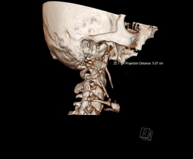
A 27-year-old man with history of tonsillectomy 3 months ago complains from foreign body sensation in the throat. The patient was referred to radiology department to undergo computed tomography (CT) examination of the neck. Three dimensional (3D) reconstructed Volume Rendered Technique (VRT) images revealed bilateral elongated calcified stylohyoid ligaments. The right side calcified stylohyoid ligament measured 5cm and left side measured 4.2 cm in length (Figure 1).
Discussion
Eagle syndrome was first described by W. Eagle in 1937. It results from elongated styloid process or calcified stylohyoid ligament. Depending on anatomical structures compressed or irritated by the elongated/calcified styloid process symptoms vary greatly, ranging from cervico- facial pain to cerebral ischemia [2].The patients mainly complain from recurrent throat pain, foreign body sensation, dysphagia, and/or facial pain [3]. Kumar et Al reported a case of eagle syndrome causing sudden death probably due to vagus-mediated cardiac inhibition [2]. The diagnosis of Eagle syndrome is based on the clinical features, digital palpation of the styloid process in the tonsillarfossa, radiological findings and lidocaine infiltration test.Three-dimensional reconstructed CT is considered as gold standard for the diagnosis [4]. It can well depict the elongated styloid process, the calcified stylohyolid ligament and the effaced adjacent structures. The only imaging feature that is diagnostic for this entity is the length of styloid process/calcified stylohyoid ligament. The normal styloid process measures approximately 2.5 cm [5] however Kaufman et al. reported 3 cm length as the upper limit for normal size [1]. The length over than this can be regarded as abnormal. Treatment can be surgical or nonsurgical, however surgical shortening of the styloid apophysis is the most satisfactory and effective treatment through either trans-oral or cervical approach [6].
References
-
Savranlar A, Uzun L, Ugur MB, Ozer T (2005) Three- dimensional CT of Eagle’s syndrome. Diagnostic and Interventional Radiology 11(4): 206-209.
-
Kumar P, Rayamane AP, Subbaramaiah M (2013) Sudden death due to Eagle syndrome: a case report. Am J Forensic Med Pathol 34(3): 231-233.
-
Murtagh RD, Caracciolo JT, Fernandez G (Murt) CT findings associated with Eagle syndrome. AJNR 22(7): 1401-1402.
-
Uludag IF, Ocek L, Zorlu Y, Uludağ B (2013) Eagle syndrome: case report. Agri 25(2): 87-89.
-
Jewett J, Moriarity R (2014) Eagle syndrome: an incidental finding in a trauma patient: a case report. J Emerg Med 46(1): e9-e12.
-
Torres AC, Guerrero JS, Silva HC (2014) A modified transoral approach for carotid artery type Eagle syndrome: technique and outcomes. Ann Otol Rhinol Laryngol 123(12): 831-834.
- Ultrasound Guided Therapeutic Nerve Blocks
- Cyclops Lesion Without ACL Reconstruction: A Rare Case in a Patient with Intact Anterior Cruciate Ligament and Tibial Plateau Fracture
- Dosimetric Comparison between Two Dose Calculation Algorithms in SBRT Treatment of Lung Cancer in Ring-based and C-arm Radiation Therapy Equipment
- Adolescent Testicular Adrenal Rest Tumors: A Case Report and Review of the Literature
- Giant Intrathoracic Lipoma: A Rare Presentation
- Image of a Right Renal Angiomyolipoma Complicated by Hemorrhage