Metaplastic Breast Carcinoma: Clinical and Imaging Features
Metaplastic breast carcinoma is a very infrequent type of malignancy (<1% of invasive breast tumors), of poor prognosis and of high histological grade. It is due to a metaplasia of the breast glandular epithelium. There are several types, among which squamous cell carcinoma is the most frequent. The age of presentation is usually in women older than 50 years who report a fast growing tumor. Radiologically they are usually well circumscribed lesions, and if they are of a large volume they can be complicated, with ulceration or fixation to the skin or even to the chest wall. They have a worse prognosis than patients with infiltrating ductal or lobular carcinoma, with an incidence of metastatic disease between 5-30%. We present the case of a 50 years old woman with a palpable mass in the left breast, ulcerated. We show the characteristics of this entity in the different imaging techniques. Finally, an ultrasound biopsy confirmed the diagnosis of metaplastic breast carcinoma.
Case Report
A 50-year-old woman was addressed to the Gynecology department for palpable mass in the left breast. Physical examination shows a mass of
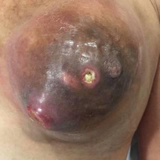
Image Article
approximately 10 cm that affects the entire breast, with areas of ulceration (Figure 1).
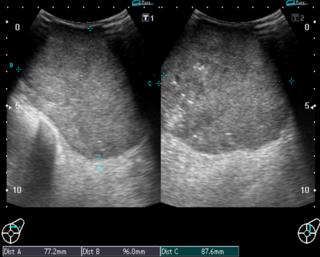
transmission (Figure 2). It presents hyperechogenic points inside, which translates the presence of micro calcifications.
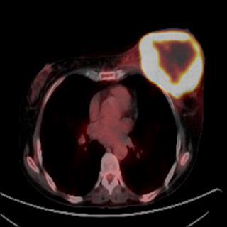
BAG was performed with ultrasound control, with a histological result of breast metaplastic carcinoma. As an extension study, magnetic resonance (not shown) and PET-CT (Figure 3) were performed, showing the presence of mass with peripheral enhancement.
Discussion
Metaplastic breast carcinoma is a very infrequent type of malignancy (<1% of invasive breast tumors), of poor prognosis and of high histological grade [1, 2]. It is due to a metaplasia of the breast glandular epithelium, although generally are a complex admixture of carcinomatous and metaplastic areas [3]. It was officially recognized as a distinct pathologic diagnosis only in 2000 [2]. Histologically, this cancer is characterized by divergent cellular differentiation and heterogeneous cells, including squamous, spindled, sarcomatoid, pleomorphic, chondroid and osseous differentiation, among which squamous cell carcinoma is the most frequent [4].
The etiology of metaplastic breast carcinoma is unclear. The majority of cases are sporadic without familiar aggregation [3]. The age of presentation is usually in women older than 50 years who report a fast growing tumor. Radiologically they are usually well circumscribed lesions, and if they are of a large volume they can be complicated, with ulceration or fixation to the skin or even to the chest wall [5, 6]. They have a worse prognosis than patients with infiltrating ductal or lobular carcinoma, with an incidence of metastatic disease between 5-30%. It is due frequently associated with other poor prognostic indicators being mostly negative for hormone receptors and Her2/neu overexpression. Its outcome has been reported worse compared with invasive ductal carcinoma or triple negative ductal carcinoma. In addition, patients with metaplastic breast carcinoma have a lower rate of pathologic complete response to neoadjuvant chemotherapy [5, 6, 7].
Conclusion
Metaplastic carcinoma of the breast, due to its low incidence and its histological variability, presents some controversy in the literature. The clinical presentation and the findings in the image can make the radiologist think of this rare entity, whose treatment and prognosis differ from the rest of invasive carcinomas in the breast.
References
-
Rakha EA, Tan PH, Varga Z, Tse GM, Shaaban AM, et al. (2015) Prognostic factors in metaplastic carcinoma of the breast: a multi-institutional study. Br J Cancer 112(2): 283-289.
-
El Zein D, Hughes M, Kumar S, Peng X, Oyasiji T, et al. (2017) Metaplastic carcinoma of the breast is more aggressive than triple negative breast cancer. A study from a single institution and review of literature. Clin Breast Cancer 17(5): 382-391.
-
Zhu J, Li K, Dong X, Zhou P, Li P, et al. (2018) Metaplastic breast carcinoma composed of epithelial- myoepithelial carcinoma and squamous cell carcinoma: A case report. Medicine 97(15): 0364.
-
Mathews AC, Verma S, Magalhaes MCF, Jeter SC, Zhang Z, et al. (2016) Clinicopathologic analysis of 45 patients with metaplastic breast carcinoma. Am J Clin Pathol 145(3): 365-372.
-
Siegelmann DN, Murphy TJ, Meschter SC, Stein ME, Prichard J (2005) Primary pure squamous cell carcinoma of the breast. Clin Breast Cancer 6(3): 270- 272.
-
Günhan-Bilgen I, Memiş A, Ustün EE, Zekioglu O, Ozdemir N (2002) Metaplastic carcinoma of the breast: clinical, mammographic, and sonographic findings with histopathologic correlation. AJR Am J Roentgenol 178(6): 1421-1425.
-
Fayaz S, Demian GA, Eissa HE, Amanguno H, Abuzalouf S (2017) Metaplastic breast carcinoma: analysis of 31 cases from a single institute. J Egypt Nati Canc Inst 29(3): 141-145.
- Ultrasound Guided Therapeutic Nerve Blocks
- Cyclops Lesion Without ACL Reconstruction: A Rare Case in a Patient with Intact Anterior Cruciate Ligament and Tibial Plateau Fracture
- Dosimetric Comparison between Two Dose Calculation Algorithms in SBRT Treatment of Lung Cancer in Ring-based and C-arm Radiation Therapy Equipment
- Adolescent Testicular Adrenal Rest Tumors: A Case Report and Review of the Literature
- Giant Intrathoracic Lipoma: A Rare Presentation
- Image of a Right Renal Angiomyolipoma Complicated by Hemorrhage