Preliminary Study of 89Zr Labeled Bevacizumab to Detect Angiogenesis in a Pre-Clinical Model of Sarcoma
The goal of the investigation was to demonstrate uptake of 89Zr labeled bevacizumab as non-invasive probe for angiogenesis in a xenograft model of sarcoma. Methods: HT-1080 human fibrosarcoma cell were established as xenografts in both athymic nude mice and BALB/c nude mice. Bevacizumab (Bev) was conjugated to 89Zr oxalate using the bifunctional chelate, p-isothiocyanatobenzyl-desferrioxamine (Df-Bz-NCS). Mice were injected with1.8 – 3.7 MBq of 89Zr-Bev and imaged over 11 days. Results: Uptake of 89Zr-Bev was clearly demonstrated in HT-1080 xenografts with peak tumor SUVMAX at 4 days post injection when normal tissue uptake had reduced. Declining levels of radioactivity persisted in the tumor for the 11-day observation period. Significant uptake was seen in bone tissue. Conclusions: These preliminary results demonstrate that 89Zr-Bev is a potential new tracer for noninvasive imaging of VEGF in the microenvironment of sarcomas.
Introduction
Soft tissue sarcomas (STSs) make up <1% of adult malignancies with an estimated 12,000 new cases of STS diagnosed in the US each year, with approximately 5,000 deaths [1]. STSs are derived from mesenchymal cells with their most common location. Involving the extremities/chest wall regions of the body [1]. At the time of initial diagnosis, distant metastases are rarely present, but blood is the most common route for the disease to spread, most frequently to the lungs [2]. Current treatments for STSs include surgery, radiation, and chemotherapy with little progress being made with respect to targeted therapeutics. TNM stage histologic grade remain the most important clinicopathological indicators of prognosis with the additional factors of surgical margins, age, anatomic site, and histologic subtype [3].
Non-invasive functional imaging involving fluorodeoxyglucose (18F-FDG)-positron emission tomography (PET)/computed tomography (CT) imaging has been assessed for its utility for discriminating benign from malignant lesions, correlation with tumor grade and histological subtype and with prognosis with variable conclusions as to its utility [4, 5, 6]. FDG is a useful non-specific probe to assess metabolic activity in a wide range of cancers but is influenced by many biological factors. Imaging probes that are directed towards more specific attributes of STS may be more informative. The metastatic rate of sarcomas is dependent on the development of angiogenesis or new blood vessels formation [7, 8]. An important mediator of angiogenesis has been identified as vascular endothelial growth factor (VEGF)–A. High levels of this growth factor, in patients with STSs, correlates with higher tumor grade, increased recurrence and metastatic rate, and decreased overall survival [8].
Antibody based positron emission tomography (immuno-PET) imaging is a relatively new field which is gaining increasing attention [9]. However, the slow binding characteristics and receptor occupancy of intact antibodies requires the use of radionuclides with longer half-lives than used in traditional PET studies. 89Zirconium (89Zr) has a half- life of 78.4 hours (h) which matches the pharmacokinetics of antibodies and it has a relatively low average positron energy of 395 keV; both characteristics making it a suitable candidate for high resolution PET imaging of slow- accumulating biomolecules [10]. Bevacizumab (Bev) is an antibody that targets all splice variants of VEGF-A, some of which are partly associated with tumor blood vessels and the extracellular matrix of tumor cells. 89Zr--Bev PET/CT imaging has already been shown to be tolerable and effective for noninvasive in vivo imaging of VEGF-A in preclinical human ovarian tumor xenografts [11]. In addition, several published preliminary studies of adult human tumors in Europe including primary breast cancers, advanced, progressive neuroendocrine tumors and renal cell carcinomas [12, 13, 14] and have demonstrated safe and accurate detection of VEGF-A with 89Zr-Bev PET/CT.
To our knowledge there are no reported studies of using 89Zr-Bev PET/CT in sarcomas. In addition, 89Zr is not yet approved as a PET radiotracer in the USA. This preliminary communication was performed to establish the utility of 89Zr- Bev to image angiogenesis in a sarcoma model to support imaging correlates in a clinical trial entitled “ A phase Ib Trial of Image-Guided Preoperative Simultaneous Integrated Boost Radiation Therapy for Patients with Locally Advanced Non-Metastatic Soft Tissue Sarcomas of the Extremity/Body Wall”.
Material and Methods
Tumor model
The HT-1080 human fibrosarcoma tumor cell line (ATCC® CCL-121™) was purchased from the American Type Culture Collection (Manassas, VA) and established in cell culture using standard conditions. Female athymic nude mice and BALB/c nude mice were obtained at 7-8 weeks old from Charles River Laboratories, Inc. (Wilmington, MA). Al experiments were approved by Animal Care Committee. Flank tumors were established on anesthetized mice by subcutaneous inoculation with 1 x 10⁶ HT-1080 cells in 100μl of Matrigel. Three days after tumor implantation mice were injected with 89Zr-Bev.
Synthesis of 89Zr-Bev
10mCi of 89Zr-oxalate solution was synthesized and dispensed by BV Cyclotron VU, (Amsterdam, Netherlands). Bevacizumab was obtained from Selleckchem (Houston, TX). Conjugation of bevacizumab to 89Zr-oxalate solution was carried out using previously established methods [15, 16]. Briefly, a 1 mL solution of 5mg bevacizumab was adjusted to pH 8.9-9.1 using 1 M Na2CO3. The bifunctional chelate, p-isothiocyanatobenzyl-desferrioxamine (Df-Bz- NCS) (Macrocyclics, Plano, TX), was dissolved in DMSO at a concentration of 3.5mM and 20 μl added to the antibody solution and incubated for 30 minutes at 37oC. The conjugation reaction was pipetted into a PD-10 column pre- rinsed with 5mg.ml-1 gentisic acid and 0.25 M sodium acetate (pH 5.4-5.6) and the Df-Bz-NCS-bevacizumab collected. Radiolabeling was achieved by dispensing 5 mCi (185MBq) of 89Zr-oxalate solution into a reaction vial and mixing with 90 μl of 2 M Na2CO3 for 3 minutes at room temperature. Subsequently 0.3 ml of 0.5 M HEPES (pH 7.1-7.3), 0.71 ml of Df-Bz-NCS-bevacizumab and 0.7 ml of 0.5 M HEPES was successively added to the vial and incubated for 60 minutes at room temperature. Radiolabeling efficiency (typically>85%) was determined by ITLC. The mixture was purified using a PD-10 column rinsed with 5mg.ml-1 gentisic acid and 0.25 M sodium acetate (pH 5.4-5.6). The purified 89Zr-bev was analyzed by ITLC, HPLC and SDS-PAGE to calculate overall yield and radiochemical purity.
Imaging studies
For the imaging studies a small animal Northridge Tri-Modality Imaging (formerly Gamma Medica-Ideas, Chatsworth, CA) FLEX Triumph PET/SPECT/CT system was used. Mice were anesthetized with 1-3% isoflurane (balance 100% O2) and the tail veins catheterized. Animals were injected with 1.8 – 3.7 MBq of 89Zr-Bev in 0.2 mL saline. Animals were imaged 1, 3, 4, 8 and 11 days after tracer injection. Animals were positioned in the center of the PET ring field of view, with video monitoring and constant monitoring of respiratory rate using integrated instrumentation (SAII, Stony Brook, NY) during PET/CT. CT was collected using 512 projections and PET was a 60-minute acquisition. PET data was reconstructed using an OSEM-
3D algorithm resulting in a voxel size of 0.5mm x 0.5mm x 0.6mm (x, y, z) and CT data was reconstructed with a voxel size of 0.15mm x 0.15mm x 0.15mm. The PBAS tool from PMOD (PMOD Technologies, Zurich, Switzerland) software was used to delineate the tumor and other tissues. Volume- of-Interest (VOI) from the CT image and standardized uptake value, (SUV), which is defined as the tumor or tissue activity divided by the injected dose per body weight of the mouse calculated at each imaging time-point.
The contrast-to-noise ratio (CNR) is a measure of the signal level in the presence of noise given by:
(Mean (lesion)-Mean (background) CNR (SD (background) =
Where the mean is calculated from a VOI that was the whole tumor volume and the background was a VOI in leg muscle in the opposite leg to the flank containing the tumor.
Results
89Zr-bev imaging
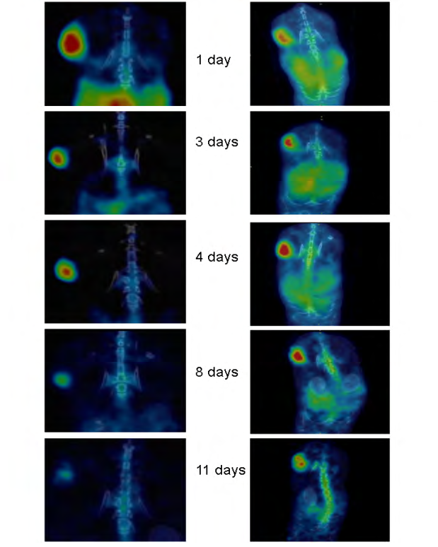
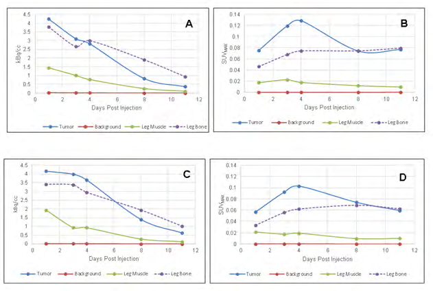
Figure 1 shows the sequential PET images from HT- 1080 xenografts in both mouse strains and Figure 2 shows the KBq/cc and SUVMAX for tumor, leg muscle, leg bone and background. Tumor visualization was excellent with the SUVMAX reaching maximum values at 4 days post-injection. There was uptake in well perfused organs such as liver and spleen which declined at later time-points. Clearance of 89Zr-Bev from internal organs was more rapid in the athymic mouse strain compared to the BALB/c mice. This was evident in Table 1 where the CNR was maximal at 4 days in the athymic mice but reached a maximum at 8 days in BALB/c mice due to persistence of radioactivity in organs and muscle.
| Mouse strain | Day 1 | Day 3 | Day 4 | Day 8 | Day 11 | |
|---|---|---|---|---|---|---|
| CNR athymic | 42.38 | 21.60 | 25.73 | 16.21 | 16.63 | |
| CNR BALB/c | 10.80 | 30.37 | 24.68 | 48.46 | 18.56 |
Table 1: The contrast to noise ratio for tumor versus leg muscle.
As has been observed previously, there was significant uptake of 89Zr-Bev in bone (Figure 2). This was analyzed by a VOI in the knee region of the opposite side to the flank implanted with tumor. The SUVMAX increased with time and plateaued at day 4 and remained at this level until day 11 (Figure 2B and 2D) despite the radioactivity counts declining with time (Figure 2A and 2C).
Discussion
In this preliminary communication we have been able to demonstrate significant uptake of 89Zr-Bev in a xenograft model of human fibrosarcoma. Previous studies have established that HT-1080 cells form aggressive tumors when injected subcutaneously in the flanks of the immuno- compromised mice [17]. Histology has shown that the tumors contain several vascular channels harboring red blood cells that are indicative of high levels of angiogenesis whilst immunohistochemical analyses reveal various angiogenic markers like PECAM, VE-Cadherin, VEGF, VEGF165, NRP-1 and VEGFR-2 [17]. This emphasizes that HT-1080 cells acquire a vascular-like phenotype in vivo making it a suitable model to study a vascular imaging probe in the context of sarcoma.
As mentioned previously 89Zr has attributes that make it an ideal radionuclide for immuno-PET imaging in terms of stability, half-life and being a residualizing radionuclide which results in improved tumor retention and enhanced tumor-to-normal tissue ratios [18]. However, it also has some disadvantages which involve chelation instability. It has been shown that the 89Zr−DFO complex is partly unstable and can result in release of 89Zr from the chelator and the subsequent accumulation in bone [19]. In this study we have used a desferrioxamine (DFO) derivative, Df-Bz-NCS, which facilitated direct conjugation to bevacizumab. Interestingly, the original publication describing the chelation method did not report any accumulation of radioactivity in bone [16] but our study did show significant uptake and retention. This may be due to the fact that we are using an antibody that targets vasculature whereas the previous study conjugated antibodies against epidermal growth factor receptor (EGFR) and CD44v6 [16]. Many new chelators with improved 89Zr coordination properties are under development [19].
This preliminary study does have several limitations. First, very few mice were studied in this proof-of-principle study, more research is now required to study different sarcoma tumor models and different stages of tumor growth. Second, we were not able to study the correlation between 89Zr-Bev imaging and assessment of vascularity; this is planned for future studies.
In summary, the uptake of 89Zr-Bev in this present study was consistent with that described in previous studies [11, 12, 14] with maximum uptake and tumor to normal tissue ratios at 3 to 4 days after injection. Although the results were qualitatively similar in the two mouse strains, the athymic nude mice seemed to clear the tracer more rapidly from normal tissues than the BALB/c. Our preliminary results demonstrate that radiolabeled bevacizumab is a potential new tracer for noninvasive imaging of VEGF in sarcomas.
References
-
Hoang NT, Acevedo LA, Mann MJ, Tolani B (2018) A review of soft-tissue sarcomas: translation of biological advances into treatment measures. Cancer Manag Res 10: 1089-1114.
-
Christie Large M, James SL, Tiessen L, Davies AM, Grimer RJ (2008) Imaging strategy for detecting lung metastases at presentation in patients with soft tissue sarcomas. Eur J Cancer 44(13): 1841-1845.
-
Zagars GK, Ballo MT, Pisters PW, Raphael E Pollock, Shreyaskumar R Patel, et al. (2003) Prognostic factors for patients with localized soft-tissue sarcoma treated with conservation surgery and radiation therapy: an analysis of 1225 patients. Cancer 97(10): 2530-2543.
-
Ioannidis JP, Lau J (2003) 18F-FDG PET for the diagnosis and grading of soft-tissue sarcoma: a meta-analysis. J Nucl Med 44(5): 717-724.
-
Roberge D, Vakilian S, Alabed YZ, Turcotte RE, Freeman CR, et al. (2012) FDG PET/CT in Initial Staging of Adult Soft-Tissue Sarcoma. Sarcom 960194.
-
Kubo T, Furuta T, Johan MP, Ochi M (2016) Prognostic significance of (18)F-FDG PET at diagnosis in patients with soft tissue sarcoma and bone sarcoma; systematic review and meta-analysis. Eur J Cancer 58: 104-111.
-
DuBois S, Demetri G (2007) Markers of angiogenesis and clinical features in patients with sarcoma. Cancer 109(5): 813-819.
-
Rocchi L, Caraffi S, Perris R, Mangieri D (2014) The angiogenic asset of soft tissue sarcomas: a new tool to discover new therapeutic targets. Biosci Rep 34(6): e00147.
-
Marik J, Junutula JR (2011) Emerging role of immunoPET in receptor targeted cancer therapy. Curr Drug Deliv 8(1):70-78.
-
Van De Watering FC, Rijpkema M, Perk L, Brinkmann U, Oyen WJ, et al. (2014) Zirconium-89 labeled antibodies: a new tool for molecular imaging in cancer patients. Biomed Res Int 2014: 203601.
-
Nagengast WB, De Vries EG, Hospers GA, Nanno H Mulder, Johan R De Jong, et al. (2007) In vivo VEGF imaging with radiolabeled bevacizumab in a human ovarian tumor xenograft. J Nucl Med 48(8): 1313-1319.
-
Gaykema SB, Brouwers AH, Lub de Hooge MN, Rick GP, Hetty TB, et al. (2013) 89Zr-bevacizumab PET imaging in primary breast cancer. J Nucl Med 54(7): 1014-1018.
-
Van Asselt SJ, Oosting SF, Brouwers AH, Alfons HH Bongaerts, Johan R de Jong, et al. (2014) Everolimus Reduces (89)Zr-Bevacizumab Tumor Uptake in Patients with Neuroendocrine Tumors. J Nucl Med 55(7): 1087- 1092.
-
Oosting SF, Brouwers AH, van Es SC, Wouter BN, Thijs H Oude Munnink ,et al. (2015) 89Zr-bevacizumab PET visualizes heterogeneous tracer accumulation in tumor lesions of renal cell carcinoma patients and differential effects of antiangiogenic treatment. J Nucl Med 56(1): 63-69.
-
Vosjan MJ, Perk LR, Visser GW, Marianne Budde, Paul Jurek, et al. (2010) Conjugation and radiolabeling of monoclonal antibodies with zirconium-89 for PET imaging using the bifunctional chelate p-isothiocyanatobenzyl-desferrioxamine. Nat Protoc 5(4): 739-743.
-
Perk LR, Vosjan MJ, Visser GW, Marianne Budde, Paul Jurek, et al. (2010) p-Isothiocyanatobenzyl-desferrioxamine: a new bifunctional chelate for facile radiolabeling of monoclonal antibodies with zirconium-89 for immuno- PET imaging. Eur J Nucl Med Mol Imaging 37(2): 250- 259.
-
Misra RM, Bajaj MS, Kale VP (2012) Vasculogenic mimicry of HT-1080 tumour cells in vivo: critical role of HIF-1alpha-neuropilin-1 axis. PLoS One 7(11): e50153.
-
Verel I, Visser GW, Boerman OC, Julliette E M van Eerd, Ron Finn, et al. ( 2003) Long-lived positron emitters zirconium-89 and iodine-124 for scouting of therapeutic radioimmunoconjugates with PET. Cancer Biother Radiopharm 18(4): 655-661.
-
Heskamp S, Raave R, Boerman O, Rijpkema M, Goncalves V, et al. (2017) (89)Zr-Immuno-Positron Emission Tomography in Oncology: State-of-the-Art (89)Zr Radiochemistry. Bioconjug Chem 28(9): 2211-2223.
- Ultrasound Guided Therapeutic Nerve Blocks
- Cyclops Lesion Without ACL Reconstruction: A Rare Case in a Patient with Intact Anterior Cruciate Ligament and Tibial Plateau Fracture
- Dosimetric Comparison between Two Dose Calculation Algorithms in SBRT Treatment of Lung Cancer in Ring-based and C-arm Radiation Therapy Equipment
- Adolescent Testicular Adrenal Rest Tumors: A Case Report and Review of the Literature
- Giant Intrathoracic Lipoma: A Rare Presentation
- Image of a Right Renal Angiomyolipoma Complicated by Hemorrhage