Accidental Ingestions of Dental Materials Visualized on Medical Radiographs in an Emergency Radiology Department
The ingestion and more rarely the aspiration of foreign bodies is not uncommon in medical radiographs, particularly in Emergency Departments. The results particularly in cases of aspiration can be life threatening to the patient. In the past there has been numerous case reports of dental materials and items ingested or aspired by patient. However there have been no studies lately to identify the characteristics of the materials and their results. The present paper is a case series of dental materials and items ingested or aspired in a large hospital, which is most cases not recorded by dentists or doctors.
Introduction
Ingestion and aspiration of foreign objects can occur during all types of dental procedures. The results may be in cases of aspiration in particular, life-threatening, and can cause all types of medical emergencies, from damage to the digestive tract up to pneumonia, mediastinitis, peritonitis or sepsis. In some cases, surgical intervention is needed to retrieve foreign objects of dental origin as is the case with many foreign objects. In cases of aspiration, dentists must be alert to the signs and symptoms of airway obstruction and, if necessary, provide immediate and appropriate treatment until the arrival of emergency support. The incidence of aspiration in the general population is around 0.004%, and in general, it is well known that ingestion is more common than aspiration [1]. The purpose of the present study was to investigate and note causes of accidental ingestion and aspiration during dental procedures as they presented to a hospital emergency department. We analyzed the details of the foreign bodies, the sites and types of dental treatment being performed when the events occurred, details of the professional experience of the practitioners, and the patients’ characteristics. The findings were also compared with previous reports of ingestion and aspiration of dental materials.
Materials and Methods
In the study, a retrospective recording of all incidentally noted dental materials and components on a series of medical radiographs was performed. Cases of accidental ingestion of foreign bodies during dental procedures that presented at the radiology department of AHEPA general hospital from 2017 to 2022 were studied. This included both cases of ingestion, that were totally 9 and one case of aspiration. Of the 10 cases there were 6 females and 4 male patients with an average age of 54 y (12-72 y). The protocol of radiographic studies that are taken depends on the emergency of the case but in all cases involves an initial plain radiograph of the patient prior to other advanced imaging modalities that may be chosen to study the cases.
Results
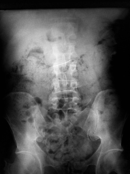
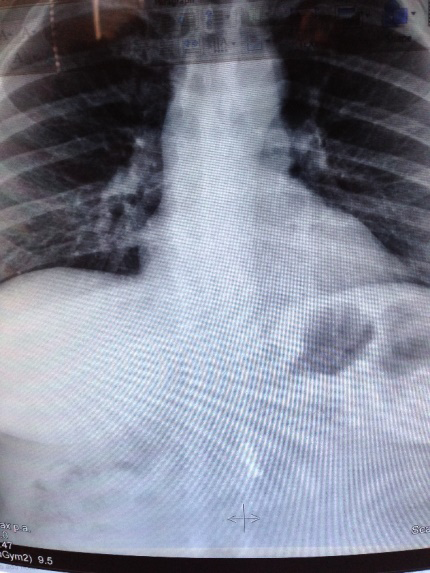
There were 9 reports of ingestion, and 1 of aspiration which will be presented separately. As previously mentioned of the patients involved, 6 were female and 4 male, with a median age of 54 y (12-72 y). The ingested objects were 1 metal inlay, 1 Metalloceramic bridge (Figure 1), 3orthodontic appliances (wires and bracets) (Figure 2), 3 fillings (Figures 3,4), 1 dental water syringe (Figure 5) and finally 1 crown that was aspirated and blocked the airway and will be described separately (Figures 6,7). The location was mostly in the oesophagus and the stomach radio graphically, while the crown was seen in the bronchial tree. One of the fillings was observed in the large intestine and made it very difficult for the radiologist to identify. This case required two radiographs, one standing and one with the patient lying down as seen in Figures 6 and 7.
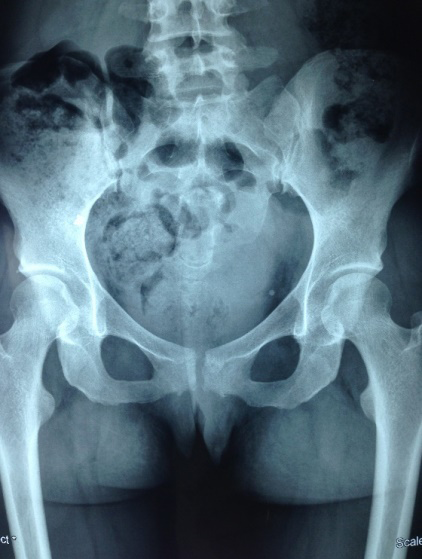
Even though the exact localization within the gastrointestinal tract was not possible, it can be presumed that the object could be possibly in the small intestine. Prospective follow-up with abdominal radiographs can be performed to monitor the progress of the object along the intestine and exclude pneumoperitoneum in the case of perforation.
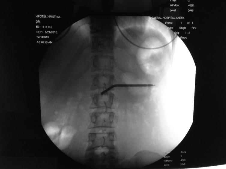
Fluoroscopic images detected the ingested water syringe in the upper left quadrant of the abdomen. This location suggested that the object was situated inside the gastric fundus.
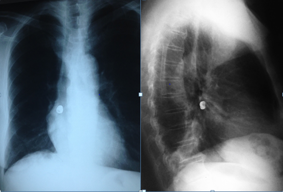
Figures 6 & 7: Frontal and lateral chest radiographs showing a radio-opaque foreign object shaped in keeping with a crown, situated in the right parahilar region. The crown projected in the retrocardiac space in the frontal radiograph and in the retrocardiac space on the lateral view, suggesting that the crown was impacted in the right lower lobe bronchus, appearing immediately inferior to the main bronchus.
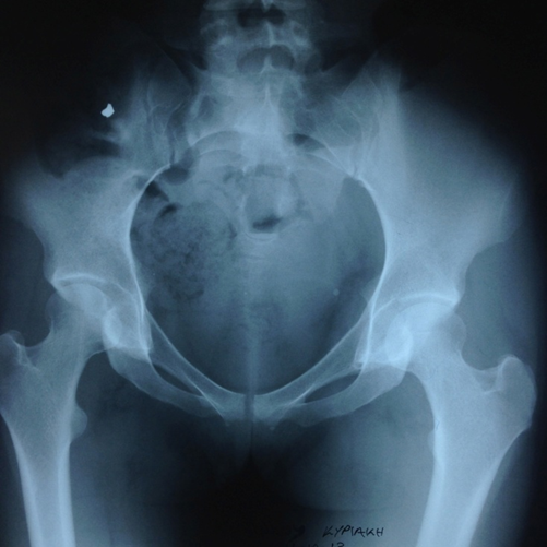
A pelvic radiograph revealed the presence of a radio- opaque foreign object shaped like a filling, situated in the right lower quadrant. This projection suggests a location of the object likely inside the cecum or the terminal ileus.
It is expected that follow-up radiographs over the following hours will demonstrate rapid progress of the object along the colon.
Discussion
The aspiration and ingestion of dental materials and components is rare, but the potential consequences can be serious for the patients, even jeopardizing their health. Accidental ingestion is more common than aspiration, and usually does not cause any clinical signs or symptoms, with the majority of reported cases having the foreign bodies being ingested passing through the gastrointestinal tract without complications [2]. The aspiration however as in the presented case, can be a life threatening complication of dental procedures. In a number of cases in the literature it is often that no direct measures are taken and the patient is advised to just ensure within three days that the material will be egested. In the literature there are reported complications such as intestinal obstruction, perforation with subsequent abscessa formation, hemorrhage, or failure of the objects to progress through the gastrointestinal tract [3]. Gastric erosion and perforation of the esophagus caused by ingestion of dental foreign objects have also been reported [2, 4, 5]. Once a foreign object has crossed the esophagus and reached the stomach, there is a more than 90% chance that it will pass through the gastrointestinal tract as a result of peristaltic movement without complications, usually after a 7-10-day period [6]. As a precaution, it is often recommended that swallowed foreign objects are assessed by serial radiography until egested. If the patient with the ingested material, develops symptoms of perforation, such as acute pain or vomiting, or if objects remain lodged longer than 2 weeks, surgical intervention is required.
Aspiration on the other hand can evolve to a life threatening situation for the patient. It requires immediate treatment, as it can lead to airway obstruction or even death [7]. In the case presented there was pulmonar blockage from a dental crown but unilateral in the main bronchi. As with the cases of emergence medicine, upon aspiration, when an object is located in the oropharynx, the patient should be placed in a reclining position, and cough vigorously to secure the airway. It is thought that foreign objects are likely to fall into the right bronchial tree because it is oriented more vertically, and in fact clinical data show that such objects become preferentially localized on the right side [8, 9]. The initial symptoms of chocking, inspiratory stridor, and reduced breathing are marked and noted as signs of airway obstruction by aspired foreign objects [10]. If the patient does not improve by coughing, the Heimlich maneuver should be performed, and attempts should be made to relieve the laryngeal obstruction [11]. This procedure needs to be performed as soon as possible after aspiration; otherwise emergency help must be summoned immediately for transferring of the patient to a hospital or an emergency unit. The practitioner and team must consider measures for emergency life support, including airway provision via a Cricothyroidotomy, if appropriate and feasible [11].
To avoid such complications a number of actions and measures can be taken to prevent their incidence. Endodontists encourage and practically use rubber dam during endodontic procedures daily and in most cases, both for prevention of ingestion or aspiration as well as for reducing stress arising from safety concerns and improved infection control. Rubber dam use has been made daily practice in many cases of dental treatment such as in cases of operative dentistry. (Rubber dam applications) its use has significantly affected the ingestion and aspiration probability of foreign bodies in dentistry. Though there are not many papers that could support this since the literature is sparce and limited to case reports or case series. They are very rare in medical emergency departments [12, 13].
Conclusion
Dentists should be alert for the probability of ingestion and even aspiration of dental materials and objects. Also it is of high significance that medical doctors are aware of such objects, since their identification is in most cases easy and in due time, but in the rarity of aspiration it can prove to be even life threatening for the patient.
References
-
Hisanaga R, Hagita K, Nojima K, Katakura A, Morinaga K, et al. (2010) Survey of accidental ingestion and aspiration at Tokyo Dental College Chiba Hospital. Bull Tokyo Dent Coll 51(2): 95-101.
-
Chuujoh T, Yokobayashi Y, Mizuno K (2002) Acci- dental swallowing of foreign bodies associated with dental treatment; Report of 5 cases. Niigata Shigakkai Zasshi 32: 69-73.
-
Susini G, Pommel L, Camps J (2007) Accidental ingestion and aspiration of root canal instruments and other dental foreign bodies in a French population. Int Endod J 40(8): 585-589.
-
Maleki M, Evans WE (1970) Foreign body perforation of the intestinal tract. Report of 12 cases, and a review of the literature. Arch Surg 101(4): 475-477.
-
Athanassiadi K, Gerazounis M, Metaxas E, Kalantzi N (2002) Management of esophageal foreign bodies: a retrospective review of 400 cases. Eur J Cardiothorac Surg 21(4): 653-656.
-
Webb WA (1988) Management of foreign bodies of the upper gastrointestinal tract. Gastroenterology 94(1): 204-216.
-
Ayed AK, Jafar AM, Owayed A (2003) Foreign body aspiration in children: diagnosis and treatment. Pediatr Surg Int 19(6): 485-488.
-
Ulku R, Baskan Z, Yavuz I (2005) Open surgical approach for a tooth aspirated during dental extraction: a case report. Aust Dent J 50(1): 49-50.
-
Pingarron ML, Moran SMJ, Sanchez BR, Burgueno GM (2010) Bronchial impaction of an implant screwdriver after accidental aspiration: report of a case and revision of the literature. Oral Maxillofac Surg 14(1): 43-47.
-
Milton TM, Hearing SD, Ireland AJ (2001) Ingested foreign bodies associated with orthodontic treatment: report of three cases and review of ingestion/ aspiration incident management. Br Dent J 190(11): 592-596.
-
Heimlich HJ (1977) The Heimlich manoeuvre: prevention of death from choking on foreign bodies. J Occup Med 19(3): 208-210.
-
Tiwana KK, Morton T, Tiwana PS (2004) Aspiration and ingestion in dental practice: a ten-year institutional review. J Am Dent Assoc 135(9): 1287- 1291.
-
Farmakakis T, Dessypris N, Alexe DM, Frangakis C, Petoussis G, et al. (2007) Magnitude and object-specific hazards of aspiration and ingestion injuries among children in Greece. Int J Pediatr Otorhinolaryngol 71(2): 317-324.
- Ultrasound Guided Therapeutic Nerve Blocks
- Cyclops Lesion Without ACL Reconstruction: A Rare Case in a Patient with Intact Anterior Cruciate Ligament and Tibial Plateau Fracture
- Dosimetric Comparison between Two Dose Calculation Algorithms in SBRT Treatment of Lung Cancer in Ring-based and C-arm Radiation Therapy Equipment
- Adolescent Testicular Adrenal Rest Tumors: A Case Report and Review of the Literature
- Giant Intrathoracic Lipoma: A Rare Presentation
- Image of a Right Renal Angiomyolipoma Complicated by Hemorrhage