Diabetic Retinopathy Screening with Artificial Intelligence: A Pivotal Experience in Italian Healthcare System – Preliminary Report
Diabetic retinopathy is the main complication of diabetes. Its prevalence is 30% and if not diagnosed in time can cause serious damages to vision, up to blindness. Screening of retinopathy is a healthy procedure with the best ratio cost/effectiveness. If done in a massive way, it allows to detect initial lesions, and so preventing or delaying evolved forms. ASL TO 5 is a Local Health District of Turin Metropolitan Area with 310.315 residents. In 2019, ASL TO5 performed an integrated care pathway dedicated to diabetic retinopathy. The clinical pathway develops technological improvements, in order to make the diagnostic and therapeutic process more effective. New knowledge and technologies, such as non- mydriatic cameras and artificial intelligence (AI) software allow to recognize elementary lesions making a first diagnosis of presence or absence of retinopathy reducing the diagnostic burden on eye care specialists and time costs for patients. Dairet, an artificial intelligence system distributed by Meteda srl (Rome, Italy) powered by Retmarker SA, demonstrated accurate sensitivity in detecting both mild and moderate retinopathy. Sensitivity ratio was 91,6% for mild retinopathy and 100% for moderate retinopathy. More over specificity was acceptable with a low false positive rate (specificity ratio 82,6%).
Introduction
Diabetes is a global eye health issue. Given the rising diabetes prevalence and aging population, this poses a significant challenge to perform retinopathy screening.
Preventing blindness due to diabetic retinopathy is possible through programs of screening performed periodically. Screening for diabetic retinopathy is necessary to detect referable cases that need timely full ophthalmic examination and treatment to avoid permanent visual loss. The new technologies based on telemedicine and artificial intelligence are changing the screening strategies and are improving the cost-effectiveness of screening.
ASL TO 5 is a Local Health District of Turin Metropolitan Area with 310.315 residents. 11598 patients refer to our Diabetic Center. For some years the screening of diabetic retinopathy has been carried out with telemedicine methods. Retinal Images were taken by nurses staff of the 4 districts of ASL To5 using digital non-mydriatic cameras and uploaded to a digital folder. One reading center in the whole Local Health District provided reports with retinopathy grading.
In 2019, ASL TO5 performed an integrated care pathway dedicated to diabetic retinopathy. The clinical pathway has been developing technological improvements in order to make the diagnostic and therapeutic processes more effective. An integrated clinical pathway for diabetic retinopathy between diabetologists and ophthalmologists enhances the quality of care by improving patient outcomes and reducing prevalence of chronic complications. Beginning from 2021, reports are provided with the aiding tool of artificial intelligence (AI). Dairet distributed by Meteda srl (Rome, Italy) is an algorithm powered by Retmarker SA, commercially available in Italy, working within the folder Smart Digital Clinic ® (Meteda, Rome, Italy) and is able to recognize elementary lesions of retinopathy making a first diagnosis of presence/absence of retinopathy.
In this manuscript we present a preliminary report of screening performance (sensitivity, specificity and predictive
values) conducted with Artificial Intelligence on patients referring to our department of diabetic disease. This is the first experience in the Italian Healthcare System.
Methodology
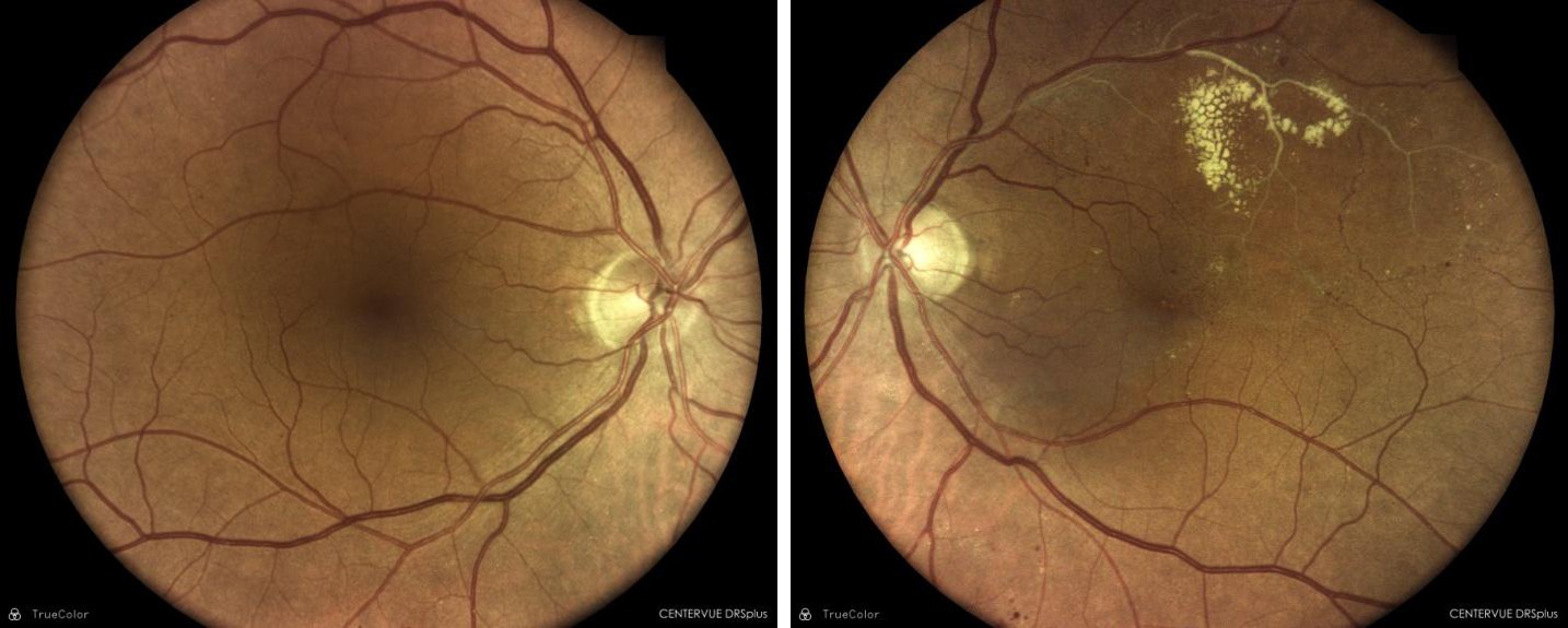
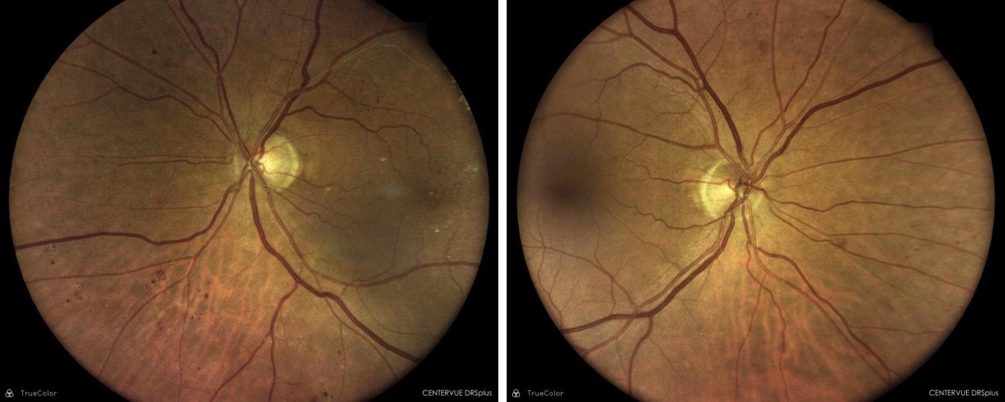
This is an observational study: participants were aged 18 years or older with diagnosis of diabetes mellitus type 1 or 2. They were enrolled for screening attending their annual visit. Main exclusion criteria were as follows: presence of myopia > 6D, presence of Age Macular Degeneration (AMD) both early (drusen) and late forms, presence of any other disorder of the macula. All patients provided written informed consent. The majority of retinal images were obtained with a true –color, confocal, fully automated non-mydriatic fundus imaging system (DRSplus ® Centervue Spa, a company of iCare Finland Oy; Vantaa, Finland) in a primary care setting (District of Moncalieri) without that participants underwent pupil dilatation. At Carmagnola Hospital, another district of the Territory of Asl To5, nurses obtained retinal images using Canon CR-2 Plus® a non-mydriatic camera, in the same way, without pupil dilation. Two photographic image field were taken of each eye, one centered on the optic disc and the other on the macula (Figure 1). Afterwards the images (the maximum capacity of the digital folder is 5 images for each visit) were uploaded to the Smart Digital Clinic through a corporate network. Dairet is able to recognize elementary lesions of diabetic retinopathy making a first diagnosis of presence or absence of complications.
Participants to study underwent ophthalmic examination to identify exclusion criteria. Diabetic Retinopathy of each patient was graded by an ophthalmologist and compared with the response of artificial intelligence. Human grader followed the ETDRS classification [1] (Table 1):
- R0 means absence of retinopathy.
- R1: Mild retinopathy with only microaneurysms,
- R2: Moderate retinopathy: microaneurysm, hemorrhages, hard exudates, cotton wools spots
- R3: Severe retinopathy: microaneurysm, hemorrhages, hard exudates, cotton wools spots, venous bending, IRMA (intraretinal microvascular abnormalities).
| Gender , female | 36 (42,4%) | |
|---|---|---|
| R0 | 52 | 8 (9,4%) |
| R1 | 12 | 77 (90,6%) |
| R2 | 20 | |
| R3 | 1 | |
Table 2: Classification of retinal images by manual grade (Ophthamologist).
R0 : No retinopathy
- R1 : Mild non proliferative retinopathy
- R2 : Moderate non proliferative retinopathy
- R3 : Severe non proliferative retinopathy Table 1: Classification of retinal images by manual grade (Ophthamologist).
Primary outcome measures included the evaluation of sensitivity in identifying mild and moderate retinopathy, in addition the evaluation of specificity and rate of FP in identifying patients without retinopathy.
Results
Eighty five patients were included in a sample calculation of sensitivity and specificity.
Mean (SD) participant age was 66,6 (11,5) years (range 38 – 92 years). Of 85 individuals, 8 (9,4%) had type 1 diabetes, 77 type 2 (90,6%). A total of 36 participants were women (42,4%) and 49 were men (57,6%). Complete demographic characteristics were reported in Table 2.
| AGE, years , mean +/- SD | 66,6 +/- 11,5 | |
|---|---|---|
| Gender, male | 49 (57,6%) | |
| Gender , female | 36 (42,4%) | |
| Diabetes type 1 | 8 (9,4 %) | |
| Diabetes type 2 | 77 (90,6%) |
Table 1: Demographic Characteristics.
Only one patient that underwent a fundus examination showed a severe retinopathy, that is evidence of both adequate metabolic control [2] and the efficacy of our integrated clinical pathway able to take care of patients since the first diagnosis of diabetes, thus preventing evolved forms of retinopathy.
A mild retinopathy was present in 12 patients (14,1% of overall sample), while a moderate retinopathy took place in 20 patients (23,5% of overall sample).
Dairet was able to detect all patients with moderate retinopathy (rate of sensitivity 100%), while recognized 11
out of 12 patients with mild retinopathy (rate of sensitivity 91,6%) (Table 3). Positive predictive value, indicating the percentage of patients with retinopathy among those with a positive AI result was 78%.
| Dairet Specificity (95% confidence interval) | FP | |
|---|---|---|
| R0 | 82,6 % (+/- 0,10) | 17,3 % |
Table 3: Sensitivity rate for dairet automated retinal image analysis systems compared with manual grade.
Specificity rate for dairet automated retinal image analysis systems compared with manual grade.
| Dairet Sensitivity (95% confidence interval) | FN | |
|---|---|---|
| R1 | 91,6 % | 8,4 % |
| R2 | 100 % | 0 % |
Table 4: Sensitivity rate for dairet automated retinal image analysis systems compared with manual grade.
In addition Dairet showed a false positive rate very low: 9 cases out of 52 graded R0, that means a rate of specificity of 82,6% (95% confidence interval: 72,6-92,6%). Dairet performance in this preliminary report seems to be better than previous data in Literature [3, 4, 5]. Negative predictive value, indicating the percentage of patients without retinopathy among those with a negative AI result was 97,7 %.
The rate of cases classified as ungradable by AI system was 15% for Canon Retinal Camera, according with Literature [6], while DRSplus confocal fundus camera didn’t have any case of ungradable image.
Discussion
In the Italian Healthcare System screening programs for detecting diabetic retinopathy are not widespread. Economical and organizational problems make difficult to implement these procedures, even though they have demonstrated efficacy in preventing diabetic-associated blindness and a very good ratio costs/effectiveness.
ASL To5, a Local Health District of Turin Metropolitan Area performed in 2019 an integrated care pathway dedicated to diabetic retinopathy with the aim of improving screening programs and taking care of patients during their clinical pathway.
Artificial Intelligence is an important tool to improve these procedures, reducing the diagnostic burden on eye care specialists and time costs for patients. Dairet achieved a high specificity in our sample: only few patients were referred accidentally to an ophthalmic examination. False positive rate is lower (17,3%) if compared with other Automated DR image assessment systems [5]. We observed that presence of foveal reflex in young patients often lead AI to a wrong evaluation. This could be a problem for young people screening.
Dairet showed an excellent sensitivity for referable retinopathy (moderate and severe forms). The true-positive ratio was 100%. A limitation of this study is a limited sample. The encouraging performances will have to be confirmed in the next report with a larger sample of diabetic population.
The very interesting performance of Dairet is possible thanks to the new generations of fundus cameras. Modern retinal cameras achieve images without pupil dilation, therefore patients can receive prompt and accurate detection of retinopathy in a primary care setting without ophthalmologist involvement. Particularly, DRSplus ®, a confocal fundus imaging system, fully automated, provides excellent and never ungradable true- color images. Nurses, minimally trained, can easily acquire retinal images and upload them to Smart Digital Clinic, a widespread digital folder used in Italy. In this way retinopathy screening can be a procedure totally managed by diabetologists.
Finally, Sensitivity rate for mild retinopathy, generally not much investigated in Literature, was 91,6% . In this early stage, patients are not referable to ophthalmologist, but their diabetological therapy can be reassessed in order to improve metabolic control in an effort to prevent or delay sight threatening retinopathy [2].
Conclusion
New Knowledge and technologies such as artificial intelligence offer important support in diabetic retinopathy screening. Among the main automated image assessment systems, Dairet demonstrated an accurate sensitivity in detecting both mild and moderate retinopathy. More over specificity was acceptable with a low false positive rate. This AI system can improve DR screening and monitoring in people with diabetes by non-eye care professionals. Nevertheless, future research is required to address several challenges of automated image detection algorithms: for example medico- legal implications or management of other ophthalmic disorders like age macular degeneration, detected in diabetic patients undergoing screening for retinopathy.
References
-
(1991) Grading diabetic retinopathy from stereoscopic color fundus photographs – an extension of the modified Airlie House Classification: ETDRS report number 10. Early Treatment Diabetic Retinopathy Study Research Group. Ophthalmol 98(5): 786-806.
-
(1987) Diabetes Control and Complications Trial (DCCT): results of feasibility study. The DCCT Research Group. Diabetes Care 10(1): 1-19.
-
Tufail A, Rudisill C, Egan C, Kapetanakis VV, Salas- Vega S, et al. (2017) Automated Diabetic Retinopathy Image Assessment Software: Diagnostic Accuracy and Cost-Effectiveness Compared with Human Graders. Ophthalmol 124(3): 343-351.
-
Vujosevic S, Aldington SJ, Silva P, Hernández C, Scanlon P, et al. (2020) Screening for diabetic retinopathy: new perspectives and challenges. Lancet Diabetes Endocrinol 8(4): 337-347.
-
Grzybowski A, Brona P, Lim G, Ruam Boonsuk P, Tan GSW, et al. (2020) Artificial Intelligence for diabetic retinopathy screening: a review. Eye (Lond) 34(3): 451- 460.
-
Ipp E, Liljenquist D, Bode B, Shah VN, Silverstein S, et al. (2021) Pivotal Evaluation of an Artificial Intelligence System for Autonomous Detection of Referrable and Vision-Threatening Diabetic Retinopathy. JAMA Netw Open 4(11): e2134254.
- Investigation of Polymorphisms in PPAR-Ɣ and TRHR Genes and their Impact on Turkish Diabetic and Obese Individuals
- The Impact of Aircraft Noise Exposure on the Efficacy of Empagliflozin Therapy in an Animal Model of Obesity
- Rooibos Mitigates Metabolic and Inflammatory Dysfunctions in Mice Fed a High-Carbohydrate Diet
- Synergistic Effect of Combined Leaf Extract of Vernonia amygdalina, Ocimum gratissimum, and Zingiber officinale Tuber on Phytochemical Profile, Antioxidant Activity, Serum Insulin, and Biochemical Parameters in Streptozotocin-Induced Diabetic Rats
- Investigation of Cardiovascular Responses to Aerobic Exercise in Obese University Students
- A Look at the Phase Angle Obtained by Electrical Bioimpedance