Orthotopic Liver Transplantation in a Patient with X-Linked Centronuclear Myopathy after Liver Hemorrhage due to Peliosis Hepatis
X-linked centronuclear myopathy (XLCNM) is a rare disease which sometimes is accompanied by extramuscular complications. We report a 15 month old boy with XLCNM and hepatic failure due to peliosis hepatis. Liver transplantation of a full size organ was performed successfully. This is the first report of liver transplantation in a patient with XLCNM and liver hemorrhage due to peliosis hepatis. Patients with XLCNM should undergo a close hepatological monitoring and corresponding treatment.
Introduction
X-linked centronuclear myopathy (XLCNM), also known as X-linked myotubular myopathy (XLMTM) is characterized by congenital mild to severe muscle weakness due to mutations in the myotubularin gene (MTM1; MIM 300415). Major histopathological finding is a high amount of small myofibres with centrally located nuclei [1]. Although the prognosis had long been considered desperate, almost two thirds of males survive nowadays infancy. Nevertheless the majority of patients require initial ventilatory support [2, 3]. Patients who survive the first year of life continue to have significant morbidity and early mortality. The cause of death is usually related to respiratory failure. Liver associated symptoms may be impaired liver function, gall stones, elevated liver enzymes and in single cases liver hemorrhage on the base of peliosis hepatis (PH) [4]. In the medical literature 8 patients with XLCNM and peliosis hepatis are described [4, 5, 6, 7, 8, 9]. The pathophysiological mechanisms of peliosis hepatis are unknown. The entity is characterized by blood filled cystic areas in the perisinusoidal space or in the sinusoids of the liver. In patients besides myotubular myopathy PH is in single cases associated with other chronical diseases like cystic fibrosis, malnutrition, fanconi anemia, tumors of the adrenal glands, Marfan syndrome, congenital heart defects, and after kidney transplantation [10]. Predominantly in adults an association with certain therapeutic drugs was observed [11], as well as infection- associated cases, especially with Bartonella henselae [12].
Case Report
A 15 month old boy with XLCNM was admitted to our hospital with abdominal pain, paleness and fatigue since the morning. He was the first child of his healthy parents. He was born at term by ceasarean section due to polyhydramnion and impaired intrauterine movement. Postnatally severe muscular hypotonia with secondary respiratory failure required ventilator support. In suspicion of a neuromuscular disease muscle biopsy was performed, followed by histopathological diagnosis of XLCNM. The clinical diagnosis was subsequently confirmed by sequence analysis of the MTM1 gene revealing a carrier status in the patient and his mother. After hospital discharge at 13 weeks of life he still required respiratory support by high flow device without additional oxygen; at daytime he was breathing spontaneously without any support. Past medical history since his initial hospital discharge included respiratory syncytial virus pneumonia, severe secondary developmental delay and bilateral maldescensus testis. Intermittent elevated transaminases and cholestasis parameters were recorded which almost normalized under a therapy with ursodeoxycholic acid. Three days prior to admission a routine check had been carried out showing normal linear growth (60th percentile, body weight < 3rd percentile, head circumference < 3rd percentile) and delayed motoric development. The cognitive stage was normal. Blood analyses were without pathological findings (wbc 6.8/nl; hemoglobine 13.5 g/dl, direct bilirubine 0,14 mg/dl, ALT 33 U/l, AST 41 U/l, GGT 20 U/l). One day before admission measles, mumps, and rubella vaccination had been carried out. On admission, the patient was in a reduced stage, pale, tachycardic with epigastric abdominal pain. Blood analysis showed wbc 6,5/nl, hemoglobine 4,8 g/dl; platelets 5/nl, total bilirubine 0.53 mg/dl, direct bilirubine 0.44 mg/dl, ALT 1390 U/l; AST 1199 U/l, GGT 20 U/l. Erythrocytes and platelets were substituted immediately. Ultrasonography showed no free fluid in the abdomen.
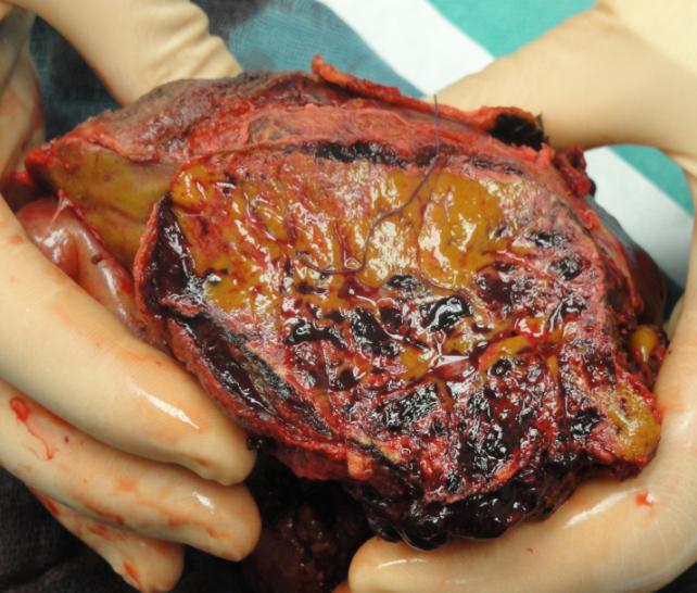
A few hours later, a rapid decrease of blood pressure, bradycardia and decrease of oxygenation occurred. The patient could be stabilized under cardio-pulmonary resuscitation with fluid therapy and katecholamins. Now, abdominal ultrasound showed extensive abdominal fluid most likely severe intraabdominal hemorrhage. After CT scan laparotomy was performed which showed a massively enlarged liver with hemorrhagic parenchyma, necrotic areas and subcapsular hematoma which had lead to rupture of the liver capsule, as well as blood in the abdomen. Histological examination of resected liver tissue afterwards led to the diagnosis of peliosis hepatis. The bleeding could be stopped and abdomen was packed and closed with a patch. After four days a second look- operation was performed. The bleeding could be stopped, but massive liver damage led to liver failure and necessity for liver transplantation as the only curative therapy. After stabilization six days after admission liver transplantation was performed (full size organ of a 15 month old donor). The explanted organ showed hematoma and extended necroses (Figure 1), histology showed extended hemorrhages into the parenchyma, surrounded by parenchyma necroses, as well as hemocongestion. In the environment of the hemorrhage there were hepatocytes with multiple nuclei and plurivascular steatotic liver epithelia with a degree of steatosis of 20-30%. On posttransplant days 6, 9 and, 13 the abdominal wall was closed stepwise. The situation was complicated by hemostasis problems, an early rejection with rising transaminases and cholestasis parameters from day 7 after transplantation, and a thrombus 2 cm in diameter in the right heart ventricle, first detected at day 7 after transplantation.
After the acute bleeding event on the day of admission until liver transplantation the patient was treated with heparin, first because of dialysis, afterwards to prevent thrombosis after transplantation. Until transplantation he received 12 PRBC, 17 platelet concentrates, once tranexamic acid, 3 factor seven concentrates and 1 fibrinogen concentrate. After liver transplantation the patient had a sufficient spontaneous diuresis. The need for platelet and erythrocyte transfusion decreased after transplantation, 5 days after transplantation substitution of blood components was no longer necessary. Due to the thrombus in the right ventricle on day 7 after transplantation we switched from unfractionated to low molecular weight heparin, anticoagulation was controlled by measuring of factor anti-Xa. Ten days after transplantation acute rejection was treated by high dose steroids for 6 days. In the course of treatment persistent elevated transaminases and cholestasis parameters were noticed. Biopsy showed signs of ischemia. As there were no signs of poor perfusion during the operation and in sonographic controls, we discussed a possible large for size “constellation after full size transplantation. In the further course cholestasis parameters decreased gradually, organ function was normal at any time. After post-transplant treatment was completed, the patient was transferred to a neuropediatric rehabilitation unit. He still needed respiratory support for several hours a day. Neurological situation improved slightly. After 5 months discharge with domestic nursing service is planned.
Discussion
This is the first report about liver transplantation in a patient with XLCNM after liver hemorrage and consecutive liver failure due to peliosis hepatis. In the literature 8 cases with liver hemorrhage in patients with XLCNM are described. In 6 cases the patients died, in one patient the bleeding could be stopped by embolization of the hepatic artery, the patient survived with sufficient liver function [8]. A further patient survived after surgery and consecutive conservative therapy [5]. However, in both patients it remains unclear, if hepatic bleeding re- occurred in the later course or not. Clinical manifestations of peliosis hepatis are variable. There are asymptomatic cases, detected only by ultrasonography routine checks, as well as lethal courses with bleeding and liver rupture. The clinical course is variable as well. Drug-induced cases can improve after withdrawal of medication; infection-related courses can be cured by intensive antibiotic therapy. The etiopathogenesis of peliosis hepatis in patients with XLCNM is poorly understood. The defect MTM1 protein in XLCNM is a main regulator of the desmin cytoskeleton as well as mitochondrial homeostasis, specifically in skeletal muscles [13]. Interestingly desmin is also expressed in the hepatic stellate cells within the liver sinusoids suggesting dysregulation of the desmin in liver cell cytoskeleton as a possible cause for hepatic damages in XLCNM patients. In support of this desmin- positive cells were detected only in the endothel of liver vessels, but not in the sinusoids of PH-affected liver of the patient with XLCNM which could elucidate the correlation between XLCNM and PH [9]. To the best of our knowledge liver transplantation in peliosis hepatis-caused liver failure has been reported three times in adults so far [14, 15, 16]. In two cases and association to oral contraceptives was suspected, in the third case a clear trigger for the liver failure could not be found. In those cases liver transplantation was performed months to years after diagnosis of hepatopathy. To our knowledge there is no case of liver transplantation in children with peliosis hepatis.
Conclusion
This is the first case of successful liver transplantation in a patient with XLCNM and hepatic failure after liver hemorrhage. Patients with XLCNM should undergo a close hepatological monitoring and corresponding treatment.
References
-
Spiro AJ, Shy GM, Gonatas NK (1966) Myotubular myopathy. Persistence of fetal muscle in an adolescent boy. Arch Neurol 14(1): 1-14.
-
McEntagart M, Parsons G, Buj-Bello A, Biancalana V, Fenton I, et al. (2002) Genotype-phenotype correlations in X-linked myotubular myopathy. Neuromuscul Disord 12(10): 939-946.
-
Jungbluth H, Wallgren-Pettersson C, Laporte J (2008) Centronuclear (myotubular) myopathy. Orphanet J Rare Dis 3: 26.
-
Herman GE, Finegold M, Zhao W, de Gouyon B, Metzenberg A (1999) Medical complications in long- term survivors with X-linked myotubular myopathy. J Pediatr 134(2): 206-214.
-
Wang SY, Ruggles S, Vade A, Newman BM, Borge, MA (2001) Hepatic Rupture Caused by Peliosis Hepatis. J Ped Surg 36(9): 1456-1459.
-
Karger B, Varchmin-Schultheiß, K, Fechner G (2005) Fatal hepatic haemorrhage in a child—peliosis hepatis versus maltreatment. Int J Legal Med 119: 44- 46.
-
Motoki T, Fukuda M, Nakano T, Matsukage S, Fukui A, et al. (2013) Fatal hepatic hemorrhage by peliosis hepatis in X-linked myotubular myopathy: A case report. Neuromuscul Disord 23(11): 917-921.
-
Terlizzi JP, Azizi R, Chow MD, Underberg-Davis S, Nosher JL, et al. (2013) Peliosis hepatis in a child with myotubular myopathy: successful treatment using hepatic artery embolization. J Pediatr Surg; 48(8): e9- e12.
-
Hagiwara S, Kubota M, Sakaguchi K, Hiwatari E, Kishimoto H, et al. (2015) Fatal Hepatic Hemorrhage from Peliosis Hepatis with X-linked Myotubular Myopathy. J Pediatr Gastroenterol Nutr; 60(5): e45- e46.
-
Samyn M, Hadzic N, Davenport M, Verma A, Karani J, et al. (2004) Peliosis Hepatis in Childhood: Case Report and Review of the Literature. J Pediatr Gastroenterol Nutr 39: 431-434.
-
Cavalcanti R, Pol S, Carnot F, Campos H, Degott C, et al. (1994) Impact and evolution of peliosis hepatis in renal transplant recipients. Transplantation 58(3): 315-316.
-
Ahsan N, Holman MJ, Riley TR, Abendroth CS, Langhoff EG, et al. (1998) Peliosis hepatis due to Bartonella henselae in transplantation: a hemato- hepato-renal syndrome. Transplantation 65(7): 1000- 1003.
-
Hnia K, Tronchère H, Tomczak KK, Amoasii L, Schultz P, et al. (2011) Myotubularin controls desmin intermediate filament architecture and mitochondrial dynamics in human and mouse skeletal muscle. J Clin Invest 121(1): 70-85.
-
van Erpecum KJ, Janssens AR, Kreuning J, Ruiter DJ, Kroon HM, et al. (1988) Generalized peliosis hepatis and cirrhosis after long-term use of oral contraceptives. Am J Gastroenterol 83(5): 572-575.
-
Muradali D, Wilson SR, Wanless IR, Greig PD, Cattral M, et al. (1996) Peliosis hepatis with intrahepatic calcifications. J Ultrasound Med 15(3): 257-260.
-
Hyodo M, Mogensen AM, Larsen PN, Wettergren A, Rasmussen A, et al. (2004) Idiopathic extensive peliosis hepatis treated with liver transplantation J Hepatobiliary Pancreat Surg 11(5): 371-374.
- Management of Gallbladder Perforations: A Review
- From The Mouth to the Gut: The Oral Microbiome's Role in Promoting Gastrointestinal Disease
- Case Report: Intraductal Papillary Mucinous Neoplasm (IPMN) Complicated by Portal Vein Plaquing and Biliary Obstruction Mimicking Pancreatic Metastatic Malignancy
- Management of Non-Cirrhotic Portal Hypertension during Pregnancy: A Review
- Effectiveness of Omeprazole versus Pantoprazole for Symptomatic Relief of Gastro-Esophageal Reflux Disease (GERD)/ Acid Peptic Disease (APD): A Real-World Evidence (RWE) Study
- Case of Splenic Infarction; A Rare Presentation of Complicated Enteric Fever in a Pediatric Patient